




Abstract
Purpose:
Coronavirus disease 2019 (COVID-19) caused by the severe acute respiratory syndrome coronavirus 2 (SARS-CoV-2) and is an acute respiratory illness. Although most infected persons are asymptomatic or have only mild symptoms, some patients progress to devastating disease; such progression is difficult to predict or identify in a timely manner. COVID-19 patients who do not require hospitalization can self-isolate at home. Calls from one disease epicenter identify the need for homebased isolation with telemedicine surveillance to monitor for impending deterioration.
Methodology:
Although the dominant approach for these asymptomatic/paucisymptomatic patients is to monitor oxygen saturation, we suggest additionally considering the potential merits and utility of home-based imaging. Chest computed tomography is clearly impractical, but ultrasound has shown comparable sensitivity for lung involvement, with major advantages of short and simple procedures, low cost, and excellent repeatability. Thoracic ultrasound may thus allow remotely identifying the development of pneumonitis at an early stage of illness and potentially averting the risk of insidious deterioration to severe pneumonia and critical illness while in home isolation.
Conclusions:
Lung sonography can be easily performed by motivated nonmedical caregivers when directed and supervised in real time by experts. Remote mentors could thus efficiently monitor, counsel, and triage multiple home-based patients from their “control center.” Authors believe that this approach deserves further attention and study to reduce delays and failures in timely hospitalization of home-isolated patients.
Purpose
Coronavirus 2019 (COVID-19) pneumonia caused by the novel coronavirus severe acute respiratory syndrome coronavirus 2 (SARS-CoV-2) has been rapidly spreading around the globe, challenging or even disrupting health care systems and human resources supporting those systems. 1 Given the rapid spread of COVID-19 caused by the SARS-CoV-2 virus to date, a crisis similar to Northern Italy's and New York's may evolve in many more locales. Sensible discrimination between patients needing hospitalization and those amenable to self-isolation allows using the limited hospital resources most effectively. However, many initially asymptomatic/paucisymptomatic patients progress to severe or critical disease. Embracing suggestions to develop telemedical adjuncts to home monitoring of patient care, 2,3 we would like to open a discourse and stimulate research interest in using simple and affordable telementored imaging procedures in monitoring of isolated patients. In addition, enhancing remotely managed in-home monitoring may reduce the number of medical providers affected. This “outside-the-box” approach might reduce the risk of delaying appropriate care of patients with insidious deterioration while in-home isolation, at the same time optimizing the use of health care resources and improving provider safety.
The TeleMedical Ultrasound Supported Medical Interventions (TMUSMI) Research Group at the University of Calgary has long sought to empower less experienced point-of-care providers, to perform beyond their expected scope with the remote assistance of experts communicating real time, with simple but powerful informatic technologies. 4 –6 This paradigm has been an iterative evolution of technology assessments that began with the need to provide care to astronauts in low earth orbit, demonstrating how motivated nonexperts guided by remote content experts can accurately assess a multitude of health care scenarios. 7,8 The International Space Station has relied on ultrasound capability since its early days without having trained ultrasonographers onboard, and will continue relying on it as the only medical imaging capability available on spacecraft. This paradigm requires the untrained subjects to perform ultrasonography on themselves or their self-isolated crewmate while being guided through every step in image acquisition by a remote ultrasound expert. This expert is responsible for determining the quality of the images generated and, if sufficient, to interpret those images. 7,9,10 Recognizing the potential value of being able to remotely guide novices to obtain meaningful ultrasound diagnoses in geographically distant locations, the TMUSMI group has since evaluated this remote guidance technique using readily available off-the-shelf informatics. 4,11 –13 .
Methodology
Home Isolation, Outreach Criteria, and Early Medical Rescue
Part of the societal problem with our response to COVID-19 has been the perception that it is an “old persons” or “sick persons” disease. This was partially vindicated by early reports out of Wuhan, China, that noted that factors associated with progression to severe COVID-19 pneumonia were advanced age, smoking history, low albumin, and male gender, with smoking having an odd's ratio of 14 for severe disease progression. 14 However, it is readily becoming apparent that this is not the case, and many of those requiring prolonged critical care are young and otherwise healthy. Statistically though, young healthy people with previously healthy lungs are still at low risk for progression to severe life-threatening disease and most will experience only mild symptoms and recover by self-isolating at home. 15 Nacoti et al., writing during the midst of their catastrophe in Lombardy, stated that the key to avoid tragedies elsewhere and in the future would be a massive deployment of outreach resources to focus on community-centered care rather than relying on hospital-based care. 2 We recognize, however, that such an approach will require being able to recognize those isolated patients who are at risk of deterioration, and “rescuing” or “safety netting” them quickly with an aggressive and comprehensive response. 15
Lung Ultrasound as a Potential Remote Diagnostic Tool Protecting Health Care Providers
A practical question regarding recommending home isolation of patients, however, is how to identify patients before clinical deterioration? Early chest computed tomography (CT) has been suggested as a screening tool for suspected COVID-19 patients with better sensitivity than polymerase chain reaction. 16 Chest CT is clearly impractical for home-isolated patients, notwithstanding the logistics there would be unacceptable risks associated with transporting and exposing such patients to all those involved in actually performing a standard CT scan. We thus propose that simple off-the-shelf telemedical self-assessment technology might be supplied to self-isolated and/or quarantined individuals with normal lungs who are at risk for progressive clinical decline to severe acute respiratory distress and death. Owing to the fact that COVID-19 typically presents as a diffuse interstitial bilateral pneumonia mainly in the lung periphery, 17,18 lung ultrasound examinations, such as the extended focused assessment with sonography for trauma, 19 may be able to quickly and remotely identify those at risk of clinical deterioration to severe pneumonia at an early stage of illness. 20 –23 Lung ultrasound may be positive even before polymerase chain reaction testing. 24 Lung ultrasound, by imaging between the ribs and focusing on the pleural interface, has shown comparable results with chest CT with markedly reduced logistical challenges, 20 sparing the time, effort, and potential exposure of medical providers. Realistically, regardless of any degree of personal protective equipment utilized, the safest medical interaction from a public health standpoint is one that is completely remote, with no physical contact.
Sonographically, disease progression from normal lungs to a simple alveolar-interstitial pattern can be seen by the appearance of single or confluent vertical artifacts (B-lines) or confluent vertical artifacts. 23 This is an ultrasound syndrome believed to correlate with thickening of the subpleural interlobular septa and increased extravascular lung water producing the comet tail reverberation artifact. 25 This may be chronic or acute, involving conditions such as infiltrative lung diseases, pulmonary edema, acute respiratory distress syndrome, or interstitial pneumonia. 26 Often sonographic findings are apparent before those seen on plain radiography. 27 Previous study has demonstrated that with remote guidance, nonexpert point of care providers can be guided to place an inexpensive ultrasound probe onto the chest to assess the visceral–parietal pleural surface ( Fig. 1, Supplementary Video S1). 28 –31 We thus suggest that assessment of this pleural surface may aid in the early diagnosis of COVID-19. If this is proven to be true by continued research, mentored lung examinations could be performed by the patient themselves analogous to an astronaut being guided to perform a self-examination, or by a family member who is isolated with them. Current recommendations for suspected COVID-19 patients are to use small probes such as wireless probes that are more easily sterilized or protected. 32 With simple off-the-shelf informatics, we also propose that the mentoring expert (or panel of experts) could be anywhere in the world, 4,12,33 assessing patients as needed, even if they were themselves isolated or in quarantine. Presumably, expert reassurance from a human voice would be reassuring to naturally anxious individuals, recognizing that the healthy but panicked survivors are typically the first wave to potentially overwhelm medical resources in any disaster scenario.
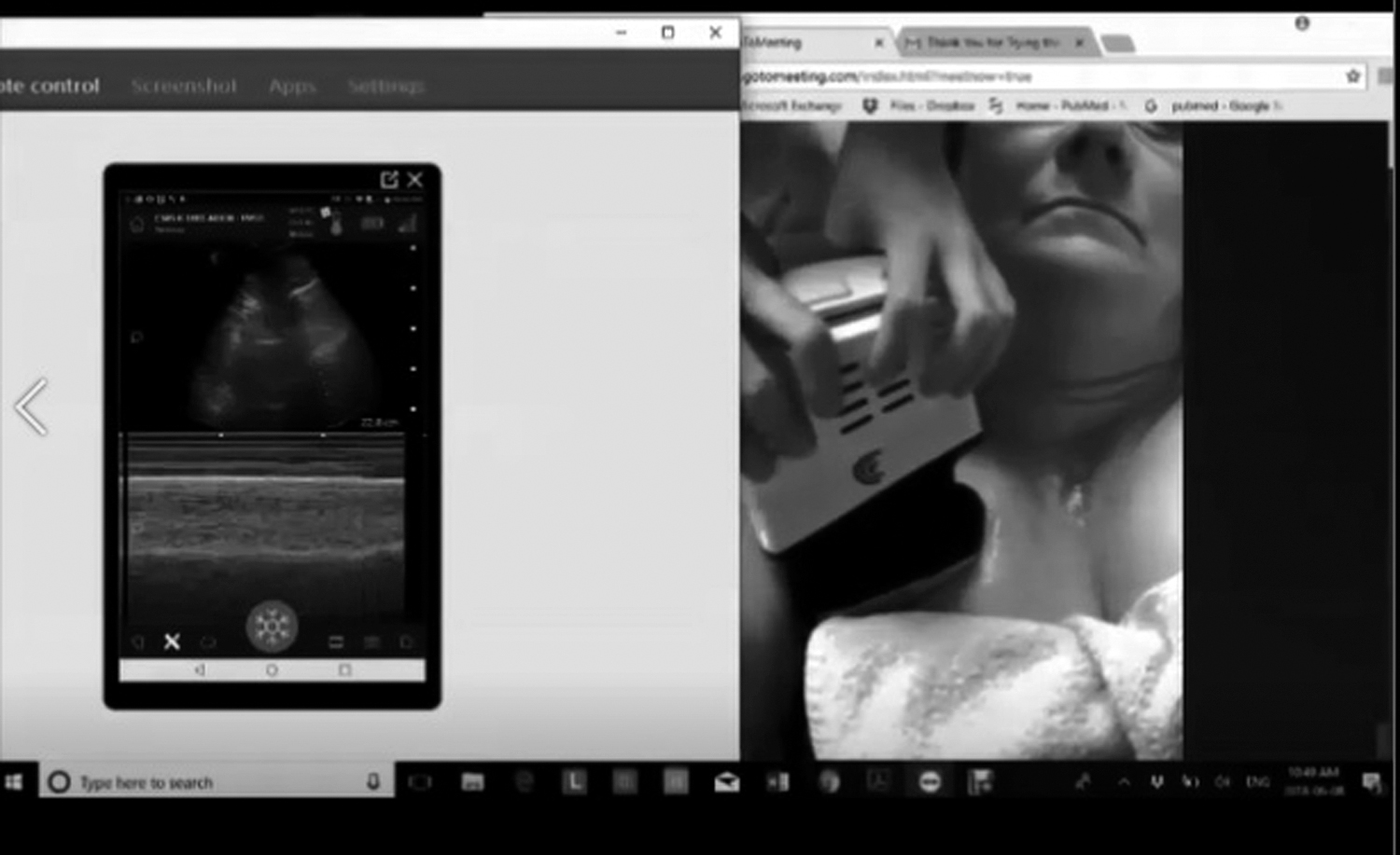
Still image of EFAST pleural interface depiction provided by a 7-year-old child examining his grandmother under real-time remote telementored guidance. EFAST, Extended Focused Assessment with Sonography for Trauma.
Conclusions
We propose that telementored ultrasound, for assessing self-isolated patients with suspected or possible COVID-19 pneumonia, at risk of deterioration, is feasible and should be urgently studied to more fully assess its practicality, accuracy, and potential utility. We suggest that such a paradigm might involve serial remote sonographic assessment of the lung, along with peripheral oxygen saturation monitoring. Lung sonographic assessment does not require complex ultrasound machines and we believe it can be done with small economical hand-held devices. Such probes could either be left at a home, or with appropriate sterilization and infection control procedures circulated to multiple households without direct human-to-human contact, potentially even by drone delivery.
Adopting such a paradigm of remote but vigilant telemedical assessment, and early diagnoses of severe cases, could be performed, while minimizing and preserving the health of expert caregivers by minimizing their exposure.
Declarations
This study was conducted as part of the ongoing project: The Marriage of Hyper-Realistic Surgical Training for First Responders with Telepresence Mentoring. The people illustrated in the figure and Supplementary Video S1, are the mother and child of the second author. Written consent for publication of their images was provided for this article. The data sets used and/or analyzed during this study are available from the corresponding author on reasonable request.
Footnotes
Authors' Contributions
A.W.K. and J.L.M. obtained the clinical images and performed the initial literature review and drafted the commentary. All authors reviewed the data, literature, and read and approved the final article.
Acknowledgments
The authors acknowledge the support of Brenda Southwick and Gavan McKee for donating their time and cooperation.
Disclosure Statement
The authors have no competing interests related to this commentary. Potential competing interests outside of this study include that A.W.K. has consulted for the Innovative Trauma Care and SAM Corporations, and is the PI of a randomized controlled trial partially supported with unrestricted funding from the Acelity Corporation (
Funding Information
No funding was provided for this study, although J.L.M. position is partially funded by a New Earth Space Technologies (NEST) grant from the University of Calgary and I.W.Y.M's work is supported by the John A. Buchanan Chair of General Internal Medicine, University of Calgary.
Supplementary Material
Supplementary Video S1
References
Supplementary Material
Please find the following supplemental material available below.
For Open Access articles published under a Creative Commons License, all supplemental material carries the same license as the article it is associated with.
For non-Open Access articles published, all supplemental material carries a non-exclusive license, and permission requests for re-use of supplemental material or any part of supplemental material shall be sent directly to the copyright owner as specified in the copyright notice associated with the article.
