




Abstract
DNA samples from four rickettsial species in Japan—R. japonica Aoki, R. asiatica IO-1, R. helvetica IP-1, and Rickettsia tamurae AT-1—were analyzed by polymerase chain reaction–restriction fragment length polymorphism (PCR-RFLP) in which a portion of the sca4 gene was amplified and restriction digested with the MspI enzyme. Each spotted fever group rickettsial species exhibited unique restriction profiles after MspI digestion. To examine the possibility of applying PCR-RFLP analysis to field samples, 36 Ixodes persulcatus ticks were collected from the Tokachi region (Hokkaido, Japan) by the flagging method and subjected to PCR-RFLP analysis. Of the ticks collected, five had identical restriction profiles to that of R. helvetica. Amplification and sequencing of the gltA gene for two of the five positive samples revealed that the sequences were identical to that of the Rickettsia sp. Tokachi-B-IP17m, which exhibits the highest similarity to R. helvetica. These results demonstrate that PCR-RFLP analysis is useful for identifying Rickettsia species in Japan and is applicable to epidemiological studies involving tick samples.
Introduction
Comparative sequence analysis of genes with validated species has been used to identify Rickettsia species; however, gene sequencing is not time and cost efficient. Species-specific polymerase chain reaction (PCR) to identify Rickettsia species has limitations, including the requirement for multiple PCR reactions, as well as the inability to detect SFG rickettsiae other than the target species. An alternative approach, PCR–restriction fragment length polymorphism (PCR-RFLP) analysis, allows the detection of SFG rickettsiae, including new species, by PCR and further distinguishes known SFG rickettsial species by their characteristic restriction profiles. The PCR-RFLP on rickettsial gltA and ompA genes using three enzymes was reported (Gage et al. 1994); however, the PCR-RFLP on gltA cannot differentiate R. asiatica from R. helvetica. The objectives of the present study were to use PCR-RFLP analysis to detect and identify Rickettsia species in tick samples from Japan.
Materials and Methods
Rickettsia species and DNA extraction
R. asiatica IO-1, R. helvetica IP-1, R. japonica Aoki, and R. tamurae AT-1 were supplied by Dr. Hiromi Fujita (Ohara Hospital, Fukushima, Japan). DNA was extracted from each rickettsial culture using the QIAamp DNA Mini Kit (Qiagen, Hilden, Germany) by following the manufacturer's protocol.
PCR-RFLP analysis
DNA samples from each rickettsial species were subjected to PCR using the primers Rj1707f (5′-CTC-TGA-ATT-AAG-CAA-TGC-GG-3′) and Rj2837r (5′-CCT-GAT-ACT-ACC-CTT-ACA-TC-3′) with annealing at 50°C, which was designed to amplify approximately 1140 bp fragment of the sca4 genes of SFG rickettsiae based on that of R. japonica (GenBank accession number AF155055). The PCR products were purified using the QIAquick PCR Purification Kit (Qiagen) by following the manufacturer's protocol, and observed on a 1% agarose gel by electrophoresis by ethidium bromide staining with the Quick-Load 100 bp DNA Ladder (New England Biolabs, Ipswich, MA). The signal intensity of the samples was compared with those of the ladder to estimate the DNA concentration. Appropriate volumes (5–17.3 μL) of purified amplicon were digested by MspI (Promega, Madison, WI) by following the manufacture's instruction, and observed on a 2% agarose gel as described above.
Sequencing analysis of the sca4 gene
The purified PCR products were also sent to Akita Prefectural University for sequencing (DNA sequencing; Sigma Aldrich Japan, Tokyo, Japan) using primers Rj1707f, Rj2837r, Rj2126r (5′-GCT-GAT-GCC-ATA-ATA-AGT-GC-3′), Rj2316f (5′-TGA-GGC-AGC-TTT-AGA-TAG-AG-3′), and Rj2335r (5′-CTC-TAT-CTA-AAG-CTG-CCT-CA-3′). The obtained sequences of R. tamurae, R. helvetica, R. asiatica, and R. japonica were deposited to GenBank with accession numbers FJ358503, FJ358501, FJ358500, and FJ358502, respectively.
PCR-RFLP analysis of tick samples
Ticks were collected by flagging in Tokachi region of Hokkaido, Japan (42°44′N–43°10′N and 143°00′E–143°20′E), and species and stages of ticks were recorded. DNA was extracted from individual ticks, and PCR-RFLP analysis was performed as described above. The positive samples were also analyzed by sequencing 480 bp of the rickettsial gltA gene (Roux et al. 1997). DNA from R. asiatica that has not been reported in Hokkaido was used as a positive control for the PCR and MspI treatment, and water template as a negative control.
Results
PCR-RFLP analysis on samples from rickettsial culture
PCR using the primers Rj1707f and Rj2837r on the extracted DNA from each rickettsial species resulted in single-amplification products that were purified, digested with MspI, and observed on a 2% agarose gel. PCR products from each rickettsial species exhibited unique restriction profiles (Fig. 1).

PCR-RFLP analysis of four SFG rickettsial species. Shown are restriction profiles of four SFG rickettsial species. Purified PCR products from each rickettsial species were treated with MspI and observed on a 2% agarose gel stained with ethidium bromide. Each SFG rickettsial species exhibited a unique restriction profile. The lengths of fragments were 148, 246, and 742 bp in Rickettsia tamurae; 81, 166, and 871 bp in Rickettsia asiatica; 81, 148, 166, and 724 bp in Rickettsia Helvetica; and 81, 166, 364, and 523 bp in Rickettsia japonica. M, marker; Rt, R. tamurae; Ra, R. asiatica; Rh, R. helvetica; Rj, R. japonica.
Sequencing analysis of the sca4 gene of each Rickettsia species
The size of sca4-PCR products of R. tamurae, R. asiatica, R. helvetica, and R. japonica were 1134, 1116, 1116, and 1131 bp, respectively. The length of fragments obtained in the PCR-RFLP was consistent with the result shown in Figure 1.
PCR-RFLP on tick samples
Thirty-six Ixodes persulcatus (24 females, 10 males, and 2 nymphs) were collected. Five female I. persulcatus were sca4 positive by PCR. PCR-RFLP analysis demonstrated that the restriction profiles of the five tick samples were identical to that of R. helvetica (Fig. 2). Sequencing of the gltA gene from two of the five PCR products suggested that they were identical and a perfect match for the “Uncultured Rickettsia sp. clone Tokachi-B-IP17m (DQ887275),” which exhibits the highest identity to R. helvetica (Inokuma et al. 2007).
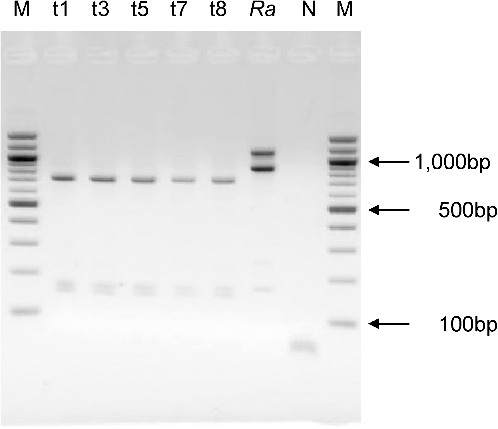
PCR-RFLP analysis of tick samples. Five DNA samples from sca4-positive ticks (t1, t3, t5, t7, and t8) have identical restriction profiles to that of Rickettsia helvetica shown in Figure 1. In the Ra lane, a part of original amplicon of the sca4 gene of Rickettsia asiatica (1116 bp) remained undigested and was observed on the gel. M, marker; Ra, R. asiatica (positive control); N, negative control.
Discussion
In this study, we demonstrate that the restriction profiles of R. japonica, R. helvetica, R. asiatica, and R. tamurae obtained by MspI digestion are unique, suggesting that PCR-RFLP is a useful method to identify Rickettsia species.
To test the potential application of PCR-RFLP for epidemiological studies, field tick samples were analyzed to identify the infecting Rickettsia species. The restriction profiles of the five sca4-positive ticks were identical to that of R. helvetica. Further, sequencing a portion of the gltA gene revealed that the SFG rickettsial species detected in tick samples were similar to R. helvetica, demonstrating that PCR-RFLP can be applied to tick samples for Rickettsia species identification. Three of the four SFG rickettsial species in Japan were isolated and/or detected in multiple tick species, including Ixodes ovatus, I. persulcatus, and Ixodes monospinosus (Fournier et al. 2002). Recently, several Rickettsia species that were first isolated from ticks and thought to be nonpathogenic to humans have been shown to be pathogenic (Parola et al. 2005). The methods described in our study can help identify the distribution of known SFG rickettsial species infecting ticks in Japan.
In conclusion, the results of our study suggest that PCR-RFLP can be used for the identification of SFG rickettsiae from patients and epidemiological studies involving tick samples.
Footnotes
Acknowledgments
This work was supported by the Grant H18-Shinkou-Ippan-014 for Research on Emerging and Re-emerging Infectious Diseases from the Japanese Ministry of Health, Labor and Welfare; Grants-in-Aid for Scientific Research from the Japan Society for the Promotion of Science (No. 18380185); and a Grant-in-Aid from the Zoonoses Control Project of the Ministry of Agriculture, Forestry and Fisheries of Japan.
Disclosure Statement
No competing financial interests exist.