
Abstract
We evaluated the serological and molecular prevalence of selected organisms in 145 dogs during late spring (May/June) of 2005 and in 88 dogs during winter (February) of 2007 from the Hopi Indian reservation. Additionally, in 2005, 442 ticks attached to dogs were collected and identified as Rhipicephalus sanguineus. Infection with or exposure to at least one organism was detected in 69% and 66% of the dogs in May/June 2005 and February 2007, respectively. Exposure to spotted fever group (SFG) rickettsiae was detected in 66.4% (2005) and 53.4% (2007) of dogs, but rickettsial DNA was not detected using polymerase chain reaction. Active Ehrlichia canis infection (by polymerase chain reaction) was identified in 36.6% (2005) and 36.3% (2007) of the dogs. E. canis infection was associated with SFG rickettsiae seroreactivity (p < 0.001). Anaplasma platys DNA was detected in 8.3% (2005) and 4.5% (2007) of the dogs. Babesia canis and Bartonella vinsonii berkhoffii seroprevalences were 6.7% and 1% in 2005, whereas in 2007 prevalences were 0% and 1.1%, respectively. No Bartonella spp., Ehrlichia chaffeensis, or Ehrlichia ewingii DNA was detected. Dogs on this Hopi Indian reservation were most frequently infected with E. canis or A. platys; however, more than half of the dogs were exposed to a SFG-Rickettsia species.
Introduction
In 2004, a human outbreak of Rocky Mountain spotted fever (RMSF) was reported in the White Mountain region of eastern Arizona (Demma et al. 2005). During this outbreak investigation, Rhipicephalus sanguineus was implicated for the first time as a vector for Ri. rickettsii in North America, and the role of local dogs as short-term reservoirs and primary hosts for the vector tick was suggested (Demma et al. 2005, Nicholson et al. 2006). This tick species is already a known vector for Babesia spp., Ehrlichia spp., and potentially Anaplasma platys and Bartonella spp. in North America (Simpson et al. 1991, Higuchi et al. 1995, Mathew et al. 1996, Wikswo et al. 2007). In light of the 2004 discovery, frequent Rh. sanguineus infestations of dogs on a Hopi Indian Reservation located in the northeastern region of Arizona raised concerns about the presence of vector-transmitted diseases in that area.
The present study used a variety of assays to evaluate the frequency of infection with and/or exposure to Anaplasma phagocytophilum, A. platys, Babesia canis, Bartonella spp., Borrelia burgdorferi, Dirofilaria immitis, Ehrlichia canis, Ehrlichia chaffeensis, Ehrlichia ewingii, and spotted fever group (SFG) rickettsiae in dogs located on the Hopi Indian Reservation, in northeastern Arizona, and indirectly to evaluate the human exposure to these organisms.
Material and Methods
Study location
This study was conducted at the Hopi Reservation, located at the northern half of the State of Arizona (Fig. 1). The Hopi land covers 6557.262 km2 (2531.773 sq. miles) and is surrounded entirely by a Navajo Reservation. Elevation ranges from approximately 1600–2000 m, and the topography is characterized by a series of broad mesas (or plateaus) separated by shallow canyons. Blood samples were collected during May/June 2005 and in February 2007 from the following villages: Moenkopi (36°6′N 111°13′W), Hotevilla-Bacavi (35°55′N 110°40′W), Oraibi (35°52′N 110°38′W) and Kykotsmovi (35°52′N 110°37′W), Mishongnovi (35°48′N 110°29′W), First Mesa (35°50′N 110°22′W), Shungopavi (35°47′N 110°30′W), and Keams Canyon (35°48′N 110°11′W). Additionally, two samples were collected from Tuba city (36°8′N 111°14′W) on the Navajo Reservation, located approximately 1.2 miles west of the Hopi Reservation. The average monthly temperatures during May/June 2005 were 67.97°F and 77.67°F, with average maximum monthly temperatures of 86.13°F and 96.04°F and average minimum monthly temperatures of 49.81°F and 59.12°F, respectively. During February 2007, average monthly temperature was 42.96°F (average monthly range 56.14°F–29.79°F). The region is semi-arid, with average monthly precipitation of 0.0″ and 0.1″ during May/June 2006 and 0.22″ during February 2007 (source:
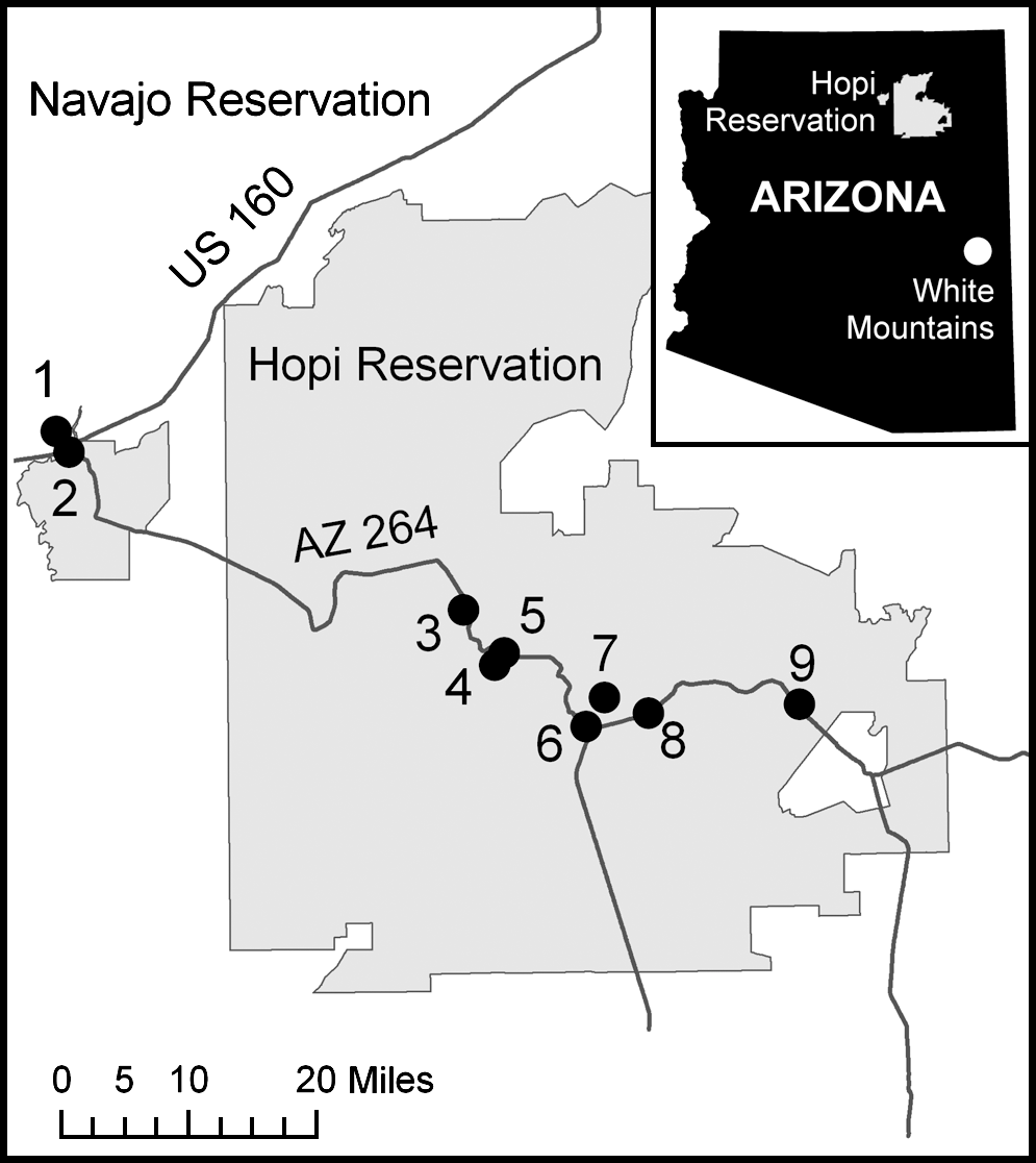
Locations of the villages from where dog samples were collected for this study. 1, Tuba City; 2, Moenkopi; 3, Hotevilla-Bacavi; 4, Kykotsmovi; 5, Oraibi; 6, Shungopavi; 7, Mishongnovi; 8, First Mesa; 9, Keams Canyon. Figure insert: locations of the Hopi Indian Reservation (light gray) and the White Mountains Park (white) in the Arizona state map. ArcGIS 9.2 (Environmental Systems Research Institute, Redlands, CA). Geographic data from December 2005 database.
Study population and data availability
A total of 233 pet and stray dogs were sampled, without specific inclusion or exclusion criteria. In May/June 2005 and in February 2007, respectively, 145 and 88 dogs were sampled. Dogs previously enrolled in 2005 were not sampled in 2007. Age was known for 68.2% (159/233) of the dogs sampled. Specific age was available for 103, whereas 56 dogs were described as older than 1 year. Adult dogs represented 86.8% (138/159) of the population, whereas dogs under 12 months of age represented 14.5% (23/159). The median age for both years was 36 months (25% quartile: 12 months; 75% quartile: 60 months; range: 3–204 months). Median age was not significantly different from 2005 to 2007 (p = 0.896, Kruskal-Wallis test). Sex information was available from 68.7% (160/233) of the dogs, in which males represented 60% (96/160) and females represented 40% (64/160). Sex distribution between years was not significantly different (p = 0.730, Fisher's exact test). Stray dogs, included in the population evaluated, represented 13.7% of the total (32/233).
Blood and tick collection
All samples were collected with oral consent from the owners. EDTA anti-coagulated blood and whole-blood samples to obtain serum were collected from the jugular or cephalic veins, aliquoted, and stored at −20°C until analysis. Additionally, in 2005, tick attachment data were available from 67.6% (98/145) of the dogs, in which 45 subjects (45.9%) had ticks at the time of the evaluation. From these dogs, 442 attached ticks were collected, and morphologically identified at the Defense Pest Management Information Analysis Center/Armed Forces Pest Management Board, Walter Reed Army Medical Center, Washington, DC. Collected ticks were not tested for the presence of pathogens in this study.
Indirect immunofluorescence assay (IFA)
Bab. canis, Bartonella vinsonii subsp. berkhoffii, and SFG rickettsiae IFA were performed as described previously (Kordick et al. 1999). Because of limited serum availability and the number of serological assays performed, starting dilution for titers was 32 and positive titers were defined as equal to or greater than 64, without determining endpoint titers. Positive samples were defined as brightly stained bacteria that could be detected on an ultraviolet microscope. IFA results were available for 82.4% (192/233) of the dogs.
ELISA-based assay (Snap® 4Dx® Test)
Serum from dogs included in the study was tested by trained technical staff using a rapid, in-clinic ELISA (Snap 4Dx Test; IDEXX Laboratories, Westbrook, ME) at VBDDL—NCSU. This test simultaneously detects IgM and IgG antibodies to three tick-borne pathogens as well as heartworm infection in dogs. Specifically, the test utilizes A. phagocytophilum, Bo. burgdorferi, and E. canis antigens and D. immitis antibodies. For antibody detection, the assay uses well-defined peptides on both the solid phase of the device as well as in the conjugate. In the case of Bo. burgdorferi, the C6 peptide is specific for exposure to the spirochete and does not cross-react with vaccinal antibodies (O'Connor et al. 2004). Additionally, experimental Bo. burgdorferi infections in dogs have demonstrated that the C6 peptide antibody response declines after antibiotic treatment (Philipp et al. 2001). Although A. phagocytophilum antigens were used on the device, the manufacturer now agrees that A. platys antibodies may also be detected.
DNA extraction, polymerase chain reaction (PCR) assay, and quality control
From 200 μL of canine EDTA-anticoagulated blood samples, genomic DNA was automatically extracted with a commercially available kit (MagAttract DNA Blood kit; Qiagen, Valencia, CA). PCR amplifications of a fragment (approximately 700 bp) of the intergenic spacer region of Bartonella spp. (Diniz et al. 2007), a fragment (approximately 400 bp) of the 16S rRNA gene of Ehrlichia spp. (Eddlestone et al. 2007), and a fragment (approximately 210 bp) of the ompA gene of SFG rickettsiae (Kidd et al. 2008) were performed on all samples. Any positive samples for Ehrlichia spp. were further tested using specific primers for a fragment of 16S rRNA (approximately 400 bp) of E. canis (Eddlestone et al. 2007), E. chaffeensis, and E. ewingii (Beall et al. 2008). For A. platys detection, a 515-bp fragment of GroEL gene was targeted as described (Beall et al. 2008). DNA amplified from clinical samples of the following organisms was used as positive controls: E. canis (similar to CP000107), E. chaffeensis (similar to AF416764), E. ewingii (similar to U96436), and A. platys (similar to AF478129). For more stringent A. phagocytophilum specificity, new primers were manually designed (primer position based on sequences U96728) to amplify a 527-bp fragment of GroEL gene at the annealing temperature of 62°C: Gro-Ap67s, 5′-TTA TGC TAC GGT TGT TTG TTC TAT TG-3′; Gro-Ap594as, 5′-TCT TAC TTC CTA TGT TCT TGT CTC CAT-3′. Amplifications were performed as described previously (Beall et al. 2008). Dog genomic DNA from a healthy subject was used as a PCR negative control. The PCR products were cloned into a plasmid vector pGem-T easy vector (Promega, Madison, WI), and Escherichia coli DH-5alpha strain was transformed according to the protocol of the supplier. Recombinants were selected by the blue-white color of colonies, and both strands of DNA insertions were sequenced by Eurofins MWG Operon (Hunstville, AL). A clone with a partial sequence of GroEL of A. phagocytophilum (CP000235) was quantified by EcoRI digestion and 2% gel eletrophoresis. This plasmid clone was diluted 10-fold in purified dog DNA, ranging from 1,000,000 to 1 plasmid/μL. The limit of detection observed in PCR amplifications was 10 copies of target gene per reaction. These A. phagocytophilum GroEL primers did not amplify DNA of the following alpha-Proteobacteria: A. platys, Bartonella henselae, Bar. vinsonii subsp. berkhofii, E. canis, E. chaffeensis, and E. ewingii or the protozoa Babesia gibsonii.
Statistical analysis
Data from age were categorized as <1 year (young dogs) and ≥1 year (adults). IFA results were categorized using a cut-off titer of 64. Univariate analysis of categorical data was performed with Fisher's exact test. Multivariate logistic regression with full models using only main effects were performed for establishing association between variables under effect of the following confounders: year of study, age, sex, and ownership status. Associations were described using adjusted odds ratio and 95% confidence intervals (CI). Cohen's kappa coefficient was used for evaluating the agreement between serology and PCR results, reported with the 95% CI. Statistical significance was set at 0.05. Statistical calculations were performed with a commercial software package (JMP v.7.0.1; SAS Institute, Cary, NC).
Results
All available samples were tested by PCR; however, a complete data set including PCR and serological test results was available for 192 of 233 dogs (104 dogs from 2005 and 88 dogs from 2007). Infection with or exposure to at least one organism was detected in 69% of the dogs on May/June 2005 (100/145) and in 65.9% on February 2007 (58/88), with no significant difference among the two years (p = 0.666, Fisher's exact test). Frequencies of exposure and infection are summarized in Table 1. Co-infections and co-exposures with the tested organisms occurred in 39.3% of the dogs on May/June of 2005 (57/145) and in 48.9% on February 2007 (43/88) (Table 2), with no significant difference between years (p = 0.173, Fisher's exact test). E. canis infection (PCR+) and exposure (seroreactive) were both associated with SFG rickettsiae seroreactivity (p < 0.001, Fisher's exact test). Seroprevalence of Bab. canis on May/June 2005 was higher than on February 2007 (p = 0.016, Fisher's exact test). Other frequencies of exposure or infection with selected organisms did not vary significantly between studied months.
Dogs positive by PCR, IFA, or Snap 4Dx to at least one organism listed above. Total does not represent sum of each column.
PCR, polymerase chain reaction; Snap 4Dx, commercially available in-house ELISA-based kit; IFA, immunofluorescence assay.
Total does not represent sum of each column.
IFA, immunofluorescence assay; SFG, spotted fever group; Snap 4Dx, commercially available in-house ELISA-based kit.
All collected ticks were Rh. sanguineus, and tick attachment at the time of sampling was associated with E. canis exposure as well as active infection (p = 0.021 and 0.016, respectively, Fisher's exact test), but was not associated with other organisms. Anaplasma sp. antibodies were also associated with E. canis exposure (p = 0.013) and with A. platys infection (p = 0.001, multivariate logistic regression). The Cohen's kappa coefficient of agreement between A. platys PCR and Anaplasma sp. Snap 4Dx was 0.465, which is considered a moderate level of agreement (CI: 0.3423 < kappa < 0.5888), whereas there was better agreement at 0.618 between E. canis PCR and Snap 4Dx results (CI: 0.4911 < kappa < 0.745).
Discussion
This is the first study to investigate exposure to multiple vector-borne pathogens in domestic dogs in Arizona. According to our results, almost 70% of the dog population was exposed to or infected with at least one tick-borne pathogen. Similar prevalences were also found in sick dogs or dog populations that are highly exposed to ticks in Rh. sanguineus endemic regions of Brazil (Diniz et al. 2007, Santos et al. 2007), Spain (Roura et al. 2005, Solano-Gallego et al. 2006), Italy, including Sardinia and Sicily Islands (Cocco et al. 2003, Torina et al. 2007), Portugal (Bacellar et al. 1995), Thailand (Suksawat et al. 2001), and Grenada Island (Yabsley et al. 2008). Contrary to the expected results, the prevalence of exposure and active infections did not vary between the summer of 2005 and the winter of 2007, with exception of Bab. canis exposure (Table 1). Seroreactivity to selected organisms, including E. canis, can be expected to persist for several months after initial exposure, but active infection with E. canis and A. platys (detected by PCR) was also verified during winter, suggesting new acquisition or chronic infection. All of the organisms evaluated by PCR or serology in this study are vectored by Rh. sanguineus, which was the sole tick species recovered and removed from 46% of the dogs evaluated for tick attachment on the Hopi Indian reservation. In contrast to most ixodid ticks, Rh. sanguineus are often endophilic; for example, they are often found indoors (Dantas-Torres 2008). This unique characteristic protects them from cold temperatures and allows year-around activity. This tick is of public health concern because of its unique ability to thrive within human homes, its potential to feed on dogs and humans during the same life cycle, and its ability to transmit zoonotic organisms such as Ehrlichia spp. and Rickettsia spp. (Demma et al. 2005, Perez et al. 2006, Dantas-Torres 2008). Further, in warmer climates Rh. sanguineus exhibits more aggressive host seeking and feeding behaviors, which results in an increased propensity for this tick species to infest humans (Parola, 2008 #128). Finally, it has been shown that Rh. sanguineus is differentially adapted for life in a dry environment, due to its xerophilic (water-conserving) capability (Yoder et al. 2006), which makes the arid domestic environments of Arizona a favorable microhabitat for multiplication and dissemination of this tick species. Arizona is ranked 7th out of 40 U.S. states with high tick infestations on dogs, as estimated by analyzing electronic medical records of more than 8 million dog visits to veterinary hospitals in the U.S. between 2002–2004 (Raghavan et al. 2007).
The recent outbreak of RMSF in humans in the White Mountain region of eastern Arizona was associated with Rh. sanguineus tick infestations for the first time in North America (Demma et al. 2005). Other ticks, especially the principal vectors for Ri. rickettsii, Dermacentor andersoni and Dermacentor variabilis, were not detected in the outbreak area (Nicholson et al. 2006). Dog exposure to SFG rickettsiae antigens was also evaluated during the outbreak investigation. From two neighbor communities, 57% to 70% of the evaluated dogs were SFG rickettsiae positive at titers ≥32 (Demma et al. 2006). In our study, using a serology cut-off of ≥64, we found 60.4% of the dog population in northeastern Arizona was exposed to SFG rickettsiae. Authors from the human outbreak suggested that RMSF may now be endemic in the eastern region of Arizona (Demma et al. 2005). Our study expanded the area evaluated, because the Hopi Indian reservation is located approximately 160 miles north of the White Mountain region. DNA of SFG rickettsiae was not detected in any dog from the Hopi Indian reservation, which was not unexpected because dogs were not selected based upon active clinical signs of RMSF. Because RMSF is an acute febrile disease in dogs and human patients, PCR techniques are more likely to amplify rickettsial DNA if specimen collection is carried out as early as possible in the course of the illness. Due to the inconsistency between the high seroprevalence found in Arizona dogs and lack of rickettsial DNA detection, we hypothesize that antibody reactivity may result from exposure to other related SFG species in addition to or instead of Ri. rickettsii. Rickettsia massiliae was isolated from a sick human from Palermo (Vitale et al. 2006), but has been detected by PCR from Rh. sanguineus ticks collected by flagging in a peridomestic environment in an area of eastern Arizona (Eremeeva et al. 2006). In Seville Province, Spain, two variants of Ri. massiliae were detected in 18% of 500 pools of Rh. sanguineus (total of 2229 ticks) collected from domestic dogs in urban and suburban areas (Marquez et al. 2008). This organism might be associated with seroreactivity to SFG rickettsiae in dogs in Arizona, but further studies are required for confirmation (Eremeeva et al. 2006). Rickettsia rhipicephali is phylogenetically close to Ri. massiliae and is transmitted by the same tick vector, Rh. sanguineus. Ri. rhipicephali has similar antigenic reactivity to Ri. rickettsii antigens (Hayes and Burgdorfer 1979, Labruna et al. 2007). In mice and guinea pigs, Ri. rhipicephali can induce cross-reactive cellular and humoral immune responses to Ri. rickettsii (Burgdorfer et al. 1975, Philip et al. 1978, Gage and Jerrells 1992). Dogs exposed to Ri. rhipicephali might develop cross-reactive immune responses to Ri. rickettsii (Breitschwerdt et al. 1988, Demma et al. 2006), but further studies are needed to establish if cross-reactivity is responsible for the SFG seroreactivity detected in this study. If actively infected with any of these SFG rickettsiae, PCR assays performed in this study should have yielded some positive results (Kidd et al. 2008).
E. canis was the second most frequent organism to which this dog population was exposed. Combining years 2005 and 2007, 43.8% (102/233) of the dogs were exposed to and 36.5% (85/233) of the dogs were actively infected with this organism. Because this organism is transmitted by Rh. sanguineus ticks, these results were not unexpected. Arizona is considered an endemic state for E. canis, with previous studies indicating seroreactivity in 12% (40/339) of dogs from Phoenix (Stephenson and Ristic 1978), and active infection in 43.8% (46/105) of dogs in another study (Wen et al. 1997). Interestingly, E. canis infection and exposure were associated with Ri. rickettsii seroreactivity, probably due to transmission of a SFG species by the same vector. A. phagocytophilum has not been detected in Arizona, and human cases of anaplasmosis have not been reported to CDC (Demma et al. 2005), probably due to the uncommon occurrence of the specific Ixodes spp. vector for A. phagocytophilum transmission in this state (Olson et al. 1992). A. platys was the only organism from this genus amplified from dog blood samples from the Hopi Indian reservation. The results of the ELISA-based commercial kit (Snap 4Dx) used for screening of seroreactive dogs for the genus Anaplasma agreed with PCR results for A. platys in 91% (212/233) of the tested samples, with only 16 seroreactive dogs negative by PCR testing and 5 PCR positive infected dogs negative by the Snap test. Similarly, E. canis PCR and Snap 4Dx results agreed in 81.5% of the samples, with 30 dogs E. canis seroreactive but PCR negative, and 13 dogs actively infected but seronegative. These discrepancies presumably reflect the phase of infection (acute, subclinical, or chronic), immunological elimination of infection before sample acquisition, level of parasitemia below the sensitivity limit for the PCR assay, as well as the cyclic behavior of A. platys rickettsemia in dogs, resulting in continued immune stimulation and antibody production (Waner et al. 1997, 2001, Harrus et al. 1998, Mylonakis et al. 2004, Eddlestone et al. 2007). It should be kept in mind that this kit was designed in its simplicity to screen for exposure rather than serve as a definitive diagnostic tool for acute phase disease diagnosis; thus, screening results should be interpreted in association with compatible clinical signs and hematological abnormalities, to achieve better positive predictive values.
In conclusion, the canine population can be used as sentinels for selected human pathogens. Dogs at the Hopi Indian Reservation are highly exposed to organisms transmitted by Rh. sanguineus, and many dogs remain actively infected even during winter months. Because Arizona is characterized by warm or hot weather for most months of the year, a factor that can increase Rh. sanguineus aggressiveness for biting people, the human population in that region might be at risk for infection with organisms vectored by this tick species.
Footnotes
Acknowledgments
We thank Julie M. Bradley, from Vector Borne Diseases Diagnostic Laboratory at NCSU, for laboratory assistance with serological testing, and Jefferson F. Essic, from Research and Information Services at NCSU, for assistance with geographic data and map design. We thank Mrs. Tonya Lee for editorial assistance. This study was supported by IDEXX® Laboratories, Inc., and by the state of North Carolina.
Disclosure Statement
Dr. Pedro Diniz's stipend was supported by IDEXX® Laboratories as part of this study.
The opinions and assertions advanced herein are those of the authors and are not to be construed as official or reflecting the views of the U.S. Departments of the Army or Defense.
*
The authors have expanded the traditional abbreviation of the genus to two letters due to the presence of so many genera with the same first letter among vector-borne pathogens.