
Abstract
Acalculous cholecystitis has been described in association with Q fever. We describe the first known case of Q fever cholecystitis diagnosed by polymerase chain reaction on gallbladder tissue. The presence of Coxiella burnetii genome in the diseased gallbladder tissue argues for direct involvement of the organism in the pathogenesis of Q fever cholecystitis.
Transaminases were raised with alanine aminotransferase 148 U/L (<45 U/L) and aspartate aminotransferase 150 U/L (<45 U/L). Alkaline phosphatase, gammaglutamyl transferase, and bilirubin were normal. C reactive protein was raised at 76 mg/L (<3 mg/L), and leucocyte count was 6.23 × 10⁹/L (3.50–11.00 × 10⁹/L) with a lymphopenia of 0.8 × 10⁹/L (1.5–4.0 × 10⁹/L). Cultures from blood, urine, and cerebrospinal fluid (CSF) were sterile. A presumptive diagnosis of Q fever was made on day 3, and the patient was commenced on doxycycline.
The patient's condition initially improved. However, on day 6 he developed severe right upper quadrant abdominal pain and fever to 38.5°C. Transaminases increased (alanine aminotransferase 325 U/L and aspartate aminotransferase 349 U/L), with evidence of cholestasis: bilirubin 61 μmol/L (0–25 μmol/L), alkaline phosphatase 287 U/L (38–126 U/L), and gammaglutamyl transferase 222 U/L (0–50 U/L). Abdominal ultrasonography revealed hepatomegaly and acalculous cholecystitis with a thickened edematous gallbladder measuring 11 mm (see Fig. 1). Intravenous ampicillin, gentamicin, and metronidazole were commenced, and the patient underwent urgent laparoscopic cholecystectomy given concern for possible gangrenous gallbladder. Histopathology showed acute cholecystitis with a diffuse infiltrate of lymphocytes, plasma cells, and occasional neutrophils. The patient defervesced postoperatively and was discharged 5 days after operation. Q fever was diagnosed by positive PCR from gallbladder tissue. Initial screening PCR assay using a Com 1–gene target was positive, and this was confirmed with a second PCR assay targeting the IS1111, a sequence in the Coxiella burnetii genome. Serological testing showed a rise in complement fixation titer from <8 to >256 after 11 days. PCR on blood from admission and day 6 were subsequently tested, and C. burnetii genome was detected on both assays.
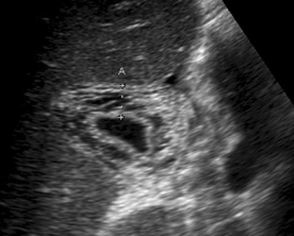
Abdominal ultrasonography of patient showing acalculous cholecystitis. Thickened edematous gallbladder wall measuring 11 mm.
Q fever is a zoonosis caused by C. burnetii. Q fever has a worldwide distribution, with the exception of New Zealand. The majority of human infections are acquired by inhalation of small particle droplets from urine, feces, milk, or bodily fluid from cattle, sheep, goats, or native animals. C. burnetii causes an initial bacteremia with widespread tissue seeding and consequently has a diverse range of clinical manifestations.
Acalculous cholecystitis has been described in association with Q fever (Rolain et al. 2003). Acalculous cholecystitis is a condition most commonly seen in patients critically unwell from a variety of conditions, including sepsis, trauma, or burns. Acalculous cholecystitis has been described as a direct complication of several microorganisms, including Salmonella spp., Mycobacterium spp., Plasmodium spp., Candida spp., cytomegalovirus, cryptosporidium, and microspora (Barie and Eachempati 2003). Such cases have been successfully managed with medical therapy directed at the specific organism.
Fifteen cases of Q fever–associated cholecystitis have been described (Kelly et al. 1986, Isaacs and Donald 1998, Modol et al. 1999, Rolain et al. 2003, Gonzalez-Delgado et al. 2005, Reina-Serrano et al. 2005, Hartzell et al. 2007). Two cases were diagnosed by liver biopsy with histopathology showing classic doughnut hole and fibrin ring surrounding a granuloma. The remainder were diagnosed by serology weeks after the initial illness. Immunohistochemistry was nondiagnostic for Q fever in all six patients who underwent cholecystectomy.
This is the first case described of Q fever cholecystitis diagnosed from gallbladder tissue. The presence of C. burnetii genome in this patient, together with the clinical presentation, acute cholecystitis, and seroconversion, suggests that C. burnetii may be directly involved in the pathogenesis of cholecystitis.
Molecular techniques are evolving as useful tools to make an early diagnosis of Q fever. New techniques utilizing htpAB-associated repetitive element of C. burnetii genome (IS1111) have been shown in an outbreak setting to have a sensitivity of 59% on serum (Turra et al. 2006). Sensitivity is highest in early infection, before immunoglobulin M rise. Molecular techniques may allow a more rapid diagnosis of Q fever cholecystitis, facilitating earlier implementation of specific treatment and reducing the likelihood of operation. Despite early medical management with doxycycline, patients may still develop complications such as secondary bacterial infection, abscess formation, and perforation necessitating surgical intervention.
The annual incidence of Q fever cases notified in Australia has decreased from 5.0 per 100,000 in 1993, to 2.2 cases per 100,000 persons in 2004 (Brotherton et al. 2007). This reduction has been attributed to implementation between 2001 and 2004 of the National Q Fever Management Program, which targeted abattoir workers, those contracted to abattoirs, sheep shearers, and sheep, dairy, and cattle farmers and their employees. The patient described in this case would not have been identified as part of the program but was in a high-risk occupational group for which immunization is recommended. Australian guidelines recommend Q fever vaccine for abattoir workers, farmers, stockyard workers, shearers, animal transporters, others exposed to cattle, sheep, goats, and kangaroos or their products, and veterinarians and laboratory staff handling C. burnetii specimens. In this instance, a recommendation was made to the employer by Work Cover New South Wales that employees working in the meat-packing industry with negative Q fever serology be vaccinated and personal protective equipment (goggles, mask, gown, and gloves) be worn when operating the band saw.
Ongoing vigilance is required to prevent Q fever in high-risk groups. Of concern is the high proportion of Q fever cases occurring in groups with no recognized risk factors. Of the 15 described cases of Q fever–associated cholecystitis, only 4 had documented animal exposure. The proportion of Australian Q fever cases in people with no direct animal contact may increase during drought, as the subsequent dusty conditions enhance dissemination of this organism.
Footnotes
Acknowledgment
Phill Cantrell, Senior Project Officer, Hazard Management Group, WorkCover, New South Wales, Australia.
Disclosure Statement
No competing financial interests exist.