
Abstract
A 23-year-old woman from Ethiopia presented with acute abdominal pain. Exploratory laparotomy revealed appendicitis, pathology of which showed numerous eggs consistent with Taenia species. Although Taenia appendicitis is rare, it should be considered in the differential diagnosis of patients presenting with appendicitis from endemic parts of the world.
Introduction
Case Report
A 23-year-old otherwise healthy Muslim woman from Ethiopia presented with 4 days of periumbilical pain, abdominal distension, nausea, and vomiting. Born in Ethiopia she lived on a farm with cattle until about 3 or 4 years of age, before immigrating to the United States. She vigorously denied any contact with pigs owing to her religious beliefs.
On examination, she was found to be febrile to 38.0°C with a markedly tender abdomen showing peritoneal signs. Her leukocyte count was 9000 with 72% neutrophils and 11% nonsegmented forms. Of note, no eosinophils were seen on her smear. A computed tomography scan of the abdomen revealed an appendicolith, fat stranding around the appendix, and possible early abscess formation. Exploratory laparotomy revealed extensive inflammation of the small bowel and terminal ileum, gangrenous appendix, and purulent fluid throughout the abdominal cavity. Acute suppurative transmural appendicitis was evident on pathologic examination of the resected appendix. Numerous eggs consistent with Taenia spp. and fragments of the helminthes body were found within the appendix (Fig. 1).
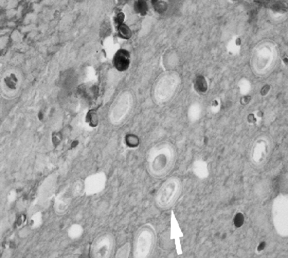
Appendiceal tissue demonstrating Taenia eggs (arrow) (H&E stain, 100 × magnification).
On the basis of the histopathologic examination it was impossible to determine which species of Taenia was present. The eggs of Taenia spp. are indistinguishable morphologically, and the proglottid segments were too disrupted to determine the number of lateral branches. However, considering her religious convictions, it was most likely T. saginatum.
Discussion
T. saginatum infection of the intestine is relatively common in sub-Saharan Africa, especially in Ethiopia. Infection follows ingestion of the cysticercus; the cyst enters the small intestine where the cyst wall is digested, freeing the worm inside. The scolex then attaches to the intestinal wall and matures over the next several months to become the adult worm. Unlike T. solium, only the adult form of T. saginatum resides in humans. The adult worm is usually 5 to 12 meters long, but can achieve a length of 25 meters.
The role of Taenia proglottids in the initiation of appendiceal inflammation is not well defined. The presence of an appendicolith raises the possibility that the patient's symptoms were simply due to mechanical obstruction and the presence of Taenia proglottids was simply an epiphenomenon. Nevertheless, it is quite possible that the Taenia proglottids contributed to the inflammatory reaction. Presumably, the proglottids were present within the appendiceal lumen prior to perforation, and it has been demonstrated that proglottids, especially T. saginata, can invade the intestinal wall and migrate through various tissues. T. saginata oncospheres possess excretory/secretory peptidases, which may facilitate the invasion of intestinal mucosa (White et al. 1996).
Parasitic infection of the appendix is a decidedly uncommon phenomenon. In a series of 1600 appendectomies performed in Sao Paulo, Brazil, over a 10-year period, parasites were found within the appendix in 24 cases. In 23 of these, Enterobius vermicularis was detected; Taenia sp. was detected in one. Pathologic analysis of the specimens showed neutrophilic inflammation of the appendix wall in about half the cases and lymphoid hyperplasia in the rest (da Silva et al. 2007). Even in countries that are endemic for Taenia spp., the association of this parasite with appendicitis remains quite rare. Prior to the case cited above, there are only four reports in the English language literature of Taenia sp. being found in the appendix (Payne 1970, Kruskowski and Miller 1977, Lejbkowicz et al. 2002, Sartorelli et al. 2005). Other parasites that have been rarely noted include Schistosoma sp. and Ascaris lumbricoides. Extraintestinal manifestations due to ascariasis are more commonly seen compared to those due to Taenia sp. In a study by Ochoa, 311 children under 12 years of age were admitted for abdominal complications caused by Entamoeba histolytica or A. lumbricoides. Of these, 145 children had abdominal symptoms produced by ascariasis, which manifested as intestinal obstruction (n = 107), migration of the parasite to the biliary tree or peritoneal cavity (n = 28), or perforation of the appendix (n = 10). In all of the patients with appendicitis, the Ascaris worms were found to be either partially extruded through the perforation or lying inside the lumen of the appendix (Ochoa 1991).
In conclusion, we present here an unusual case of appendicitis associated with T. saginatum. The role of this organism in the pathogenesis of appendicitis remains uncertain but may contribute to the development of this process in endemic regions.
Footnotes
Disclosure Statement
No competing financial interests exist.