
Abstract
Ixodes scapularis ticks are clinically important hematophagous vectors. A single tick bite can lead to a polymicrobial infection. We determined the prevalence of polymicrobial infection with Borrelia burgdorferi, Anaplasma phagocytophilum, Babesia microti, Borrelia miyamotoi, and Powassan virus in 286 adult ticks from the two counties in New York State where Lyme disease is endemic, utilizing a MassTag multiplex polymerase chain reaction assay. Seventy-one percent of the ticks harbored at least one organism; 30% had a polymicrobial infection. Infections with three microbes were detected in 5% of the ticks. One tick was infected with four organisms. Our results show that coinfection is a frequent occurrence in ticks in the two counties surveyed.
Introduction
We recently described a multiplex method for rapid and economic screening of tick-borne pathogens (Tokarz et al. 2009). This method, MassTag polymerase chain reaction (PCR), enables efficient, sensitive screening of individual ticks for polymicrobial infection. Here we report its application for tick surveillance in Lyme disease endemic areas in New York State.
Materials and Methods
All PCRs were performed using cDNA generated from individual adult ticks. Live ticks were collected from vegetation in five separate locations in the two counties (Westchester and Suffolk) of New York State in the fall of 2008 (Fig. 1). Locations 1 and 2 are in Westchester County, whereas locations 3, 4, and 5 are in Suffolk County. Prior to nucleic acid extraction, ticks were immersed in 75% ethanol for 15 min, washed twice with phosphate-buffered saline, and then homogenized in Tri-reagent LS (Molecular Research Center, Cincinnati, OH). Total RNA was resuspended in 20 μL of H2O. cDNA was generated in a 20 μL reaction with Superscript II Reverse Transcriptase (Invitrogen, Carlsbad, CA) using 10 μL of total RNA. MassTag PCR was performed using the tick panel (Tokarz et al. 2009), with additional primers for the detection of Powassan virus and relapsing fever Borrelia species (Table 1). All MassTag PCR assays utilized 3 μL of cDNA. Reaction conditions were 95°C for 5 min, one cycle at 95°C for 20 s, 65°C for 20 s (annealing), 72°C for 30 s, followed by 11 cycles with annealing temperature decreased by −1°C at each cycle. The final PCR was run for 37 cycles at the annealing temperature of 54°C. For sequencing, PCR products were size-fractionated on ethidium bromide–stained 1% agarose gel, observed under ultraviolet illumination, excised, and gel purified for dideoxy sequencing.
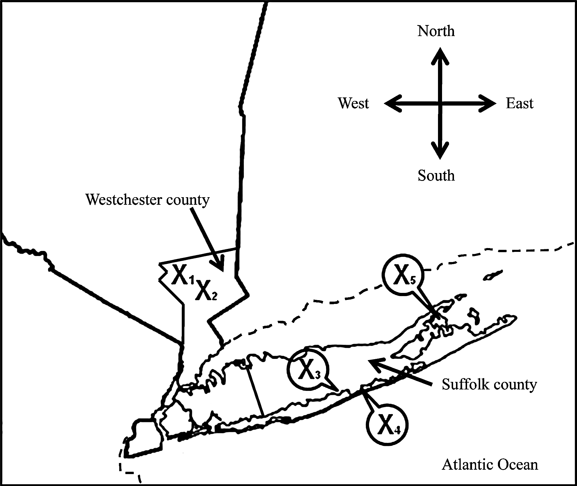
Map indicating tick collection sites within New York State.
Used for confirmatory polymerase chain reaction.
Fwd, forward; Rev, reverse.
Results
MassTag PCR was used to screen adult I. scapularis ticks collected from five locations in two different counties surrounding New York City (Fig. 1) for polymicrobial infection. The number of ticks screened from each location ranged from 24 to 98, with a total of 286 ticks analyzed. We detected at least one organism in 204 ticks (71%), of which 85 (30% of total) had a polymicrobial infection (Table 2). About 82 ticks (29%) did not have an infection with any of the microbes surveyed.
B. burgdorferi coinfection with A. phagocytophilum and B. microti
B. burgdorferi was the most common pathogen detected at all five sites, with infection rates of >50% ticks at each site (Table 3). Overall, we detected this organism in 182 out of the 286 ticks (64%). We detected A. phagocytophilum in >20% of the ticks at four out of the five sites; one notable difference being site 1, where only two out of the 56 ticks harbored the pathogen. Overall, a total of the 56 ticks (20%) were positive for A. phagocytophilum, of which 45 (80%) were also infected with B. burgdorferi. Conversely, 24% of the B. burgdorferi–positive ticks were also A. phagocytophilum positive. Ticks with a coinfection of A. phagocytophilum and B. burgdorferi accounted for 16% of total ticks analyzed. We detected B. microti in 58 ticks (20% of total), of which 48 were also infected with B. burgdorferi (83%).
B. burgdorferi coinfection with B. miyamotoi and Powassan virus
B. miyamotoi is a relapsing fever-like Borrelia species. This organism was much less frequently detected; only seven ticks (2%) were positive for this organism but all were also coinfected with B. burgdorferi. Infection with Powassan virus was also infrequent. We detected the virus in seven ticks (2% of total). Five of these were collected at location 2 and a single Powassan virus–positive tick each was collected from locations 1 and 3. The majority of these did not exhibit a coinfection; only two of the Powassan virus–positive ticks were coinfected with B. burgdorferi, and another was coinfected with A. phagocytophilum, whereas the remaining four did not harbor any of the other infecting agents we screened for. For confirmation of Powassan virus–positive samples, we amplified by PCR and sequenced a 395-base pair fragment of Powassan virus (Fig. 2A) from each positive tick. All sequences represented genotype II of Powassan virus.
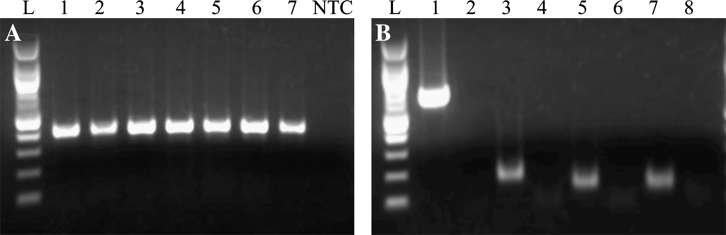
Confirmation of MassTag polymerase chain reaction (PCR) results. (
Triple infections
We detected ticks harboring a triple infection with B. burgdorferi, A. phagocytophilum, and B. microti at four sites. A total of 14 ticks (5% of total) had such a polymicrobial infection; we also detected a triple infection of B. burgdorferi, A. phagocytophilum, and B. miyamotoi in one tick. Additionally, we detected a single tick from area 5 infected with four pathogens (Fig. 2B).
Amblyomma and Dermacentor ticks
Along with I. scapularis, we also attempted to assess infection rates in other human biting ticks collected at two sites. We collected and screened 108 and 32 A. americanum ticks from locations 3 and 4, respectively. We detected B. lonestari in two ticks at each location. The only other organisms detected by our assay were Ehrlichia species, found in 17 ticks from area 3 and 2 ticks from area 4. Sequencing indicated that 10 ticks hosted Ehrlichia ewingii, 6 Ehrlichia chaffeensis, and a single tick the Panola Mountain Ehrlichia variant. Coinfections were not detected. Finally, we screened 13 Dermacentor variabilis ticks collected at area 4. We did detect Francisella tularensis in a single case. For confirmation, we amplified and sequenced fragments of the IglC and 16S rRNA genes that were >99% similar to F. tularensis subspecies tularensis.
Discussion
We screened adult I. scapularis ticks collected from five different areas for pathogen infections. We found that 71% of the ticks were infected with at least one organism. In addition, we also detected a large number of coinfections; the number of ticks coinfected with at least two microbes was higher than the number of uninfected ticks. We also detected that 5% of the ticks were infected with triple infections and a single tick with four pathogens. Our results indicate that in the areas surveyed, coinfection of adult I. scapularis may be just as common as the lack of infection.
A. phagocytophilum and B. microti were each detected in 20% of the ticks. Surveillance studies indicated that the prevalence of these organisms in ticks varies greatly, though infection rates of >20% have been shown (Piesman et al. 1986, Schwartz et al. 1997, Schauber et al. 1998). High coinfection rates can increase the likelihood of multiple infections in humans following a single tick bite, a phenomenon already documented (Benach et al. 1985, Magnarelli et al. 1998, Belongia et al. 1999, De Martino et al. 2001, Krause et al. 2002). Lyme disease and HGA are the two most common diseases reported resulting from a bite of I. scapularis, although reported Lyme disease cases outnumber HGA by >30 to 1 (CDC 2008). In our analysis, tick infection by these pathogens was not dramatically different, with a ratio of approximately 3:1. Nearly a quarter of ticks infected with B. burgdorferi were also coinfected with A. phagocytophilum, implying that in the areas sampled, B. burgdorferi transmission may be accompanied by A. phagocytophilum transmission in up to 25% of the cases. Similar rates of coinfection with B. burgdorferi and B. microti also indicate high probability of cotransmission of these two organisms as well. The large discrepancy in HGA and Lyme disease cases may reflect failure in diagnosis, underreporting of HGA cases, or lower rates of A. phagocytophilum prevalence in some geographic areas. HGA may also be mild or subclinical. Further, little is known about the transmission dynamics of coinfecting microbes. In ticks hosting multiple pathogens, the microbial interaction may result in variable microbial loads, and such differences could lead to alteration in the transmission efficiency for a given microbe.
In addition, we screened for Powassan virus and B. miyamotoi, two microbes of much lower prevalence and not detected at all surveyed sites. Powassan virus has been reported to infect 1–5% of the ticks in the North Central United States, but prevalence studies of other areas are lacking (Ebel et al. 1999, Brackney et al. 2008). We detected Powassan virus and B. miyamotoi in 2% of the ticks we screened. The two microbes had different frequency of coinfection with other pathogens. All seven I. scapularis infected with B. miyamotoi were also coinfected with B. burgdorferi. This is in contrast to Powassan virus–positive ticks, where in four out of the seven infected ticks we did not detect a coinfection. Whether this was a chance result or whether those ticks picked up the virus by feeding on an animal refractory to B. burgdorferi infection is unclear.
Polymicrobial infection following tick bites is an occurrence of which clinically much is still unknown. Our study indicates high coinfection prevalence in ticks within the areas surveyed. How these organisms interact during transmission and disease remains to be determined, but may have important impact on diagnosis and treatment of tick-borne diseases.
Footnotes
Acknowledgments
Work reported here was supported by the National Institutes of Health (AI57158-05, Northeast Biodefense Center-Lipkin) and
Disclosure Statement
No competing financial interests exist.