
Abstract
A quantitative real-time polymerase chain reaction assay to detect and quantify a portion of the outer membrane protein B gene (ompB) of Rickettsia amblyommii was employed to assess the threat of R. amblyommii exposure to humans parasitized by Amblyomma americanum (the lone star tick). A total of 72 pools of lone star ticks removed from humans were acquired from two collections and used in this study: 44 pools of A. americanum submitted to the Department of Defense Human Tick Test Kit Program in 2003 collected from 220 individuals from 14 states, and 28 pools of A. americanum representing 120 ticks obtained from boy scouts and adult leaders at the Boy Scouts of America National Jamboree held at Fort A.P. Hill, Virginia, in 2005. Of the 72 lone star tick pools representing 340 lone star ticks, 58 pools (80.5%) were positive for R. amblyommii. In addition, individual A. americanum ticks parasitizing humans collected as part of the Department of Defense Human Tick Test Kit Program in 2002 and 2003 from 17 states were evaluated. It was found that 244 of 367 (66.5%) individual A. americanum ticks tested positive for the presence of R. amblyommii DNA. These results clearly show that lone star ticks parasitizing humans are highly infected with R. amblyommii, which may potentiate rickettsial infection of and possibly disease in humans.
Introduction
A role for R. amblyommii in human pathogenesis has been suggested in individuals exposed to lone star ticks in Virginia and Arkansas (Kardatzke et al. 1992, Sanchez et al. 1992, Dasch et al. 1993). Others have also suggested that A. americanum transmits an SFG rickettsial agent, not R. rickettsii that causes disease/infection in humans (Childs and Paddock 2003, Paddock et al. 2004). More recently, Stromdahl et al. (2008) and Apperson et al. (2008) have suggested the high prevalence of R. amblyommii in the lone star tick, and its predilection for feeding on humans in all stages is related to the high prevalence of antibodies in people not reporting RMSF. In addition, Stromdahl et al. (2008) have shown the presence of R. amblyommii in the larvae of A. americanum found clustering on vegetation in eastern Maryland. This characteristic aggregation of the larvae and the ensuing mass biting that occurs after transfer of these clusters from vegetation to humans may be an important mechanism for transmission of the rickettsial agent to humans. Moreover, Apperson et al. (2008) showed sera from febrile patients classified as probable RMSF cases that had antibodies more reactive to R. amblyommii antigen than to R. rickettsii antigens. A more direct relationship between R. amblyommii and human infection comes from a recent report of the detection of R. amblyommii in an A. americanum tick removed from a patient with a bite-site rash similar to that reported with southern tick-associated rash illness (STARI). This suggests a causal relationship between the rickettsia, and human infection and disease. The removed tick did not have evidence of Borellia lonestari (the putative causative agent of STARI) or the lone star tick human pathogens Ehrlichia chaffenesis (human monocytic ehrlichiosis [HME]) and Ehrlichia ewingii (E. ewingii ehrlichiosis [EWE]) (Billeter et al. 2007). Although R. amblyommii may be infectious to humans, it has not yet been isolated from humans. This situation of proposed pathogenicity associated with a presumed tick commensal is not dissimilar to that of Rickettsia parkeri, a previously described endosymbiont of Amblyomma maculatum (Parker et al. 1939) that has recently been determined to cause human disease (Paddock et al. 2004).
To assess the potential threat of R. amblyommii transmission to humans, we conducted a study in which we evaluated the prevalence of rickettsial infection in lone star ticks parasitizing humans in the United States utilizing a newly developed quantitative real-time polymerase chain reaction (qPCR) assay described in this paper. We report herein a high prevalence (66.5%) of R. amblyommii infecting A. americanum ticks collected from humans.
Materials and Methods
Tick collections
Boy Scout National Jamboree 2005
Fine-tipped forceps were used to remove ticks from Boy Scouts or other personnel at clinics manned by volunteer physicians on the Jamboree site, Fort A.P. Hill, Virginia. Ticks were placed in individual marked vials and transported to an on-site laboratory facility for identification and processing. After identifying, the A. americanum ticks were pooled together, approximately five individuals per test tube.
Department of Defense Human Tick Test Kit Program in 2002 and 2003
The Tick-Borne Disease Laboratory (TBDL) of the Entomological Sciences Program of the U.S. Army Center for Health Promotion and Preventive Medicine (CHPPM), Aberdeen Proving Ground, Maryland, provides tick identification and testing service for ticks attached to military personnel, military dependents, and Department of Defense (DoD) civilian employees. Ticks are mailed from military medical treatment facilities in the continental United States to the TBDL for identification and analysis by PCR. In 2002, the TBDL tested 2146 A. americanum from 105 DoD installations, and in 2003, about 1627 A. americanum from 116 installations were tested.
DNA extraction protocol
For the 2005 Boy Scout National Jamboree tick crude lysates were prepared by individually dissecting each tick in 500 μL of tissue lysis buffer (Qiagen Tissue Kit®, Qiagen, Inc., Valencia, CA). Crude lysate pools of 180 μL total were prepared by combining 36 μL of lysate from each tick in a 1.5 mL microcentrifuge tube. The 28 pool samples contained a total of 120 ticks consisting of 3 larvae, 77 nymphs, and 29 female and 11 male adults. Tick pools were created separately by species but without regard to stage or sex. Tick lysate pools were incubated with 20 μL of proteinase K for a minimum of 1 h at 56°C. Nucleic acids were subsequently purified from each tick lysate pool in accordance with the Qiagen Tissue Kit protocol. One negative control was processed per DNA extraction procedure for every run of 30 samples to ensure there was no cross contamination between the samples. The knives used to cut the ticks were swished in the negative control to assess carry over. To sterilize the knife between tick processing it was washed once with detergent, twice with distilled water, and flamed with alcohol.
For the DoD Human Tick Test Kit Program (2002 and 2003) DNA extractions from tick triturates of individual adult A. americanum were performed using the IsoQuick Nucleic Acid Extraction Kit (ORCA Research, Bothell, WA) according to the manufacturer's instructions. For the first part of this study, nucleic acid from five individuals were pooled together for evaluation of rickettsial infection of ticks collected from people reporting to a DoD healthcare clinic as part of the DoD Tick Test Kit Program (Stromdahl et al. 2001) in 2003. In the second part of the study, nucleic acid from individual ticks obtained from the DoD Tick Test Kit Program in 2002 and 2003 was assessed for the presence of R. amblyommii DNA. Ninety-one of the 220 2003 tick DNA samples pooled were also assessed individually without the operator knowing the results of the pooled experiment. Both 2003 sample sets were chosen randomly and evaluated by different individuals at different times. All 2002 tick DNA preparations were tested individually.
Assessment of A. americanum for evidence of Rickettsia spp. and R. amblyommii nucleic acid
Two microliters of individual or pooled DNA extracted from A. americanum ticks was evaluated by qPCR assays for the presence of rickettsiae utilizing the Rickettsia-specific 17 kDa common antigen gene (Rick17) qPCR assay (Jiang et al. 2004) and R. amblyommii utilizing the species-specific outer membrane protein B gene (ompB) (Rambl) qPCR assay. Initially, 2003 pool tick samples were screened for rickettsial DNA using the Rick17 qPCR assay, but due to the high prevalence of R. amblyommii in the ticks, only the Rambl qPCR assay was used for subsequent analyses (i.e., 2005 pooled tick samples and individual ticks from 2002 and 2003).
Development of Rambl qPCR assay
Primers (Ra477F: GGTGCTGCGGCTTCTACATTAG; Ra618R: CTGAAACTTGAATAAATC CATTAGTAACAT) and probe (Ra532Probe: FAM-CGCGATCTCCTCTTACACTTGGACAGAATGCTTATCGCG-BHQ-1) sequences were selected from ompB based on the GenBank sequence AY375164, which was derived from the Brazilian isolate R. amblyommii, Aca3 utilizing the Beacon Designer software (Premier Biosoft, Palo Alto, CA). The primers successfully amplified a 142 bp fragment. Rambl qPCR assays were performed using Platinum Quantitative PCR Supermix UDG (Invitrogen, Carlsbad, CA) with the following final concentrations: primer 0.5 μM, probe 0.4 μM, and magnesium chloride 6 mM. Thermal cycling parameters were 95°C for 2 min followed by 45 two-step cycles of 94°C for 5 s and 60°C for 30 s using a SmartCycler II System (Cepheid, Sunnyvale, CA). The threshold level for nonspecific fluorescence was selected to be 30 fluorescence units. Fluorescence above this level indicated a positive reaction, and the number of cycles necessary to reach this level of fluorescence is referred to as the cycle threshold (CT) value for crossing the threshold. The CT value is directly proportional to the copy number of the target gene in the sample. CT values are also referred to as crossing point, take-off point (TOP), and quantification cycle.
Analytical specificity of the Rambl qPCR assay was evaluated by testing the optimized assay with nucleic acid preparations from 11 different strains of Rickettsia, 10 species of nonrickettsial bacteria, and 3 negative control samples.
A 142 bp ompB fragment amplified from R. amblyommii 85-1084 was spliced into a TOPO plasmid using the TOPO TA Cloning Kit (Invitrogen) as per manufacturer's instructions. The concentration of the plasmid DNA was ascertained using an Agilent 8453E Spectrophotometer (Agilent Technologies, Foster City, CA). A standard curve utilizing 100 to 108 copies of the plasmid containing a single copy of the target sequence was created to determine the assay's limit of detection.
Sequencing and phylogenetic analysis of R. amblyommii 85-1084
The genetic information for R. amblyommii was very limited at the time of the development of Rambl qPCR assay, though the species was first isolated in 1974. Therefore, we characterized the sequence and conducted a phylogenetic analysis of R. amblyommii 85-1084 for the ompB and ompA genes. Fragments of ompB and ompA from R. amblyommii 85-1084 were amplified by standard PCR and nested PCR using Platinum PCR SuperMix High Fidelity (Invitrogen) and run on a T-Gradient Thermocycler (Biometra, Goettingen, Germany). Primers used in this study are listed in Table 1. PCR amplicons were purified and sequenced in both directions on an automated ABI Prism 3100 gene analyzer (Applied Biosystems, Foster City, CA) similar to that described previously (Jiang et al. 2005). The sequences were assembled with Vector NTI advance 10 software (Invitrogen). Phylogenetic analyses were performed with MacVector 8.1 software (Accelrys, San Diego, CA).
Primers used for PCR amplification.
Primers used for nested PCR amplification.
Primers used for sequencing.
ORF, open reading frame.
Nucleotide sequence accession number
The sequences of R. amblyommii 85-1084 have been deposited in GenBank with accession numbers FJ455414 for ompA and FJ455415 for ompB.
Results
Analytical specificity of Rambl qPCR assay
To determine the specificity of the Rambl qPCR assay we evaluated the assay using DNA preparations from R. amblyommii and 21 near and far neighbors. We found that only DNA preparations from R. amblyommii isolates, 85-1084 (Fig. 1) and Darkwater and WB-8-2 (K. Macaluso, pers. comm.), reacted with the Rambl qPCR assay and not DNA preparations from Rickettsia montanensis 85-930, R. rickettsii Bitterroot, R. rickettsii Shelia Smith, R. parkeri Maculatum C, Rickettsia slovaca D, Rickettsia conorii ITT, Rickettsia akari 29, Rickettsia prowazekii Breinl, R. prowazekii Ananiev, Rickettsia typhi Wilmington, Rickettsia canadensis MCK-29, Neorickettsia risticii, Neorickettsia sennetsu, Francisella persica, Salmonella enterica, Proteus mirabilis, Escherichia coli, Corynebacterium sp., Legionella pneumophila, Bartonella vinsonii, and Bartonella quintana (Fig. 1). DNA integrity was assured for the specificity panel of nucleic acid preparations by successfully performing 16s rRNA gene detection on all DNA preparations by PCR (Jiang et al. 2004).
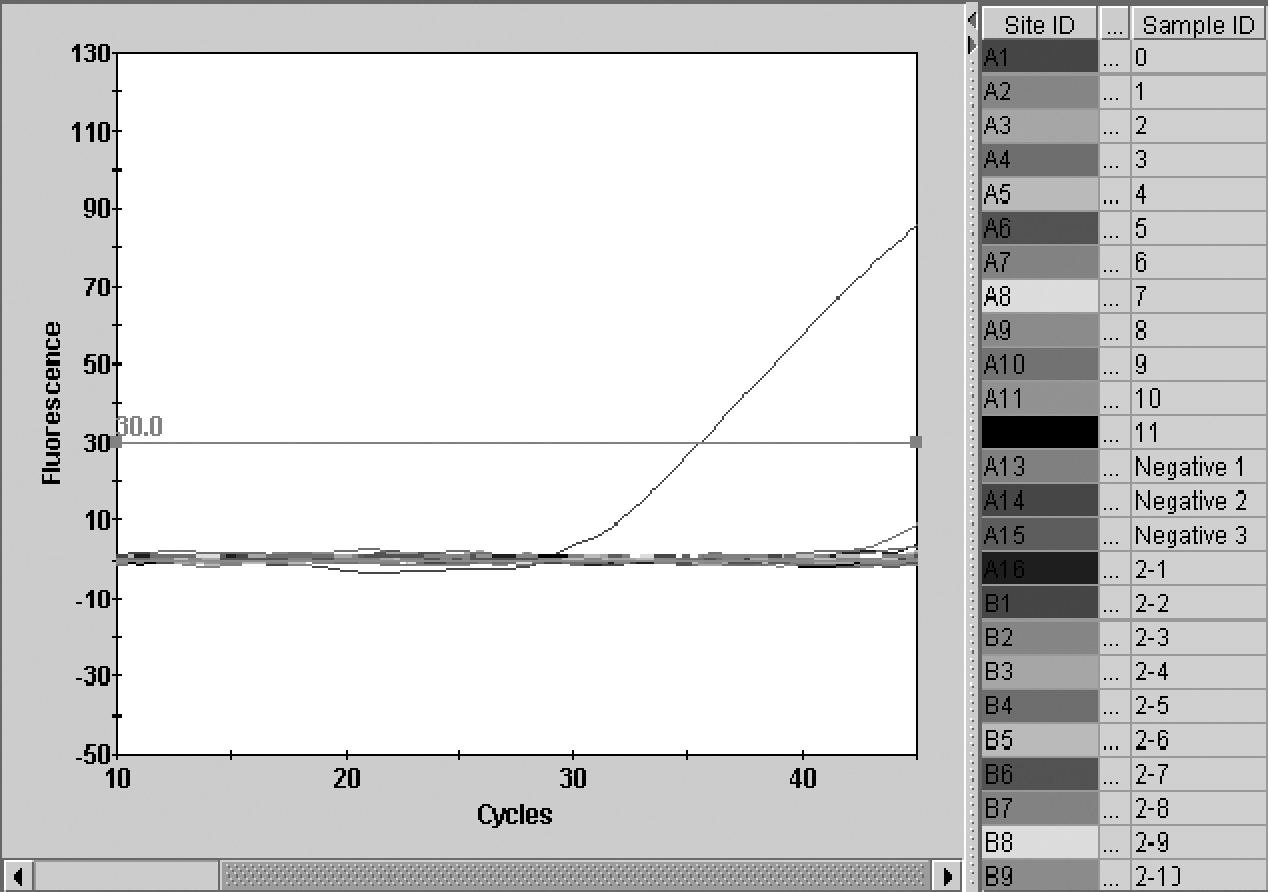
Rambl quantitative real-time polymerase chain reaction assay specificity. Sample identification of bacterial nucleic acid preparations utilized (0) Rickettsia amblyommii 85-1084, (1) Rickettsia montanensis, (2) Rickettsia rickettsii Shelia Smith, (3) Rickettsia parkeri, (4) Rickettsia slovaka, (5) Rickettsia prowazekii Breinl, (6) Rickettsia canadensis, (7) R. rickettsii Bitterroot, (8) Rickettsia conorii, (9) Rickettsia akari, (10) R. prowazekii Ananiev, (11) Rickettsia typhi, (2-1) Salmonella enterica, (2-2) Proteus mirabilis, (2-3) Escherichia coli, (2-4) Corynebacterium sp., (2-5) Legionella pneumophila, (2-6) Bartonella vinsonii, (2-7) Bartonella quintana, (2-8) Neorickettsia risticii, (2-9) Neorickettsia sennetsu, and (2-10) Francisella persica.
Limit of detection of Rambl qPCR assay
To determine the limit of detection of the Rambl qPCR assay, standard concentrations of target sequence containing plasmid DNA from 100 to 108 copies/μL were plotted against CT values. It was ascertained that the lowest copy number of the target sequence that was consistently detected was three copies per reaction (Fig. 2).
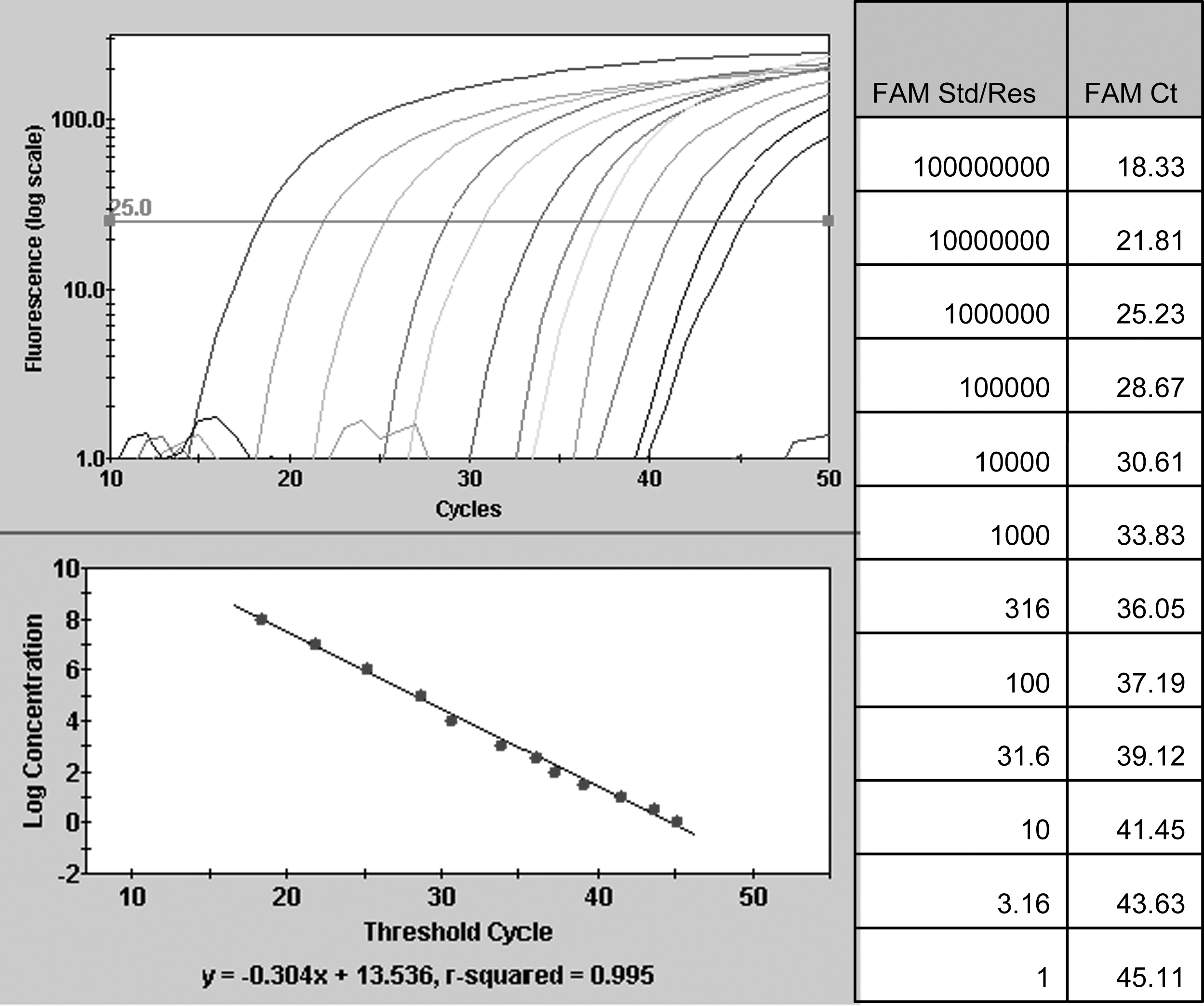
Standard curve: log graph of standard concentrations versus cycle threshold (CT) values determined for Rambl quantitative real-time polymerase chain reaction assay. Figure is representative of three experiments.
Phylogenetic analysis of R. amblyommii 85-1084
The sequences of 3188 bp of ompA and 4844 bp of ompB from R. amblyommii 85-1084 were obtained and compared to those of known Rickettsia species in GenBank. The phylogenic trees were constructed using Neighbor-Joining Best tree (Fig. 3a, b), and the trees were confirmed by Bootstrap 1000 methods for both genes (Fig. 3c, d). R. amblyommii 85-1084 was found to be in the same clade as most of the SFG members with a bootstrap value of 100%. Within this group, R. amblyommii 85-1084 was closely related to R. raoultii and a new SFG member Candidatus Rickettsia andeanae. The similarity between R. amblyommii 85-1084 and these two other rickettsiae was found to be both 97.2% for ompB, and 98.4% and 98% for ompA, respectively (Table 2).
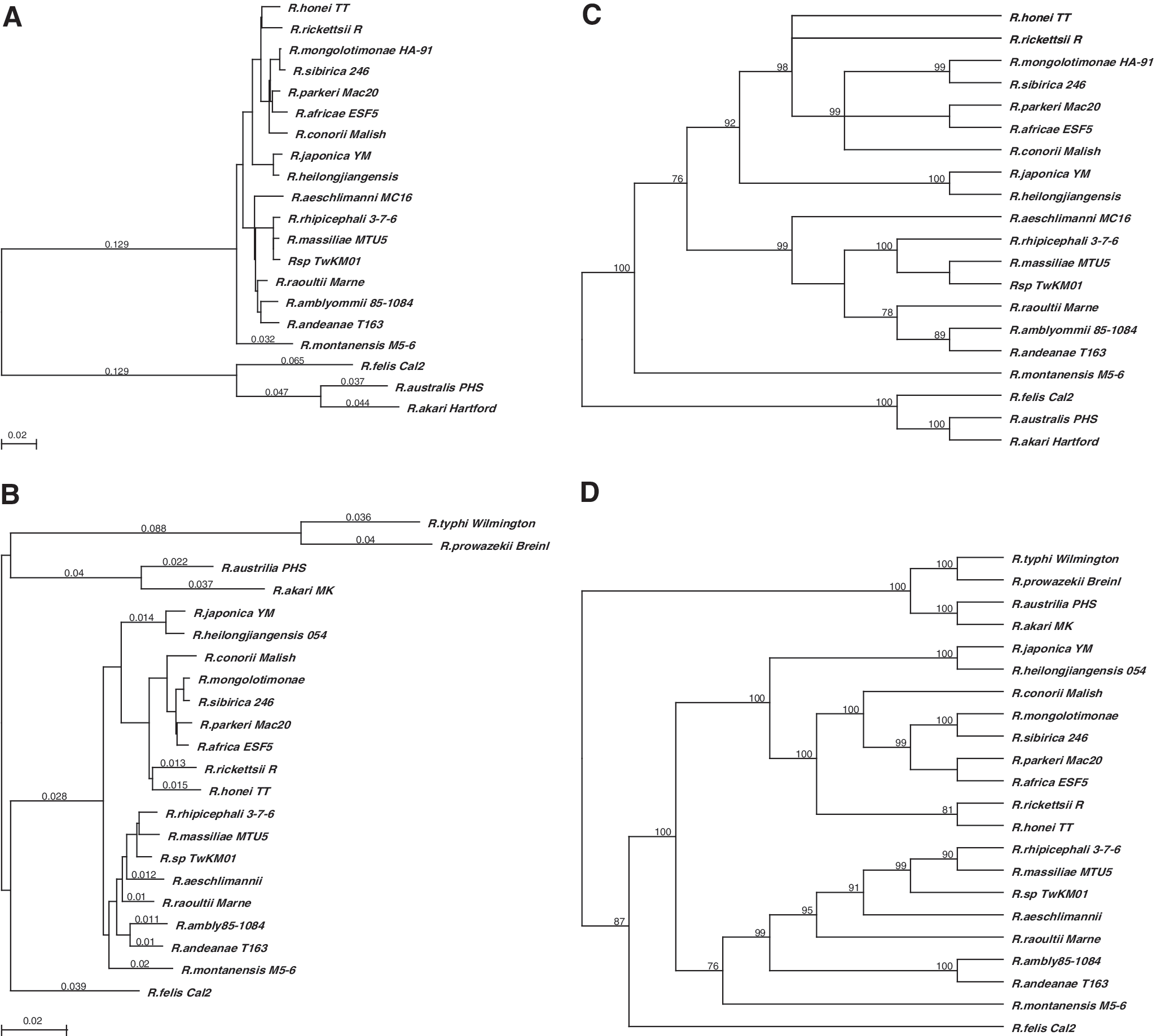
Phylogenetic best trees based on 1581 bp ompA sequence (
Pairwise identity (%) matrix of ompA was calculated based on a 1581 bp fragment (3567–5147 to R. rickettsii ORF).
Evaluation of pools of A. americanum for R. amblyommii
Evaluation of 44 pools of A. americanum ticks (220 individual ticks collected from patients presenting to DoD healthcare clinics from 14 states: AL, AR, DE, FL, GA, KY, MD, MS, NC, NJ, OK, SC, TN, and VA) for the presence of rickettsiae determined that 36 pools were Rick17 positive. Of these 36 pooled DNA preparations assessed for the presence of R. amblyommii, 33 were found to be Rambl positive (91.7%; 75% of the total 44 pools assessed). The 8 of 44 pools that were Rick17 negative were also Rambl negative (Table 3a). Evaluation of 28 pools of A. americanum nucleic acid preparations (representing 120 ticks obtained from boy scouts and adult leaders at the Boy Scouts of America National Jamboree held in July–August 2005 at Fort A.P. Hill, VA) by Rambl found that 25 of 28 (89.3%) pool preparations were positive. These pools were not assessed by the Rick17 qPCR assay. Collectively, the two pooled sample sets showed a prevalence of R. amblyommii in pooled A. americanum nucleic acid preparations of 80.5% (58 of 72 pools) (Table 3a).
Ticks obtained from the DoD Human Tick Test Kit Program year 2003.
Ticks obtained from the Boy Scout National Jamboree, Fort A.P. Hill, 2005.
Ticks obtained from the DoD Human Tick Test Kit Program year 2002.
qPCR, quantitative polymerase chain reaction.
Not determined.
Evaluation of individual A. americanum for presence of R. amblyommii
Individual ticks were assessed for the presence of R. amblyommii target DNA sequences from two sample groups from the DoD Human Tick Test Kit Program, years 2002 and 2003. For the 2002 year group 128 of 183 (69.9%) individual A. americanum tick nucleic acid preparations were positive by Rambl qPCR assay. For the 2003 year group 116 of 184 (63.0%) individual tick nucleic acid preparations were positive by Rambl qPCR assay. Collectively, 244 of 367 (66.5%) individual adult A. americanum ticks were positive for R. amblyommii DNA (Table 3b).
Distribution of individual R. amblyommii–positive A. americanum ticks
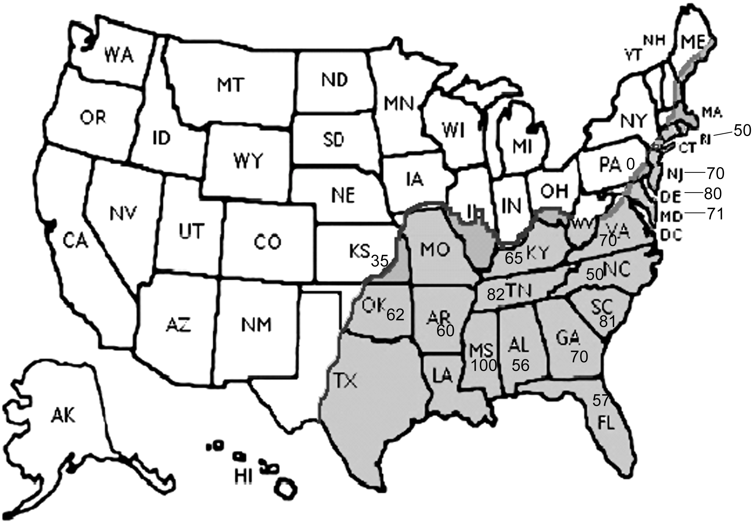
Ticks from 17 states (AL, AR, DE, FL, GA, KS, KY, MD, MS, NC, NJ, OK, PA, RI, SC, TN, and VA) were sent to the CHPPM as part of the DoD Human Tick Test Kit Program in 2002 and 2003. A. americanum ticks collected from humans and containing R. amblyommii came from 16 of the 17 states (Fig. 4). The only state that was negative for R. amblyommii of those evaluated was Pennsylvania, but only two ticks were assessed. The states providing A. americanum ticks are included in the present distribution within the United States of the lone star tick (Fig. 5).
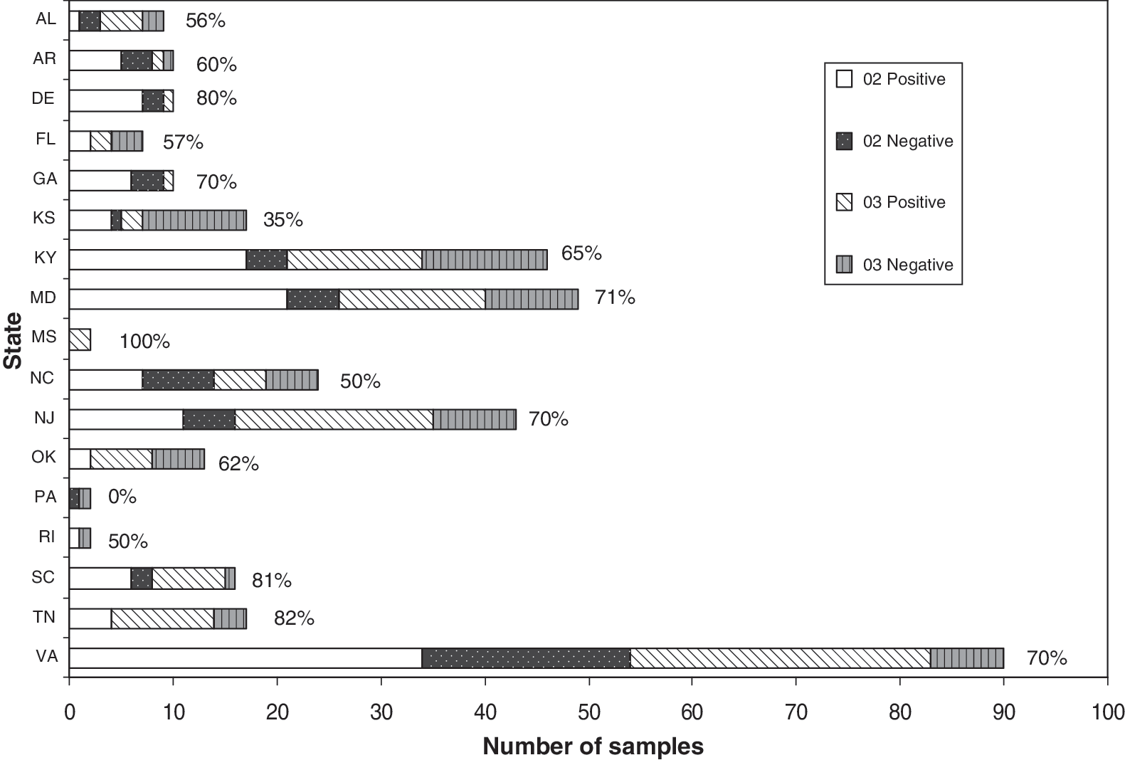
Prevalence of R. amblyommii–infected individual A. americanum ticks by state. Positive percentages are shown next to the bars.
Distribution of Amblyomma americanum and percent positivity of R. amblyommii–infected A. americanum ticks collected from humans. Gray shading indicates the currently known distribution of A. americanum ticks in the United States.
Discussion
The lone star tick, A. americanum, was first identified by Linnaeus in 1784 (Barker and Murrell 2004). Interestingly, lone star tick–transmitted human diseases have only recently been described (HME 1987, EWE 1997, and STARI 1984) (Schulze et al. 1984, Maeda et al. 1987, Buller et al. 1999). It is believed that these infectious diseases and their agents have been around as long as the tick has, but due to the previous low exposure of humans to the ticks and thus the agents, their diseases were rarely encountered, misdiagnosed, or overlooked (Anigstein and Bader 1942, Schulze and Bosler 1996, Childs and Paddock 2003). However, with the expansion of the major lone star tick host (e.g., white tail deer, coyotes, and wild turkeys) populations and, therefore, the expansion of tick numbers, the exposure rate of humans to A. americanum has increased (Mount et al. 1993, Mock et al. 2001, Paddock and Yabsley 2007). Moreover, the increase in host populations into new regions has led to the expansion of the endemic region for Amblyomma ticks (i.e., from southcentral and southeastern United States to the more populated midwestern and northeastern United States) (Ginsberg et al. 1991, Mount et al. 1993, Felz et al. 1996, Means and White 1997, Childs and Paddock 2003, Paddock and Yabsley 2007). Not only are the lone star ticks becoming more numerous in human-frequented areas, but also their behavior is such that the risk of encountering one or more individual ticks is great. This is because A. americanum ticks are very aggressive, all stages feed on humans, and the clustering behavior of larval ticks put humans at a high risk for contracting A. americanum–transmitted human diseases (Stromdahl et al. 2008).
Reports of HME (E. chaffenesis) and human granulocytic (E. ewingii) ehrlichiosis have increased in prevalence since their discovery in 1987 and 1997, respectively, from 203 cases of EWE and 99 cases of HME in 1999 (Groseclose et al. 2001) to 646 cases of EWE and 578 cases of HME in 2006 (McNabb et al. 2008). Another newly described lone star tick–transmitted human disease is the STARI (Schulze et al. 1984, Masters et al. 2008). Due to the molecular detection of B. lonestari in the erythema migrans (EM)-like rash, it was initially thought to be the causative agent of STARI (Barbour et al. 1996). However, recent investigations have shown an inability to detect B. lonestari (or B. burgdorferi) in STARI patients, leading the authors to suggest that another/other agent(s) transmitted by A. americanum may be responsible for the disease (James et al. 2001, Stromdahl et al. 2003, Wormser et al. 2005).
Before the discovery of these lone star tick–associated human diseases, it was discerned that these ticks were infected with the rickettsial agent, R. amblyommii (Burgdorfer et al. 1981). Subsequent investigations have shown that the infection rate was very common in A. americanum collected in nature (Burgdorfer et al. 1981, Kelly et al. 2005, Mixson et al. 2006, Apperson et al. 2008, Stromdahl et al. 2008). Although R. ambylommii has not been isolated from a human, three observations suggest that it may be the cause of a rickettsiosis. First, many populations assessed in the United States have been found to contain antibodies (2.5% to 28.7%) to SFG rickettsiae (Wilfert et al. 1985, Dasch et al. 1993, Yevich et al. 1995, McCall et al. 2001, Marshall et al. 2003, Graf et al. 2008). Since it is believed that the high prevalence of group-specific antibodies is not due to infection with R. rickettsii, the causative agent of RMSF, it must be due to another SFG rickettsial agent(s) (Taylor et al. 1985, Dasch et al. 1993, Marshall et al. 2003, Parola et al. 2005a, Graf et al. 2008, Paddock et al. 2008, Raoult and Parola 2008), and the most frequent rickettsia-infected tick that bites humans in many regions of the United States is the lone star tick (Merten and Durden 2000, Mixson et al. 2006, Goddard and Varela-Stokes 2009). Moreover, there is positive association with the presence of antibodies to SFG rickettsiae and areas of high RMSF incidence (Graf et al. 2008), which are areas where A. americanum is also a problematic human biter, that is, southeast and central United States. Second, human disease has been associated with recent A. americanum tick bites, including both a mild febrile disease (Sanchez et al. 1992) and a tick bite rash (Billeter et al. 2007). Third, other rickettsiae initially isolated from ticks and considered to be nonpathogenic were subsequently isolated from humans with disease. R. parkeri, which infects A. maculatum, was initially thought to be only a tick commensal (Parker et al. 1939, Bell et al. 1963). Recently, R. parkeri has been isolated from patients presenting with mild febrile disease, rash, and one or more eschars (Paddock et al. 2004, 2008, Whitman et al. 2007). This recent discovery is important to consider because another risk of disease transmission is the ability of the causative agent to be transferred from tick to human and the ability of that agent to produce an infection/disease in humans. In the case of R. parkeri it was 65 years before it was determined that indeed this agent could be passed to humans and that it could cause infection and disease. There have been several other tick-borne rickettsial agents that were initially considered to be tick commensals but have recently emerged as human pathogens. They include R. slovaca, Rickettsia heilongjiangensis, and Rickettsia massiliae among others (Parola et al. 2005a, 2005b, 2009). R. slovaca was first isolated from Dermacentor marginatus ticks in Slovakia in 1968, but the organism was not isolated from humans until 1997 (Řeháček 1984, Raoult et al. 1997). R. heilongjiangensis was first identified in 1982, but it took another 10 years before it was associated with disease (Parola et al. 2005b). It was 13 years after its identification in 1992 that R. massiliae was confirmed as a pathogen (Parola et al. 2009).
Due to the increasing incidence of human diseases transmitted by and high prevalence of infection with R. amblyommii of A. americanum, we wanted to determine the risk to people of coming into contact with lone star ticks infected with R. amblyommii and therefore potentially developing a rickettsiosis. To make this determination we utilized a faster, more sensitive, and specific means of detecting the agent then the previous procedure of PCR and restriction fragment length polymorphism/sequencing (Roux et al. 1996, Sekeyova et al. 2001, Stromdahl et al. 2008). We developed the Rambl qPCR assay, which was found to be specific for R. amblyommii based upon the ompB sequence from isolate R. amblyommii Aca3, the only sequence initially available from GenBank. We later determined that this target sequence was the same as that derived from R. amblyommii isolates 85-1084 (this article), and WB-8-2 and Darkwater (K. Macaulso, pers. comm.). Moreover, we confirmed that the R. amblyommii 85-1084 is phenotypically unique and belongs to the SFG rickettsiae based upon sequences of ompB and ompA fragments. In addition, Rambl qPCR did not recognize near and far neighbor DNA preparations. Thus, the Rambl qPCR assay specifically detects R. amblyommii but not other closely related SFG Rickettsia species. Another characteristic of this assay is that it has a limit of detection that was consistently shown to be at the level of three copies/reaction and at times as low as a single copy per reaction. Further, by the use of ticks from the DoD Human Tick Test Kit Program year 2003, we calculated that the ticks had from 313 to 129,452,375 genome equivalents/tick with a median of 63,319 genome equivalents/tick. Zanetti et al. (2008) showed comparable levels of R. amblyommii (i.e., ompB copy number) in tick tissues of 58,000, 29,000, and 5600 copies/tissue for salivary gland, gut, and ovary from adult A. americanum, which reaches the level of approximately 1 million rickettsiae/tick when you combine the results of the three tissues. These infection levels were also shown to be relatively constant during transmission and feeding events of the A. americanum (Zanettii et al. 2008). Utilizing the Rambl qPCR assay, we detected R. amblyommii in 80% of pooled lone star tick DNA samples derived from ticks removed from individuals presenting to DoD healthcare facilities in 14 states throughout the country and from scouts and scout leaders attending a National Jamboree at Fort A.P. Hill, Virginia. Since high infection prevalence in pools of ticks does not necessarily correlate to as high infection prevalence in individual ticks, we determined the infection prevalence of DNA preparations from individual ticks obtained from DoD civilian and military personnel or their dependents presenting to DoD healthcare facilities in 2002 (70%) and 2003 (63%). These prevalence levels are similar to or greater than those reported for A. americanum obtained in nature (Burgdorfer et al. 1981, Mixson et al. 2006, Apperson et al. 2008, Stromdahl et al. 2008). Thus, the results herein support the contention that people throughout the lone star tick–endemic region are at risk of infection with R. amblyommii due to the very high level of human parasitism by infected A. americanum.
The risk of rickettsiosis is amplified by groups of people attending events at places where large numbers of infected lone star ticks are located. These locations are not easily predicted because lone star ticks are not evenly distributed in nature. A. americanum ticks may not be found at all in some locations, and in other locations the tick counts can be as high as 24,000 ticks per drag cloth hour (Goddard and Varela-Stokes 2009) and high-attack rates among human and other vertebrate hosts of A. americanum have occurred (Anigstein and Bader 1942, Kardatzke et al. 1992, Sanchez et al. 1992, Goddard and Varela-Stokes 2009). In addition, there is a lack of an even distribution of R. amblyommii–infected ticks, which in one study ranged from 0% to 84% (Mixson et al. 2006). We report herein that R. amblyommii–infected lone star ticks were removed from boy scouts and scout leaders attending the Boy Scout National Jamboree in Fort A.P. Hill, Virginia, during July and August of 2005 (Table 3a).
Similarly, infected tick attacks of human groups have been described elsewhere. At Camp Bullis, Texas, a fever outbreak occurred among U.S. troops training there between 1941 and 1944. Over 1000 troops presented to the posthospital with a self-limiting disease characterized by fever, rash, adenopathy, and cytopenia. The lone star tick was implicated as the vector and a rickettsial agent, not R. rickettsii, R. akair, or Coxiella burnetii was presumed to be the causative agent. Due to the downturn in training at the end of the war and the decrease presence of ticks (due to pesticide use, draught, and introduction of predatory fire ants) Bullis fever disappeared as quickly as it appeared. Unfortunately, the clinical and tick specimens are no longer extant, and thus determining the exact identification of etiological agent is still unknown (Zapor 2006).
Likewise cases of tick-borne diseases among groups in Africa have been reported, especially among tourists (Jensenius et al. 2006). On numerous occasions multiple people at one time in one location were infected with Rickettsia africae and were found to have African tick-bite fever (ATBF) (Smoak et al. 1996, Fournier et al. 1998, Caruso et al. 2002, Jensenius et al. 2003). These reports of high attack rates usually involve the aggressive Amblyomma ticks A. hebraeum, A. variegatum, and A. lepidum, and tourists to sub-Saharan Africa (Raoult et al. 2001, Caruso et al. 2002, Jensenius et al. 2003, 2006, McQuiston et al. 2004, Cazorla et al. 2008). A comparable situation occurred among a group of U.S. troops training in Botswana, where approximately 23% (39 of 169) of individuals reported ill with symptoms of ATBF (Smoak et al. 1996).
Interestingly, people living in the ATBF-endemic region do not report to hospitals with tick-bite fever, as it appears to be a disease of tourists and travelers (Cohen et al. 1996, Raoult et al. 2001, Jensenius et al. 2003, McQuiston et al. 2004). This may very well be the situation in the United States, where most people living in lone star tick–endemic regions have already come in contact with R. amblyommii and/or similar agents as children (Taylor et al. 1985, Marshall et al. 2003), and either have an innate resistance to infection or produce a relatively mild disease that is not recognized as a rickettsiosis but that produces an antibody response (Mixson et al. 2006). This concept has been suggested by Taylor et al. (1985) in their report describing sixth-graders in an area of Texas with high RMSF incidence. Thirty-two of 352 (9.1%) had indirect fluorescence antibody assay (IFA) titers of at least 1:64; eight of the seropositive children had experienced fever with rash or headache during the previous year, but none had been hospitalized (Wilfert et al. 1985). Lack of hospitalization in RMSF cases is unexpected and suggests infection with a less virulent rickettsiosis.
As the lone star ticks move into new regions and previously nonexposed individuals come into contact with rickettsial agents, they may become infected and produce disease. It has been suggested that rickettsial diseases are more severe in older individuals (Walker 2007); therefore, nonimmune older people coming into contact with R. amblyommii for the first time may be a susceptible sentinel population like the tourists and travelers (mean age = 45 years) who contracted ATBF while visiting tick-bite fever–endemic regions of Africa (Raoult et al. 2001). This will only be determined once R. amblyommii is detected in patients with a rickettsiosis. Thus, American clinicians should be aware of a mild rickettsiosis that follows a tick bite that is unlike RMSF.
Footnotes
Acknowledgments
We greatly appreciate the efforts of Chen (Shirley) Chen, Michael J. Fryauff, and Lauren M. Klee during their participation in this project. This work was supported by the DoD GEIS program work unit # 847705.82000.25GB.A0074. We appreciate the Boy Scouts of America for providing the 2005 Boy Scout National Jamboree ticks evaluated in this study.
Disclaimer
The views expressed in this article are those of the authors and do not necessarily reflect the official policy or position of the Department of the Navy, Department of the Army, Department of Defense, or the U.S. Government.
Authors, as employees of the U.S. Government, conducted the work as part of their official duties. Title 17 U.S.C. § 105 provides that “Copyright protection under this title is not available for any work of the United States Government.” Title 17 U.S.C § 101 defines a U.S. Government work as a work prepared by an employee of the U.S. Government as part of the person's official duties.
Disclosure Statement
There is no conflict of interest for all authors.