
Abstract
Twenty-nine domestic piglets from pig farms located in three provinces of Thailand between 2003 and 2004 were used as sentinel animals for Japanese encephalitis virus (JEV) circulation. Piglets were used as sentinel to underline, on one hand, the role of domestic pigs as JEV amplifying host and, on another hand, to point out the interest of using sentinel animals for Japanese encephalitis surveillance. JEV activity was demonstrated through i/ antibody detection using a specific ELISA test for the identification of Immunoglobulins of class M and G, ii/ virus isolation on cell culture, after experimental mosquito inoculation for virus amplification. Almost 100% and 83% of the piglets, respectively, had specific IgG and IgM JEV antibodies and 35% yielded a virus isolate. Piglets of the growing farm industry act as virus amplifier increasing the risk of transmission for the human community. Conclusively, since piglets JEV infection appears early in life and is generally clinically unnoticed, it represents an exceptional sentinel model for human health threats, which has to be considered by health authorities.
Introduction
More than half of recently developed pig herds occurred in livestock husbandries from central Thailand (Ministry of Agriculture, Department of Livestock Development, Thailand 2008). Therefore, it has prompted to assess pigs' role as virus reservoir and amplifying host of JEV. Indeed, JEV infection in pigs occurs early in life without major damage (van den Hurk et al. 2008) and is generally unapparent, except for stillbirths and abortions when pregnant sows are infected. In addition, nonsuppurative encephalitis has been experimentally induced in piglets (Yamada et al. 2004). However, their viremia, as a major source of infection for mosquito vectors, appears poorly documented.
Our study aimed at assessing the age of piglets at the early seroconversion and virus isolation and confirming that pigs are efficient JEV maintenance and amplifying hosts which have to be used as sentinels to prevent epidemiological risks in areas of well-developed livestock husbandry.
Materials and Methods
Three pig farms were chosen among JE endemic provinces of Thailand where human cases were registered less than a year before the study and having an important pig husbandry: one at Chum Phae district of Khon Kaen Province and one at Kamphaeng Saen district of Nakhon Pathom Province, respectively, in the north-eastern and in the western parts of Bangkok metropolitan area; the third one is situated on the farthest south at Lang Suan District of Chumphon Province (Fig. 1). Within each study site, main agricultural practices consist of paddy fields, pig and chicken husbandries with different levels of intensification. Also, climatic conditions on the one northeast province are characterized by a monsoon season of about 4 months, from November through February, and on the two southwest provinces, by a monsoon season of about 5–6 months, from mid-May through mid-October, with dry seasons stretching over the remaining months (Khedari et al. 2002).

Study site locations, Thailand.
Characteristics of the survey in each study site are presented in Table 1.
Blood samples were collected every week from randomly selected piglets and carried on ice to the laboratory (Research Center for Emerging Viral Diseases, RCEVD, Mahidol University at Salaya) to be centrifuged (3000 rpm/4°C/10 min), to be processed, and the serum to be collected and stored at −80°C. Sera were used for virus isolation (1:2 in phosphate-buffered saline [PBS]; pH 7.4 + 30% fetal calf sera) and injected (0.34 μL/mosquito) by intra-thoracic method as previously described (Jirakanjanakit et al. 1999) into 3–5-day-old Toxorynchites splendens mosquito colony. JEV antigen was detected by head squash method using Indirect Immuno-Fluorescent Antibody Test with JEV polyclonal antibodies and 4G2 monoclonal antibodies (Rosen 1981, Sithiprasasna et al. 1994).
JEV IgM and IgG antibodies were detected by ELISA, a highly sensitive and specific test (Burke and Nisalak 1982) previously described (Jirakanjanakit et al. 1999). Briefly, microtiter plates (NUNC) were coated with 100 μL of goat anti-swine IgM or IgG and stored at 4°C. Serum specimens were diluted at 1:100 (PBS, 5% normal goat serum), then added, and incubated (37°C, 2 h). After washing, 50 μL of JEV antigen diluted at 1:40 (0.1% PBS-T; 4% non-fat dry milk (NDM)-T; 4% skim milk) was put into each tested well, incubated for 2 h at 37°C, and washed before adding 50 μL of anti JEV-4G2 mouse monoclonal antibody diluted at 1:8000. After 1 h at 37°C, plates were washed before adding 50 μL of the goat anti-mouse conjugate. Plates reading was performed at 492 nm (Elisa reader Titertex Multiskan Plus), and samples were considered positive at the 0.500 DOD.
Results
JEV was detected in 10 instances mostly from 12 to 18 week-old piglets (Fig. 2). Although the specimen RCEVD03/1 yielded two virus isolations, separated by a couple of weeks, in the absence of molecular identification of the strains it was not possible to conclude on a long-lasting viremia or a secondary infection.
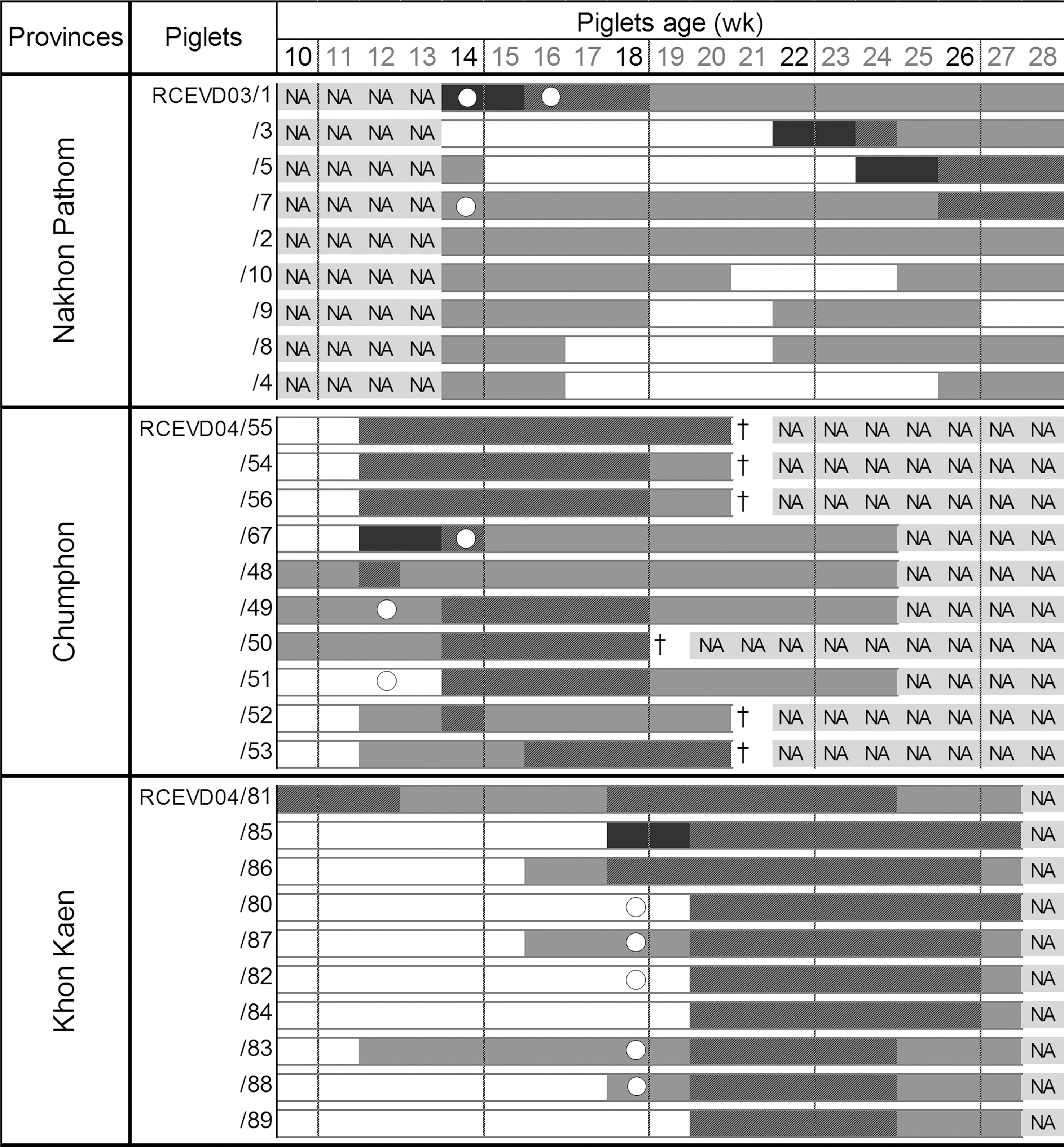
Serosurvey and virus isolation among sentinel piglets of targeted study sites enzootic for Japanese Encephalitis virus in Thailand, 2003–2004. IgM positive (black); IgG positive (dark-gray); IgM and IgG positive (black and dark-gray); negative (white); no data available (NA; light-gray); JEV isolation empty circle; dead piglets (†).
In Nakhon Pathom Province, IgM were detected in 44.4% piglets from January through May 2003. In one case (RCEVD03/1), IgM were detectable on the first virus isolation (14 weeks old) and lasted for 4 weeks; and in the other case, IgM appear 12 weeks after virus detection (RCEVD03/7). IgG were detectable among all the 10 piglets. In five instances (71.4%), piglets (RCEVD03/5, 10, 9, 8, 4) had detectable IgG since the first week of the survey (14 weeks old), without IgM or virus isolation; in four instances, IgG disappeared after 2–6 weeks, showing their potential of maternal origin. Altogether, IgG were detectable among all specimens before they were 24 weeks old.
All 10 piglets from Chumphon Province had an IgM and IgG response; however, 6 of them died of overheat (“heat death in pig” occurs regularly due to the seasonal high temperature) at 19–20 weeks of age without yielding a virus isolate.
JEV IgG and IgM reacting antibodies were observed among all sentinel piglets of Khon Kaen Province. JEV was detected among 5 piglets of 18 weeks of age (RCEVD04/80, 82, 83, 87, 88). All piglets tested positive for JEV detection were found IgM positive after 2 weeks. In one case (RCEVD03/7), IgM were detected after a long period of >10 weeks, suggesting an undetectable or nonimmune response to the infection during that time. Also, regarding piglet RCEVD04/67, we were able to detect an IgM response before virus isolation, suggesting an undetected viremia during the previous weeks, eventually due to a low virus load. IgM were not detected after 2 weeks of infection in piglets RCEVD03/7 and RCEVD04/67. No data were collected in piglets from Nakhon Pathon (weeks 10–13), Chumphon (weeks 25–28), and Khon Kaen (week 28) due to logistical problems.
Discussion
JEV was detected several times among piglets 12–18 weeks old. These results indicate JEV infection in piglets from 12-week-old piglets with a manifest viremic period that may last up to 2 weeks (RCEVD03/1), whereas experimentally reported viremia in pigs lasts for only 1 to 3 days (Williams et al. 2001).
As stated by Salmon (1984) and Salmon et al. (2009), immunoglobulins do not cross the placenta barrier in pregnant sows and neonatal piglets are agammaglobulinemic. They will then acquire passive maternal immunity via colostrum intake providing a high concentration of IgG (85% of the total amount of Ig). Then, before piglet weaning at 4 weeks old, sow milk provides a lactogenic immunity with lower concentrations of IgG and IgM; whereas IgA is the dominant immunoglobulin; also IgG and IgM are progressively degraded by piglets, respectively, with a half time of 15 and 3 days (Salmon 1984, Salmon et al. 2009). Consequently, our results suggest that piglets can be infected at at least 12 weeks of age or potentially earlier despite maternal antibodies. The apparent failure of protective effects of passively transferred maternal antibodies against JEV infection in piglets needs to be investigated.
Moreover, a cross sectional study of pigs from Chiang Mai Province (N. Nitatpattana, unpublished data) pointed out the early response of JEV IgM antibody that appears always in piglets as early as 10 weeks of age. IgM antibodies appear 2–3 days after virus infection and decrease after 2 weeks (Burke and Leake 1988). In our experience, virus isolation often occurs with the presence of IgM (maternal origin) detectable for at least 1 week.
As early noticed (Burke and Leake 1988), intense circulation of JEV occurs in pigs independently of clinical manifestations demonstrating the complex fundamentals involved for disease emergence including a viremic reservoir (piglets), an anthropophilic vector temperature, and rainfall dependent and a limited human population immunity (by natural infection or immunization campaign). During the last decade, the high industrialization of livestock production has occasioned an increase of pig farms in Thailand, favoring high densities of JEV nonimmune amplifying host (piglets). Further, high manure production may favor suitable environmental conditions for the anthropophilic Culex quinquefasciatus, which have specific preference for highly polluted organic matter oviposition sites and have been newly described as efficient vector of the JEV with a tendency to vicariate other vectors (Cx. tritaeniorhynchus) (Nitatpattana et al. 2005). Altogether, the expending pig farm industry and increasing domain of Cx. quinquefasciatus could play an important role in JE epidemics and epidemiological pattern of JEV emergence.
In conclusion, as mentioned by others (Halliday et al. 2007), piglets, as permanent animal sentinels for JEV transmission, are of importance with regard to their high sensitivity to the infection and for a constant follow up of the variability of JE dynamics mostly associated to the vector (rainfall and breeding sites). Moreover, although piglets JEV infection is generally clinically unnoticed, piglets represent an exceptional sentinel model for human health threats (Rabinowitz et al. 2009). Consequently, a practical implementation of pig sentinel surveillance in Thailand should be considered by the health authorities.
Footnotes
Acknowledgments
This study was supported by the Thai Department of Technical and Economic Cooperation and IRD, France. We thank Mr. Teerawut Vongvipirote, Vongveerakij for supplying piglets in this study; Dr. Tawal Poblap, Deputy of Nakhon Pathom Provincial Public Health Office, and his team for their cooperation.
Dr. Nitatpattana is a researcher at the Center for Vaccine development at Mahidol University Bangkok, Thailand. His main research interests are in flavivirus vaccine development, principally against Dengue and JEV.
Disclosure Statement
No competing financial interests exist.