
Abstract
This short communication describes a case of human conjunctival dirofilariosis by Dirofilaria immitis. A 51-year-old man from the Emilia-Romagna region of northeastern Italy referred for ocular foreign body sensation in his right eye. A slight swelling of the bulbar conjunctiva was observed. A live, whitish, 10-cm-long nematode was extracted from the subconjunctival space. Histology revealed typical features of a filarioid nematode belonging to the genus Dirofilaria sp. Molecular analysis with polymerase chain reaction confirmed that the extracted nematode was a male of D. immitis. The removal of the parasite leads to complete resolution of symptoms. Ocular human dirofilariosis caused by infection with the filarioid nematode D. immitis is extremely rare, but should be considered in humans living in Italian endemic areas.
Introduction
Case Report
A 51-year-old Caucasian man from the city of Carpi, located in the Emilia-Romagna region of northeastern Italy, visited the emergency room of the local hospital. He complained of a foreign body sensation in his right eye. Visual acuity, anterior and posterior segment of the eye, and the fundus were all normal. Slit lamp examination showed a swelling of the conjunctiva located in the temporal bulbar portion of the right eye, without significant hyperemia. Initial diagnosis was of a subconjunctival foreign body (possibly plastic), and the ophthalmologist proceeded to identify a possible entrance wound. During this examination, it was noted that direct contact with the swelling caused movement within the tumefaction. Examination at higher magnification revealed the presence of a thread-like object twisted around itself. The conjunctiva was incised and a live, whitish nematode was removed from the subconjunctival space. A part of the worm was fixed in 10% buffered formalin for histology and a part was frozen at −80°C for molecular analysis.
Morphological examination showed a thread-like worm ∼100 mm in length and 330 μm thick. Microscopically, the cuticular surface appeared smooth without longitudinal ridges and the lateral chords showed prominent traverse striations. A single reproductive tube was present, containing degenerated epithelial cells, along with an adjacent tubular intestine. The morphological features were suggestive of a male D. immitis (Fig. 1). Polymerase chain reaction analysis was carried out according to Favia et al. (1996) and the nematode was definitively identified as D. immitis.
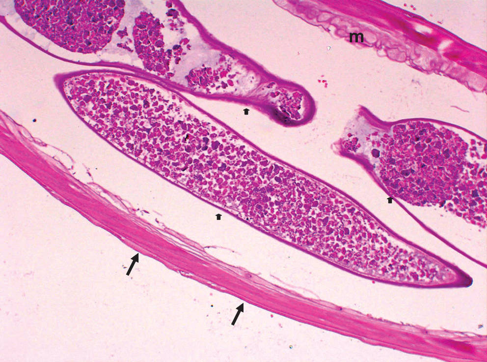
Histology of a cross section of the extracted worm. Note the smooth cuticular surface (arrows), robust muscle layer (“m”), and tubular testis (arrow heads) (H/E, [heamatoxylin/eosin] × 10).
The patient was negative for circulating microfilariae and for the presence of subcutaneous nodules and lymphadenopathy. Thoracic radiography was negative for pulmonary nodules. Removal of the parasite resulted in complete remission of symptoms.
Discussion
As far as the authors are aware, this is the first published case of autochthonous human conjunctival dirofilariosis by D. immitis in Italy. The patient had not traveled outside of the country in the previous 8 years. Orsoni et al. (1985) reported a case of conjunctival D. immitis infection in young boy from Tunisia, which was, however, likely an imported infection.
D. immitis is endemic in many countries, including Italy, but zoonotic infections by this nematode are not common and are almost always pulmonary, and ocular localization is very rare (Simón et al. 2009). Conjunctival infection probably results from the normal migration of the developing worms in the subcutaneous tissues before their entry into the venous circulation, but the exact route of entry into the eye is still unclear. Even though D. repens is responsible for the vast majority of reported cases of HD, there is ample serological evidence that humans living in endemic areas are exposed to infection by D. immitis. One survey reported a mean seroprevalence of 22% in a human population living in a D. immitis–endemic area in Spain, where over 30% of the dogs were infected with the parasite. Age was a significant risk factor and individuals over 60 years of age were over-represented (Simón et al. 1991). Most seropositive humans are asymptomatic.
In this particular case, morphological identification of the nematode as D. immitis was confirmed by polymerase chain reaction analysis. Molecular identification is particularly important when diagnosing HD to decide correct follow-up and preventive measures. Further, it can be very useful when the morphology of the parasite has been altered by surgical excision or inflammatory/degenerative processes.
Surgery is resolving for conjunctival dirofilariosis and there is no published evidence that anthelmintic treatment is either necessary or useful (Mannino et al. 2009). HD is increasing in many European countries and is currently considered an emerging parasitic zoonosis (Kramer et al. 2007). The only effective measure against infection in humans is preventive or curative treatment of the natural, definitive hosts for Dirofilaria sp. The case reported here was clearly autochthonous and it is likely that dogs living in the area have not been adequately controlled. Indeed, dogs are the main reservoir host of D. immitis, while cats play no role in the transmission of the parasite.
In conclusion, D. immitis and D. repens infections in animals and humans are spreading into previously unaffected areas. Further, in many geographical areas where HD is currently endemic, there has been a change in the climatic conditions (temperature, relative humidity, rainfall, and evaporation) that favor the development of the vector mosquitoes and thus the risk of human infection. Medical awareness of infection risk is essential for a correct diagnostic work-up of conjunctival dirofilariosis by D. immitis. This infection often presents as a soft conjunctival nodule, lasting for weeks, with poor inflammatory reaction. The direct contact with the nodule can induce spontaneous movements.
Footnotes
Disclosure Statement
No competing financial interests exist.