
Abstract
Visceral and cutaneous leishmaniases are an important public health problem in endemic geographic regions in 88 countries worldwide, with around 12 million infected people. Treatment options are limited due to toxicity and teratogenicity of the available drugs, response problems in HIV/Leishmania co-infections, and upcoming resistances. In this study, we investigated the anti-leishmanial activity of 13 plant-derived compounds in vitro aiming to find new drug candidates. Toxicity of the compounds was evaluated in human primary hepatocytes, and hemolytic activity was examined in freshly isolated erythrocytes. Two acridones, 5-hydroxynoracronycine and yukocitrine, two flavaglines, aglafoline and rocaglamide, and the sulfur-containing amide methyldambullin showed promising anti-leishmanial activities with 50% effective concentrations (EC50s) of 34.84, 29.76, 7.45, 16.45, and 6.29 μM, respectively. Hepatotoxic activities of 5-hydroxynoracronycine, yukocitrine, and methyldambullin were significantly lower compared to miltefosine and lower or equal compared to artesunate, whereas the ones of rocaglamide and aglafoline were slightly higher compared to miltefosine and significantly higher compared to artesunate. None of the compounds showed hemolytic activity.
Introduction
The classical standard drugs are pentavalent antimony compounds like sodium stibogluconate (Pentostam®) or meglumine antimoniate (Glucantime®), but resistances are increasing and severe side effects have been reported, particularly high rates of cardiotoxicity (Saldanha et al. 2000). Amphotericin B (Fungizone®) became valuable in the treatment of severe mucocutaneous leishmaniasis and visceral leishmaniasis and in cases of pentavalent antimony drug resistance. Cure rates of up to 90% were achieved; nevertheless, amphotericin B resistances have been reported in HIV/Leishmania infantum co-infections (Di Giorgio et al. 1999). Serious acute side effects can occur after infusion, for example, high fever, chills, hypotension, anorexia, nausea, vomiting, headache, dyspnea, tachypnea, drowsiness, generalized weakness, and exceptionally anaphylactic shock or cardiogenic shock. Further, amphotericin B is toxic for hematologic cell lines and for renal function. Liposomal amphotericin B (AmBisome®) is a less toxic alternative to conventional amphotericin B; however, liposomal applications are prohibitively expensive, and therefore it is difficult to increase their use in developing countries. Since 2002, the oral anti-neoplastic agent miltefosine (Impavido®), an alkylphosphocholine, is available on the market for the treatment of leishmaniasis (Sundar et al. 2002). Cure rates of up to 94% have been documented (Olliaro et al. 2005), which are the highest for any available anti-leishmanial drug; however, concerns about developing resistances have been raised, because in vitro resistant strains can be established easily (Seifert et al. 2003). Moreover, miltefosine has been reported to be teratogenic and to affect the digestive system, resulting in vomiting, diarrhea, and motion sickness (Soto and Soto 2006). In the absence of a vaccine, there is an urgent need for effective drugs to replace and supplement those in current use.
The aim of this study was to screen compounds extracted and purified from tropical plants for their anti-leishmanial activity to find new drug candidates. Drugs extracted from plants or their semi-synthetic analogs have been successfully used against other protozoal diseases; for example, quinine (Kyle and Shampe 1974) and artemisinin (Q.A.C.R.G. 1979) are employed in the treatment of malaria. It is estimated that 25% of all drugs prescribed today are derived from plants (Farnsworth and Morris 1976, Newman and Cragg 2007).
In this study, we evaluated the anti-leishmanial activity of 13 plant-derived compounds of four different plant families, namely, Rutaceae, Meliaceae, Stemonaceae, and Asteraceae by in vitro screening. The known antineoplastic activity of some of the substances precluded amastigote testing in human macrophage cell lines (behaving like cancer cells). Also, results obtained from amastigote testing, including tests in primary macrophages, may be misleading because they are highly host cell dependent (Seifert et al. 2010). Thus, we decided to perform the screening with promastigotes. Toxic and hemolytic activities of the most effective compounds were then assessed in separate assays, for which human primary liver parenchymal cells and fresh human blood, respectively, were used. In vitro assays for toxicological assessment of drug candidates reduce the need of in vivo tests, save time, and enable the use of human cell lines or tissues, thus giving results corresponding to the toxicity in humans (Runge et al. 2000, Van de Bovenkamp et al. 2007). Primary hepatocyte cultures have been reported to express most of the functions of an intact liver and are therefore used to evaluate the hepatotoxic activity of new drug candidates (Battle and Stacey 2001, Nussler et al. 2001). Hemolysis can be a side effect of chemotherapy leading to anemia.
Materials and Methods
Parasites
Promastigote L. infantum MCAN/ES/89/IPZ 229/1/89, zymodem MON 1 were cultured in axenic medium for culturing promastigotes (MKP) (pH 7.4; containing 10% fetal bovine serum) at 26°C in 25 cm2 tissue culture flasks (Iwaki) under aerobic conditions (Grimm et al. 1991).
Compounds
Thirteen compounds (Fig. 1)—the flavaglines aglafoline and rocaglamide, the furoquinolines dictamnine and iso-gamma-fagarine, the pyranoquinoline zanthobungeanine, the acridones yukocitrine and 5-hydroxynoracronycine, the coumarins microminutine and methyllacarol, the Stemona alkaloid tuberostemonine, the amide dehydrothalebanin-B, and the sulfur-containing amides methyldambullin and dehydroniranin-A—were extracted from leaves, stem, or rootbark of mostly tropical plants (Table 1). Extraction and purification were achieved by preparative column chromatography and thin layer chromatography, and purity was assessed to be >99% by high performance liquid chromatography and nuclear magnetic resonance spectroscopy. Stock solutions of the compounds were prepared in dimethyl sulfoxide (DMSO; Sigma-Aldrich).
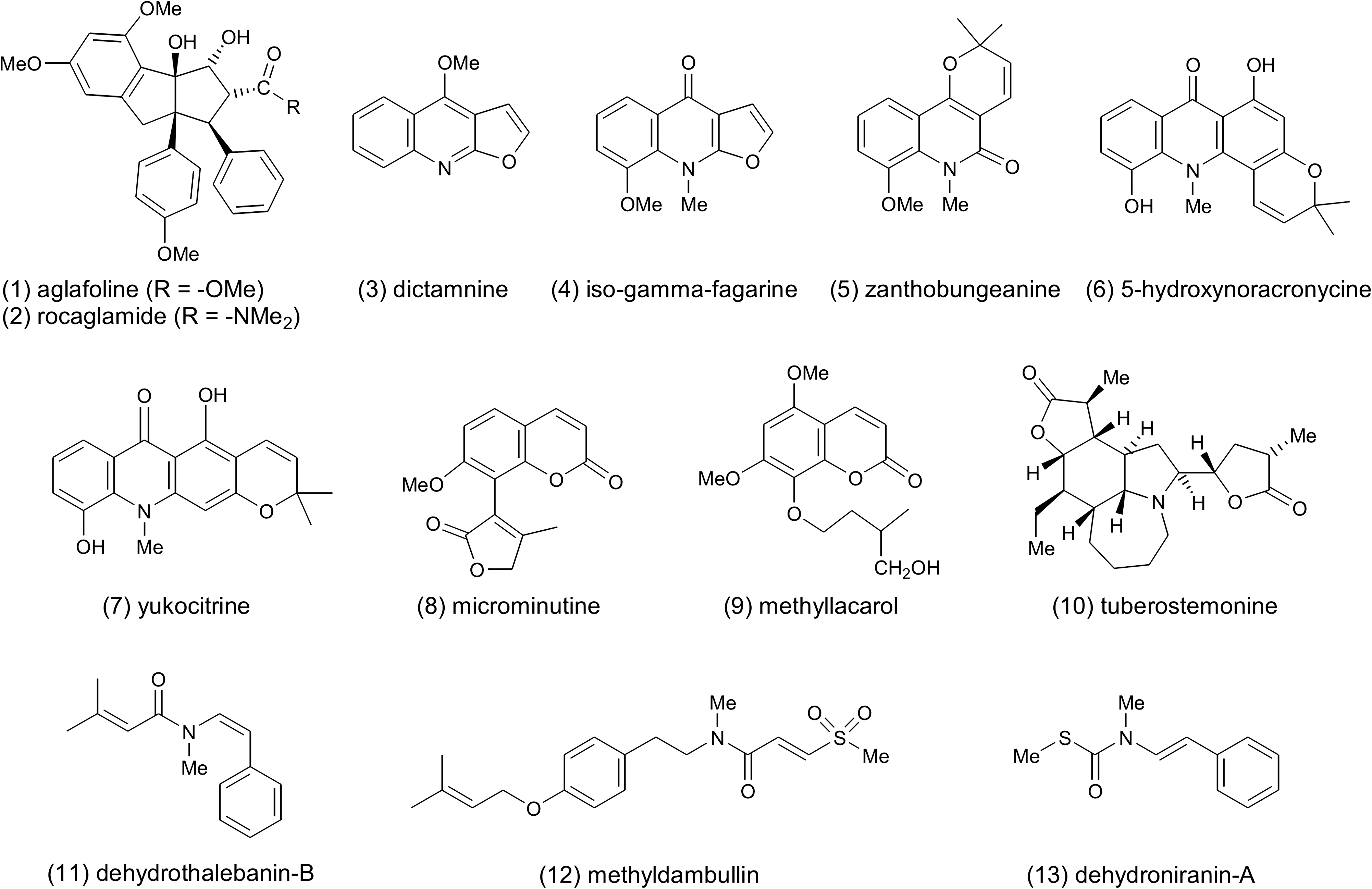
Investigated compounds.
In vitro tests and evaluation
All compounds were tested in 96-well microtiter plates. Wells were inoculated with 105 promastigotes/mL in MKP medium at 26°C. The applied concentrations of the compounds were 0.31–80 μM. Miltefosine was used as control compound. The final DMSO concentration never exceeded 0.2% (v/v) to avoid cell death by the solvent.
Viability of L. infantum cells was determined after 24 and 48 h treatment with a Buerker hemocytometer. All experiments were carried out in triplicates and repeated twice. Fifty percent and 90% effective concentrations (EC50s and EC90s) were determined via log-probit regression using SPSS 15.0 (SPSS Inc., now IBM).
Hepatocyte toxicity assay
Toxicity was investigated via 3-(4,5-dimethylthiazol-2-yl)-2,5-diphenyltetrazolium bromide (MTT) assay, which is based on the reduction of yellow MTT to purple formazan in living cells (Mosmann 1983). After informed consent human primary liver parenchymal cells were isolated from hepatic resection material of a female patient by three-step collagenase perfusion (Strom et al. 1982). The viability of the hepatocytes was 89% before seeding of 96-well microtiter plates and determined by trypan blue exclusion. About 5 × 10⁴ hepatocytes per well were plated out in 100 μL minimum essential medium containing 500 ng/mL insulin and 500 μg/mL gentamycin and cultured at 37°C with 5% CO2. After 3.5 h adherence time, the medium was changed to human hepatocyte maintenance medium (HHMM) (Runge et al. 2000) containing 10 ng/mL hepatocyte growth factor and 20 ng/mL epidermal growth factor. Hepatocytes were pre-cultured for 24 h, then HHMM was removed and hepatocytes were incubated with 50 μL fresh HHMM. The test compounds were applied in concentrations of 7.5, 30, 75, 150, 225, and 300 μM, respectively. The anti-leishmanial drug miltefosine and, because of the toxicity of miltefosine to hepatocytes, the anti-malarial drug artesunate were used as controls. A solvent control with 1% DMSO (v/v), a negative control with HHMM, hepatocyte growth factor, and epidermal growth factor as well as a positive control with 2% Triton X-100 (Sigma-Aldrich) were included. The assays were carried out in triplicates. After 24 h of treatment with the respective compound, 20 μL of 5 mg/mL MTT in phosphate-buffered saline (PBS; GIBCO) was added per well. The hepatocytes were further incubated for 1 h, HHMM was removed, and intracellular formazan was dissolved by adding 130 μL 0.7% sodium dodecyl sulfate in isopropanol for 15 min with gentle shaking. Viability of cells was determined via microplate reader (680 Microplate Reader from Bio-Rad Laboratories) by investigating the reduction of absorption of treated compared to untreated hepatocytes at 595 nm. EC50s were determined via log-probit regression using SPSS 15.0.
Hemolytic activity assay
Fresh human blood was isolated into 10 mL Vacutainer tubes containing potassium–EDTA as an anticoagulant (Widmer et al. 2006), transferred to a 50 mL centrifuge tube, and pelleted by centrifugation at 2000 g for 10 min. Erythrocytes were washed three times with 30 mL of calcium- and magnesium-free PBS (GIBCO) until the supernatant was clear. The final erythrocyte concentration was adjusted to 0.5 × 10⁹ cells/mL. About 0.5 mL of the erythrocyte suspension was mixed with 0.5 mL of the test compound to get final concentrations of 150, 100, 25, 6.25, and 3.125 μM, respectively. The mixtures were incubated at 37°C for 1 h with gentle shaking, centrifuged at 2000 g for 10 min, the supernatant was diluted 10-fold with PBS, and the optical density was measured at 540 nm. The assays were carried out in triplicates.
The values for 0% and 100% lysis were determined by incubating the cells with PBS or 0.1% (wt/volume) Triton X-100 in water, respectively. EC50s were determined via log-probit regression using SPSS 15.0.
Results
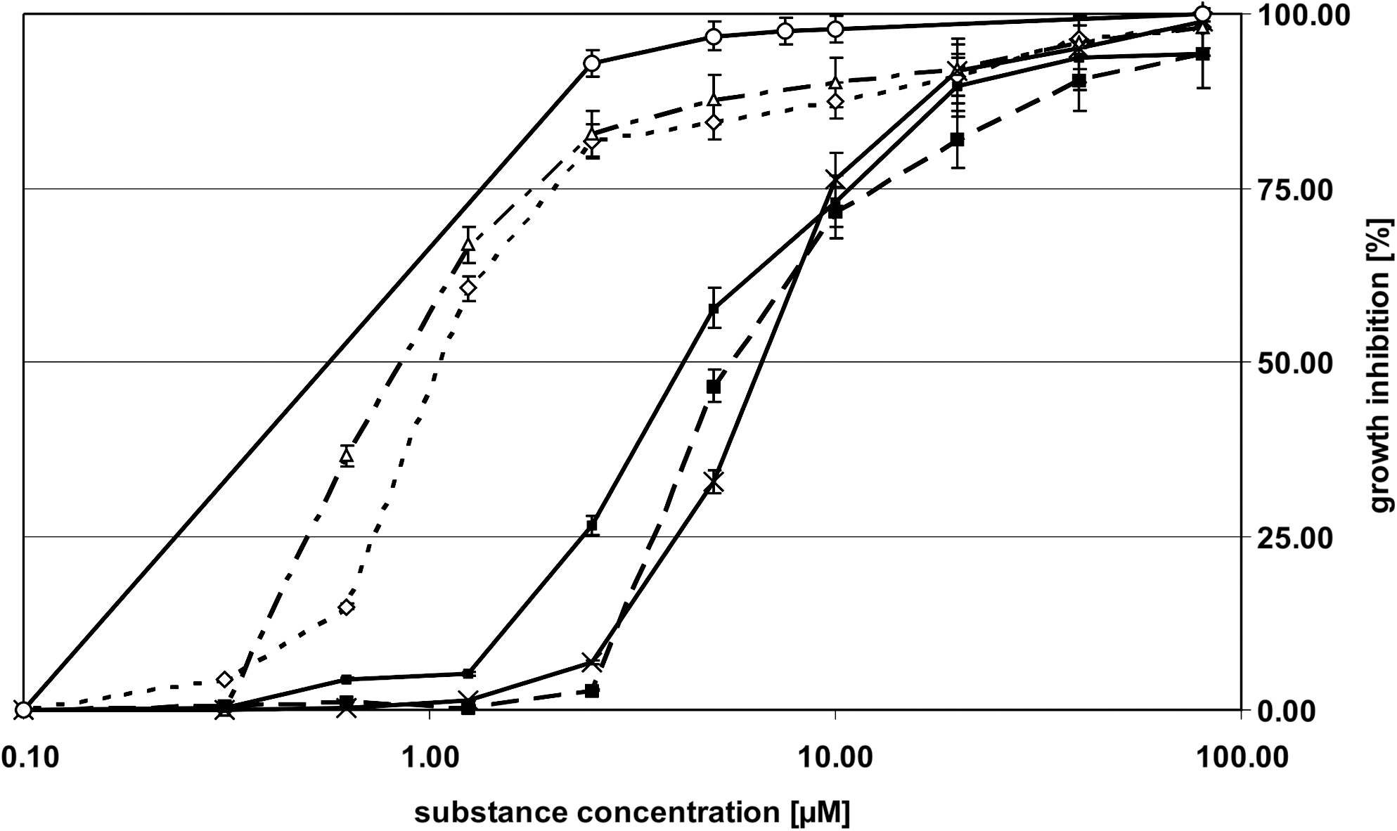
Out of the 13 compounds tested, only 5 compounds—2 acridones, 5-hydroxynoracronycine and yukocitrine, 2 flavaglines, aglafoline and rocaglamide, and 1 sulfur-containing amide methyldambullin—showed anti-leishmanial activity (Table 2). The results after 24 h of exposure are shown in Figure 2. The obtained EC50s for 5-hydroxynoracronycine, yukocitrine, aglafoline, rocaglamide, and methyldambullin were 34.84, 29.76, 7.45, 16.45, and 6.29 μM and the EC90 values were 447.58, 327.58, 63.41, 178.54, and 122.42 μM, respectively. The EC50 for miltefosine was 1.78 μM and the EC90, 9.44 μM.

Growth inhibition of Leishmania infantum MCAN/ES/89/IPZ 229/1/89, zymodeme MON 1 after 24 h treatment with 5-hydroxynoracronycine (solid-square), yukocitrine (chain-dotted-triangle), aglafoline (solid-cross), rocaglamide (dashed-square), and methyldambullin (dotted-diamond) in comparison to standard treatment miltefosine (solid-circle). Standard error of the mean is indicated.
The results after exposure for 48 h are shown in Figure 3. The EC50s for 5-hydroxynoracronycine, yukocitrine, aglafoline, rocaglamide, and methyldambullin were 4.42, 0.88, 6.99, 5.76, and 1.10 μM, respectively compared to miltefosine with an EC50 of 1.27 μM. The obtained EC90s were 20.77, 9.94, 18.81, 38.45, and 16.80 μM, respectively, and the EC90 of the control compound miltefosine was 2.31 μM.
Growth inhibition of L. infantum MCAN/ES/89/IPZ 229/1/89, zymodeme MON 1 after 48 h treatment with 5-hydroxynoracronycine (solid-square), yukocitrine (chain-dotted-triangle), aglafoline (solid-cross), rocaglamide (dashed-square), and methyldambullin (dotted-diamond) in comparison to standard treatment miltefosine (solid-circle). Standard error of the mean is indicated.
The obtained EC50 for 5-hydroxynoracronycine, yukocitrine, aglafoline, rocaglamide, and methyldambullin anti hepatocytes were 201.87, 147.06, 5.07, 4.94, and 48.43 μM, respectively. The EC50s for miltefosine and artesunate were 6.13 and 58.60 μM, respectively.
No hemolytic activity was obtained for 5-hydroxynoracronycine, yukocitrine, aglafoline, rocaglamide, and methyldambullin in concentrations ranging from 150 to 1.56 μM in contrast to miltefosine, for which an EC50 of 119.03 μM was obtained.
Discussion
Two acridones, 5-hydroxynoracronycine and yukocitrine, two flavaglines, aglafoline and rocaglamide, and the sulfur-containing amide methyldambullin showed high anti-leishmanial activity in vitro. Yukocitrine and methyldambullin even had lower EC50s than miltefosine after 48 h of treatment, namely, 0.88 and 1.27 μM, compared to 1.1 μM for miltefosine. Hepatotoxic activities of 5-hydroxynoracronycine, yukocitrine, and methyldambullin, with EC50s of 201.87, 147.06, and 48.43 μM, respectively, were significantly lower and the ones of rocaglamide (4.94 μM) and aglafoline (5.07 μM) slightly higher compared to miltefosine, with an EC50 of 6.13 μM. Compared to artesunate, methyldambullin was slightly more toxic, whereas 5-hydroxynoracronycine was ∼3.5-times and yukocitrine about 2.5-times less toxic. Moreover, none of these compounds showed hemolytic activity. Thus, all five compounds are of interest for further investigations.
The acridones 5-hydroxynoracronycine and yukocitrine were extracted from Glycosmis trichanthera. 5-Hydroxynoracronycine has already been shown to have anti-plasmodial activity (Weniger et al. 2001). An anti-allergic activity has been reported for yukocitrine (Chukaew et al. 2008).
The flavaglines aglafoline and rocaglamide have so far only been detected in Aglaia species and have been mainly explored for their cytostatic activity against tumour cell lines (Wu et al. 1997, Hausott et al. 2004, Proksch et al. 2005), but are also known to have insecticidal activity (Schneider et al. 2000, Greger et al. 2001). In preliminary experiments, aglafoline and rocaglamide showed significant activity against Plasmodium falciparum (Astelbauer et al. submitted).
Methyldambullin is a sulfur-containing amide that has until now only been extracted from the genus Glycosmis, family Rutaceae. Interestingly, a related sulfur-containing amide has recently also been reported for the genus Raphanus, family Brassicaceae (Moon et al. 2010). The sulfur-containing acid moieties are most likely derived from the amino acid cysteine, whereas the amine parts are mostly characterized by phenethyl or phenethenyl groups that additionally can be linked to various prenyloxy structures, for example, methyldambullin and sakambullin, or geranyloxy groups, for example, methylgerambullin. These prenylated amides are mostly oxidized to sulfones or sulfoxides (Hofer et al. 2000). Apart from their chemotaxonomic importance, some sulfur-containing amides show pronounced anti-fungal and insecticidal (Greger et al. 1996) or anti-cancer activity (Mohamed et al. 2004). Interestingly, methyldambullin also shows high efficacy against Trypanosoma cruzi (Astelbauer et al. 2010). However, no further anti-microbial or cytotoxic activities against human cell lines have been reported for methyldambullin.
The compounds 5-hydroxynoracronycine, yukocitrine, and methyldambullin can be chemically synthesized with high yield (Obwaller et al., unpublished), which is a great advantage for in vivo trials in a murine model of Leishmania spp. infection.
Footnotes
Acknowledgments
This study was supported by Grant 814280 from the “Österreichische Forschungsförderungsgesellschaft” (FFG). The authors wish to thank Jacek Pietrzak and Maria Gruber from the Institute of Specific Prophylaxis and Tropical Medicine, Center for Pathophysiology, Infectiology and Immunology, Medical University of Vienna, for excellent technical assistance.
Disclosure Statement
No competing financial interests exist.