
Abstract
Leptospirosis is an important bacterial zoonotic disease globally and one of the notifiable diseases in Sri Lanka. Other than human leptospirosis, little information is available on leptospirosis in domestic and feral animals in Sri Lanka. Thus, this study attempted to determine the prevalence and carrier status of leptospirosis in smallholder dairy cattle and peridomestic rodents to understand the impact of the disease on public health in Kandy, Sri Lanka. Cattle and rodent samples were collected from the Yatinuwara and Udunuwara divisional secretaries in Kandy. Serum samples were analyzed for the presence of antileptospiral antibodies using microscopic agglutination test. DNA was extracted from cattle urine and rodent kidney tissue samples, in which polymerase chain reaction was carried out to detect the Leptospira flaB gene. The cattle in 19 (38.8%) of the 49 farms harbored antileptospiral antibodies. Out of 113 cattle serum samples, 23 (20.3%) were positive; 17 (73.9%) and 6 (26.1%) reacted with serogroups Sejroe and Hebdomadis, respectively. Out of the 74 rodent samples, 13 (17.5%) were positive; 8 (61.5%) and 4 (30.8%) had reactions to serogroups Javanica and Icterohaemorrhagiae, respectively. Leptospiral DNA was detected in one cattle urine sample and identified as Leptospira interrogans. This study revealed a high prevalence of leptospirosis in cattle and rodents in Kandy. These animals were infected with a wide array of leptospiral serogroups, which are consistent with the research findings observed in humans in Kandy. Overall, serological data indicate that relative to rodents, cattle may be a more significant reservoir for human transmission and a greater source of potential risk to local agricultural communities.
Introduction
In animals, clinical presentations after acute infection can range from transient febrile episodes to life-threatening acute hemolytic disease, which depends on the Leptospira serovar–animal relationship. Where maintenance host of a serovar is involved, acute infection may be mild and can result in the establishment of chronic infection followed by an asymptomatic carrier status harboring virulent leptospires in the renal tubules for extended periods and shedding infectious leptospires into the environment (Levett 2001, O'Keefe 2002). Any mammal can be a carrier and shedder of leptospires. Important reservoir animals in human transmission are rats and mice living near human habitats; domestic animals such as cattle and swine; companion animals, especially dogs; and wild animals, especially rodents (Faine et al. 1999). Bovine leptospirosis causes severe economical losses due to abortion, stillbirth, loss of milk production, deaths, and infertility (Ellis 1984). Further, leptospirosis in cattle has been identified as an occupational zoonotic disease in agricultural communities in many countries (Talpada et al. 2003, Belmaker et al. 2004). People who have close contact with tissues or urine of infected bovine face a potential risk of infection, particularly if they have skin lesions or abrasions (Leal-Castellanos et al. 2003).
In Sri Lanka, leptospirosis is one of the notifiable diseases; the first human leptospirosis case was reported in 1953 (Nityananda et al. 1970). One to two thousand cases of human leptospirosis were recorded annually in the past 10 years, and 7421 cases (incidence rate: 36.7/100,000 population) and 207 deaths (case fatality rate: 2.8%) were reported in 2008 throughout the country (Epidemiology Unit Sri Lanka 2009). In Kandy district, the Central Province of Sri Lanka, an increasing trend was also noted since 2004; in particular, a threefold increase (151 in 2007 to 537 in 2008) was reported (Epidemiology Unit Sri Lanka 2008, 2009). Two independent studies that were undertaken in Kandy district indicated that antibodies against serogroup Sejroe were prominent among febrile patients at the local university teaching hospitals (Agampodi et al. 2008, Koizumi et al. 2009a). Another local study detected Leptospira-genus-specific antibodies in 62 of the 123 acute febrile patients from Kandy (Dassanayake et al. 2009). This Leptospira serogroup Sejroe, which consists of 24 serovars, has been identified as an infection agent for humans, bovine, mice, and rats in various regions throughout the world (The Leptospira Molecular Genetics Server 2010). Although human leptospirosis has been recognized as an endemic disease (Epidemiology Unit Sri Lanka 2009), little information is available on leptospirosis in domestic animals in Sri Lanka. According to Nityananda (1970), antileptospiral antibodies were detected in 30% of cattle (prominent serogroup was Sejroe), 22.1% of swine (serogroup was Pomona), and 8.8% of dogs (serogroup involved Canicola). Peiris and Wettimuny (1972) also reported that 8.5% of cattle serum samples reacted with leptospiral antigens whose serogroups were Grippotyphosa, Pomona, and Sejroe, whereas Wijewardana et al. (1995) reported a high prevalence of antileptospiral antibody among buffaloes (41.9%), whose prominent serogroups were Autumnalis followed by Sejroe. However, there is no recent information on the status and burden of leptospirosis among smallholder dairy cattle in Sri Lanka, particularly in Kandy. Thus, this study attempted to determine the local prevalence and carrier status of leptospirosis in the smallholder dairy cattle and peridomestic rodents, and the potential of these animals as reservoir for human transmission. Circulating serogroups and leptospiral species were identified using microscopic agglutination test (MAT) and polymerase chain reaction (PCR), respectively.
Materials and Methods
Site description
The present study was conducted in Yatinuwara and Udunawara divisional secretariats located in Kandy district in the Central Province of Sri Lanka (Fig. 1). A high number of suspected leptospirosis deaths were reported in these secretariats in 2008 (Epidemiology Unit Sri Lanka 2009). These sites, which cover 7.2% of Kandy's land area, lie 465 m above sea level with an annual rainfall of 1840 mm and an average temperature between 22°C and 25°C. Of the total population of the secretariats (209,700), 98% live in rural areas; about one-third (35%) of the inhabitants gain their income from agricultural activities, including dairy. Yatinuwara and Udunuwara consist of 219 Grama Niladhari (GN) divisions, of which 30 were randomly selected for this study. Smallholder dairy cattle and rodent samples were collected in these GN areas. Cattle were sampled from farms that were managed either intensively (i.e., cattle movements and feeding are confined to the farm premises) or semi-intensively (i.e., cattle are both farm-fed and free-roaming).
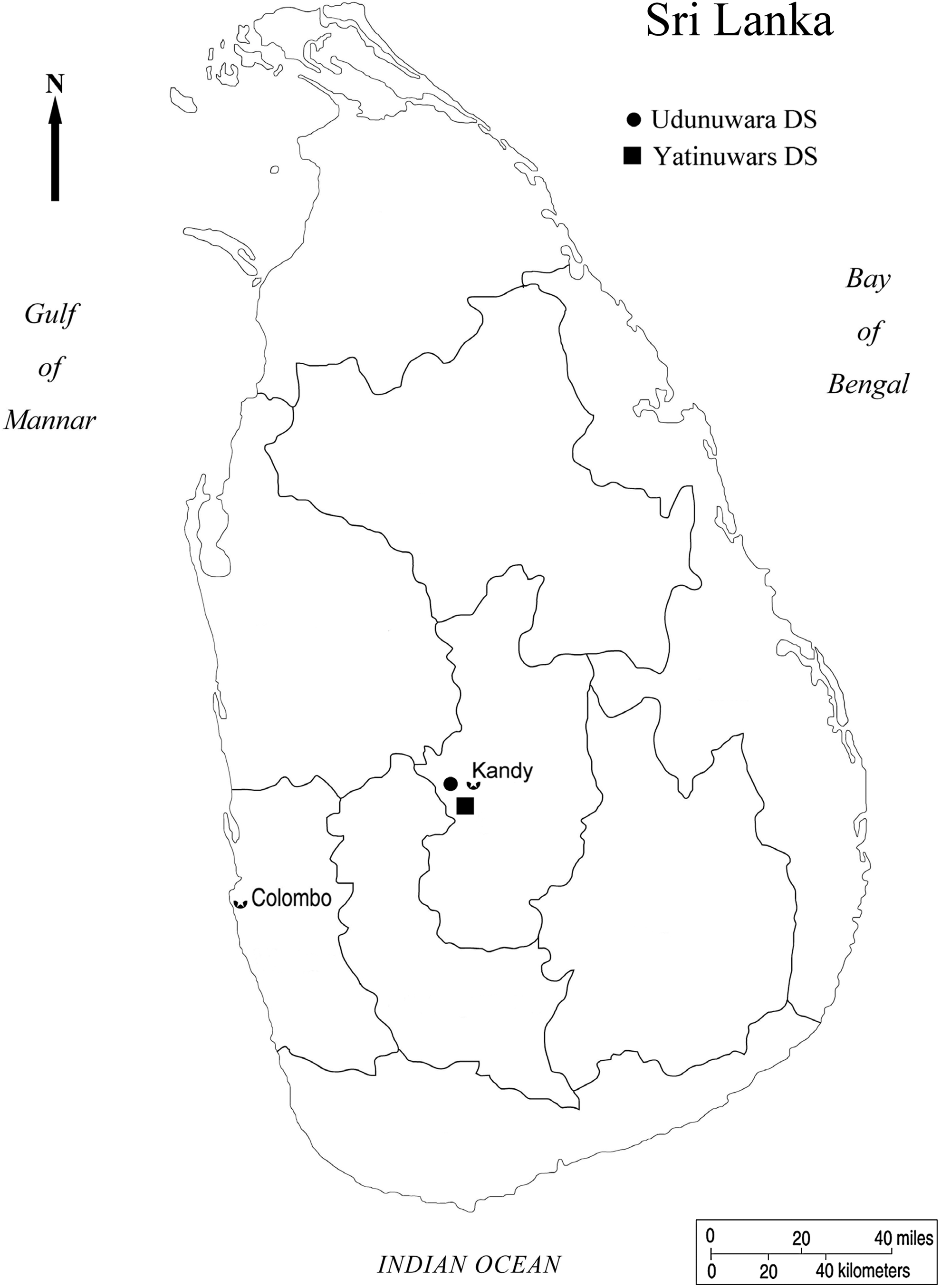
Map of Sri Lanka. Divisional secretariats of Yatinuwara and Udunuwara in Kandy district are indicated.
Sample collection from cattle and rodents
Sampling was carried out between September 5 and October 22, 2009. Two to three cattle samples were collected in each GN area. Samples were taken from apparently healthy, fully grown milking cattle with no stratification to age and dairy management practice. Permission was obtained from the farm owners of the cattle that were included in this study. Livestock development inspectors drew blood from the jugular vein of the cattle and collected 50 mL of their voided urine. Information on dairy practice and animal health was compiled for individual cattle. Rodents were captured using live traps around the farms where the cattle samples were collected. Under anesthetic condition, the heart blood of rodents was aseptically collected and their kidneys were excised. Sera were separated from the blood and stored at −80°C until serodiagnosis. Urine samples were centrifuged (5000 g, 20 min), and the resulting pellets and kidney tissues were kept at −80°C before DNA extraction.
Microscopic agglutination test
The MAT for detecting anti-Leptospira antibodies in cattle and rodent serum samples was performed using a battery of pathogenic reference strains (15 out of 26 pathogenic serogroups) described previously (Koizumi et al. 2009a). These reference strains were cultivated in liquid-modified Korthof's medium with 10% rabbit serum at 30°C (Faine et al. 1999).
Polymerase chain reaction
DNA was extracted from the kidney tissues of rodents and urine pellets of cattle using the DNeasy Blood and Tissue Kit (Qiagen, Hilden, Germany). Extracted DNAs were subjected to PCR to detect the Leptospira flaB gene (nested flaB-PCR; Koizumi et al. 2009b). Sequencing of the amplicon was performed by the dideoxynycleotide chain termination method using the BigDye Terminator V1.1 Cycle Sequencing Kit (Applied Biosystems, Foster City, CA). The flaB sequences were aligned in MEGA4 (Tamura et al. 2007) using CLUSTALW, and phylogenetic distance was calculated in MEGA4 using neighbor-joining method.
Statistical analysis
Using the Statistical Package for Social Sciences version 13.0 (SPSS Inc., Chicago, IL), bivariate analysis was performed to establish the association between the presence of antileptospiral antibodies in cattle as the dependent variable and the sample collection sites, farming management systems, allowing cattle to graze in paddy fields and presence of peridomestic animals surrounding the cattle shed as independent variables (α set at 0.05).
Results
Of the 49 smallholder daily farmhouses, 113 serum and 52 voided urine specimens, and 74 serum and kidney specimens were collected from physically healthy dairy cattle and peridomestic rodents, respectively. Among the 49 farms, 31 (63.2%) were located in Yatinuwara and the rest were located in Udunuwara. Eighteen farms (36.7%) practiced an intensive farm management, whereas the others had a semi-intensive farm management. Regardless of their management system, half of the farms allowed their cattle to graze in paddy fields. Rodents infested the majority of the farms (37, 75.5%), whereas wild boars were seen around the premises in half of the farms (25, 51.0%, Table 1). In 19 (38.8%) of the 49 farms, the cattle harbored antileptospiral antibodies. The presence of antileptospiral antibodies in cattle was not statistically significantly associated with sample collection sites, farming management systems, and allowing cattle to graze in paddy fields, and the presence of peridomestic rodents surrounding the cattle shed (Table 1).
CI, confidence interval.
Antileptospiral antibodies (reciprocal MAT titer ≥100) were detected in 23 of the 113 (20.3%) serum samples of the cattle (Table 2). The predominant reactive serogroups were Sejroe (17/23, 73.9%) and Hebdomadis (6/23, 26.1%). Eight of the 23 positive cattle serum reacted equally to multiple serovars. Among the peridomestic rodents that belong to Bandicota bengalensis (n = 54) and Rattus rattus (n = 20), antileptospiral antibodies (reciprocal MAT titer ≥25) were detected in 13 of the 74 (17.5%) serum samples (11/54, 20.3% of B. bengalensis and 2/20, 10.0% of R. rattus) (Table 3). The predominant reactive serogroups were Javanica (8/13, 61.5%) and Icterohaemorrhagiae (4/13, 30.7%). Serogroups Sejroe, Cynopteri, Javanica, Icterohaemorrhagiae, and Ballum were found in both cattle and rodents (Fig. 2). Leptospiral flaB was detected in 1 of the 52 cattle urine samples and none from rodent kidney tissues. After comparing the nucleotide sequence of the flaB gene derived from the bovine urine against the reference strains, the leptospiral species was deduced to be Leptospira interrogans (Fig. 3). The nucleotide sequence was different from those of human samples in a previous study (Fig. 3 and Koizumi et al. 2009a).

Distribution of Leptospira serogroups among cattle and rodents. Twenty-three of 113 cattle sera and 13 of 74 rodent sera possessed either one or more antileptospiral antibodies. The percentage of each serogroup detected in cattle and rodent sera is plotted.
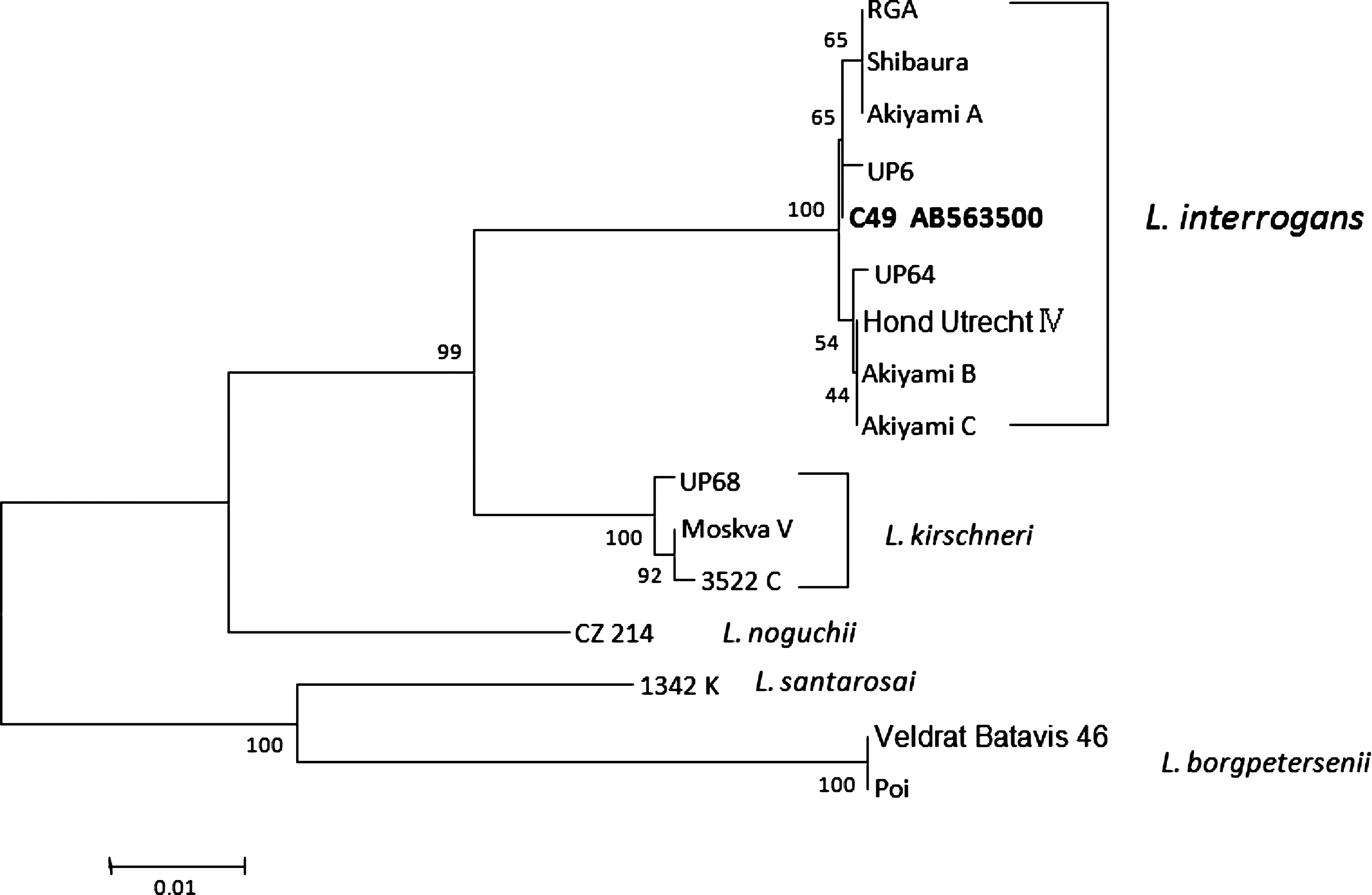
Phylogenetic tree based on the Leptospira flaB gene sequence. The sequences obtained in this study are indicated in bold type and have been deposited in DDBJ/GenBank/EMBL with accession number indicated. The sequences of UP6, 64, and 68 are derived from human serum samples in our previous study (Koizumi et al. 2009a). The sequences were aligned in MEGA4 using CLUSTALW, and phylogenetic distances were calculated in MEGA4 using the neighbor-joining method. Numbers on nodes are bootstrap support after 100 replicates.
Highest reactive serovars of each positive sample are described.
Eight of 23 samples reacted equally with multiple serovars.
MAT, microscopic agglutination test.
Highest reactive serovars of each positive sample are described.
Four of 13 samples reacted equally with multiple serovars.
Those antibodies were detected in 2 Rattus rattus and the others were 11 Bandicota bengalensis.
The serum sample from one R. rattus reacted equally with these three strains.
Discussion
The present study aimed to determine the prevalence and carrier status of leptospirosis in smallholder dairy cattle and peridomestic rodents in Kandy, and their potential role as reservoir for human infection. Biological samples of cattle and rodents were collected and examined by MAT and PCR.
In the study area, 20.3% of cattle were infected predominantly with serogroup Sejroe and 17.5% of peridomestic rodents with serogroups Javanica and Icterohaemorrhagiae. Since the dairy cattle in Sri Lanka are not vaccinated against leptospirosis, the detected antibodies are attributed to natural infection. However, because MAT titers persist for a long time (5–10 years) after the infection (Levett 2001), it is difficult to differentiate acute infection from chronic one by single sample analysis as used in this study. Further, the actual seroprevalence of cattle and rodents might be higher than that found in the present study, because MAT detects serogroup-specific antibody and only 15 serogroup strains out of 26 pathogenic serogroups were employed in the current research. Previous local studies reported rates as low as 8.5% (Peiris and Wettimuny 1972) and as high as 30.0% (Nityananda 1970) among cattle harboring antileptospiral antibodies in different parts of Sri Lanka. Other studies have reported similar results in several regions around the world (Talpada et al. 2003, Odontsetseg et al. 2005, Leon et al. 2008, Hesterberg et al. 2009).
The high prevalence of antileptospiral antibodies was seen in peridomestic rodents and similar Leptospira serogroups were detected in both cattle and rodent serum samples (Tables 2 and 3 and Fig. 2). These results imply that peridomestic rodents are potential reservoirs of leptospires for dairy cattle infection in this area. The role of rodents to transmit chronic leptospirosis to humans, livestock animals, and wildlife has been reported (Bunnag et al. 1983, Witmer et al. 2004). In addition to rodent infestation, half of the cattle farms were contaminated by wild boar's excreta. Previous studies suggested that infected wild boars could infect humans and domestic animals (Ebani et al. 2003, Jansen et al. 2007, Koizumi et al. 2008). Therefore, farm management practices, such as reducing the rodent population and preventing wild boars access to farm premises, are important for preventing leptospirosis in humans and cattle.
The present study was able to discover the carrier status of dairy cattle in the area by detecting leptospiral DNA in cattle urine using flaB-PCR (Fig. 3). Thus, infected cattle could be a source of infection for the rest of the herd. Further, half of the dairy farmers allowed their cattle to free-roam in the area, suggesting that cattle could also contaminate the environment.
In this study, antibodies against 12 and 7 Leptospira serogroups were detected in cattle and rodents, respectively. Previous investigations identified 13 (Koizumi et al. 2009a) and 8 serogroups (Agampodi et al. 2008) in human patients in the same area. All these studies revealed Sejroe as the most predominant serogroup in both dairy cattle and humans, suggesting that smallholder dairy cattle are an important reservoir for human leptospirosis in Kandy.
Other than its zoonotic nature, bovine leptospirosis has financial implications for the dairy industry as the disease could result in infertility, abortions, higher culling rates, and decrease in milk production of the cattle (Langoni et al. 1999). The smallholder dairy industry is one of the major components of the rural economy in Sri Lanka (Ranaweera 2009); therefore, it is vital to reduce leptospiral infection among dairy cattle.
Conclusion
This study revealed the existence of leptospirosis in cattle and peridomestic rodents in Kandy. These animals were found to have been infected with a wide array of leptospiral serogroups. These results suggest that the cattle may act as a significant reservoir of leptospirosis in the area and pose a potential risk to local agricultural communities. Dairy cattle should be prevented from being contaminated by the excreta from peridomestic rodents and wild boars. To identify important reservoirs, future studies must isolate and characterize leptospires from domestic and feral animals, and humans.
Footnotes
Acknowledgments
We are grateful to Yasuda S.P. and Suzuki H. for their advice on the identification of rodents by mitochondrial cytb sequencing. We also thank the support of the Global Center of Excellence program “Establishment of International Collaboration Centers for Zoonoses Control,” Ministry of Education, Culture, Sports, Science and Technology, Japan. The authors appreciate the cooperation and participation of farmers, field enumerators, and technical assistants.
Ethical Clearance
This study, which was a part of “An Ecological and Molecular Epidemiological Study of Leptospirosis in Sri Lanka,” was approved by the Committee on Research and Ethical Review, Faculty of Medicine, University of Peradeniya, Kandy, Sri Lanka (2009/EC/26).
Disclosure Statement
No competing financial interests exist.