
Abstract
Flea and tick specimens (5–10 fleas or ticks) on dogs and cats from various sites in Bangkok were tested by polymerase chain reaction and DNA sequencing to detect DNA of bacteria Rickettsia (gltA and 17 kDa genes), Anaplasmataceae (16S rRNA gene), and Bartonella (pap31 and its genes). We confirmed that Rickettsia sp. related to Rickettsia felis was detected in 66 of 98 (67.4%) flea specimens from dogs, whereas 8 Bartonella henselae and 2 Bartonella clarridgeiae were detected in 10 of 54 (18.5%) flea specimens from cats. Further, this work provides the first evidence of 10 Ehrlichia canis (3.3%), 7 Anaplasma platys (2.3%), and 2 Wolbachia spp. (0.66%) in 304 Rhipicephalus sanguineus tick specimens in Thailand.
Introduction
In Thailand, the first cases of Spotted Fever Group (SFG) rickettsioses were identified serologically in three people in 1994 (Sirisanthana et al. 1994). In 2003, Parola and colleagues reported the presence of rickettsioses in rural residents of the Thai-Myanmar border region. They tested the blood of 46 patients with fever. Four patients had murine typhus, three had scrub typhus, and eight had SFG rickettsioses including the first case of Rickettsia felis infection in Asia (Parola et al. 2003a). They also reported two Rickettsia spp. in Dermacentor ticks, three Ehrlichia spp. in Boophilus microplus and Hemaphysalis hystricis ticks, and three Anaplasma spp. in Dermacentor auratus, Amblyomma javanense, and Hemaphysalis lagrangei ticks (Parola et al. 2003b). The same year, rickettsial DNA (GenBank accession numbers AF483196, AF483198, and AF483199) was detected in 30% (9/30) of Amblyomma testudinarium ticks and 16.84% (16/35) of Hemaphysalis ornithophila ticks collected from a national park (Hirunkanokpun et al. 2003).
The first report of Bartonella infection in Thailand was the seroprevalence of Bartonella henselae in healthy Thai individuals. A total of 5.5% (9/163) were found to be positive for B. henselae IgG and 1.2% (2/163) for B. henselae IgM (Maruyama et al. 2000). The prevalence of Bartonella infection in 275 cats from four geographical regions was 27.6% (Maruyama et al. 2001). In 2003, Parola and colleagues found B. henselae in 2 Ctenocephalides felis fleas, Bartonella clarridgeiae in 3 C. felis fleas, and two genotypes related to R. felis in 3 Ctenocephalides canis, and one C. felis in 90 fleas collected in the Thai-Myanmar border (Parola et al. 2003b).
Although several studies of tick-borne and flea-borne bacterial diseases in Thailand have been conducted, the knowledge of these zoonoses is insufficient. Therefore, the potential vectors and animal reservoirs should be studied to provide crucial information about these zoonoses. Recently, the first human case of SFG rickettsiosis in urban Bangkok was reported by serological and molecular diagnosis (Sangkasuwan et al. 2007). Thus, Bangkok may be an emerging area of rickettsial pathogens.
The aim of this study was to examine the presence of Rickettsia, Ehrlichia, Anaplasma, and Bartonella spp. in ticks and fleas from dogs and cats from various sites in Bangkok.
Materials and Methods
Tick and flea samples
Rhipicephalus tick specimens (5–10 ticks/dog) were collected on dogs and flea specimens (5–10 fleas/dog or cat) were collected on dogs and cats from veterinary clinics and hospitals or dogs present in public places (i.e., stray dogs) in 45 districts of Bangkok. Dogs and cats used in this study were not from the same house or village. Sampling at these sites was conducted from June 2006 to December 2007. Ticks and fleas were identified by their morphology using microscopy according to standard taxonomic keys (Ruedisueli and Manship 2006, Titz 2007). They were stored in 70% ethanol at −20°C until processing.
Preparation of DNA from ticks and fleas
Ticks and fleas were washed in 0.1% bleach–Tween 80 and in benzalkonium chloride, then rinsed in distilled water, and dried. DNA was extracted from ticks or flea specimens using the High-Pure PCR Template Preparation Kit (Roche Applied Science) according to the manufacturer's instructions. Samples were crushed in a clean 1.5-mL microcentrifuge tube using a tissue micropestle. DNA was extracted from ticks and fleas by detergent lysis in the presence of proteinase K for 3 h at 55°C. DNA samples were stored at −20°C until use in polymerase chain reaction (PCR) or as sequencing templates.
Detection of Rickettsia spp., Bartonella spp., and Anaplasmataceae
Bacterial DNA was detected by PCR with genus-specific primers from two different target genes for each organism (Table 1). Extracted DNA from ticks was assayed for Rickettsia, Ehrlichia, and Anaplasma species. Extracted DNA from fleas was assayed for Rickettsia and Bartonella species.
PCR assay
All PCR assays were performed in 25 μL of reaction mixture containing 5 μL of extracted DNA, 0.2 pmol of each primer, and 12.5 μL of 2×Illustra Hot Start master mix (GE Healthcare). At the final 1×concentration, each reaction contained 2.5 U of Taq DNA polymerase, 200 μM of each dNTP, and 1.5 mM MgCl2. The amplification reaction was performed in a Mastercycler Gradient (Eppendorf). For the inner primer reactions, 2 μL of the outer PCR products were added into the reaction mixture. To avoid false-positive results from carry-over contamination of amplified product, the areas where PCR work was performed was separated into pre-PCR, PCR, and post-PCR areas with dedicated sets of automatic pipettes and filter tips. Positive and negative quality controls were included in each experiment. Negative controls included extraction controls as well as controls using distilled water as the template.
PCR detection of Rickettsia-specific DNA
We used PCR targeting two rickettsial specific genes: citrate synthase (gltA) and the 17 kDa gene. The primers RpCS.877p and RpCS.1258n were used to amplify a 381-bp fragment of the citrate synthase (gltA) gene, whereas 434- and 320-bp fragments of the 17 kDa gene were amplified from seminested PCR utilizing primers Rr17.61–Rr17.492 and F-Jari-17-FDW-Rr17.492, respectively, and amplification conditions described elsewhere (Schriefer et al. 1994, Noda et al. 1997). Rickettsia typhi DNA was used as a positive control.
PCR detection of Bartonella DNA
Bartonella DNA was detected by PCR targeting heme-binding phage-associated protein (pap31) and the 16S–23S rRNA intergenic spacer region (its) genes, as previously described (Rolain et al. 2003). The primers PAPn1 and PAPn2 were used to amplify a 274-bp fragment of the heme-binding pap31 gene, whereas primers URBarto1 and URBarto2 were used to amplify a 750-bp fragment of 16S–23S rRNA its gene (Rolain et al. 2003). B. henselae DNA (a clinical isolate identified by 16S rRNA sequencing) was used as a positive control. The positive control DNA samples were amplified using species-specific primers BartogltAForward and Bartohenselae for amplification of a 246-bp fragment of the gltA gene of B. henselae (Rolain et al. 2003).
PCR detection of Anaplasma and Ehrlichia-specific DNA
For Anaplasma/Ehrlichia detection, DNA was amplified by PCR targeting of the 16S rRNA gene. The primers EHR16SD and EHR16SR were used to amplify a 345-bp fragment of the 16S rRNA gene from all members within the family Anaplasmataceae, as previously described (Parola and Raoult 2001). Ehrlichia DNA isolated from blood samples of a dog with clinical signs of ehrlichiosis confirmed by microscopic examination was used as a positive control. The positive DNA samples were amplified using species-specific primers HE1 and HE3 to amplify a 389-bp fragment of the 16S rRNA of Ehrlichia chaffeensis and GE9f and GE10r to amplify a 919-bp fragment of the 16S rRNA of Anaplasma phagocytophilum under previously described conditions (Anderson et al. 1993, Chen et al. 1994).
PCR product analysis
PCR products were resolved by electrophoresis in 1% agarose gels and were purified by using Nucleospin® Extract II (Macherey Nagel) before sequencing. PCR products of the correct size were sequenced using the d-rhodamine terminator cycle-sequencing ready reaction kit (PEApplied Biosystems) according to the manufacturer's protocol. Sequences obtained were compared with those in the GenBank DNA database using the program BLAST (version 2.0, National Center for Biotechnology Information; available from
The partial nucleotide sequence of the identical 17 kDa gene from samples, named BKK20, was analyzed for phylogenetic relationship with the corresponding Rickettsia from GenBank. Phylogenetic analysis of 16S rRNA gene of Wolbachia spp. 31T and 95T detected in this work were compared with those downloaded from GenBank. Multiple-sequence alignments with the corresponding sequences, phylogenetic and molecular evolutionary analyses were conducted using MEGA version 4 (Tamura et al. 2007). The evolutionary distances were computed using the Kimura two-parameter method. One thousand bootstrap replicates were conducted for the reliabilities of the nodes on the phylogenetic trees.
Results
A total of 152 flea specimens were collected from 98 dogs and 54 cats. Seventy-eight specimens were identified as C. canis and 74 as C. felis. Three hundred four tick specimens (5–10 ticks/dog) of Rhipicephalus sanguineus were collected from 304 dogs.
Rickettsia DNA detection in ticks and fleas
Rickettsia DNA was detected in 66 of 152 (43.42%) flea specimens, including 49 of 78 (62.82%) from C. canis and 17 of 74 (22.97%) from C. felis as shown in Table 2. The percentage of positive specimens detected for C. canis (62.82%) was higher than that for C. felis (22.97%) (Fisher's exact test, p<0.0001). Eighty animal sources had clinical histories available regarding the presence of fever (rectal temperature: >102.5°F). The number of flea specimens containing rickettsial DNA from dogs with fever (17 of 26, 65.38%) was not significantly greater than that of healthy dogs without fever (30 of 54, 55.56%) (p>0.05). Rickettsia was not detected in any flea specimens collected from cats, or from tick specimens. All positive and internal controls gave the expected PCR products 100% of the time, and all negative controls were consistently negative for detectable PCR products.
The sequences of the 39 PCR products from the 17 kDa gene were determined. All sequences from these samples showed the same genotype, named Rickettsia sp. BKK2007. They were identical to those of Rickettsia sp. cf1and5 (GenBank accession number: AY953286) and Rickettsia sp. SE313 (GenBank accession number: DQ166937). They were closely related to Rickettsia sp. FS27 (99.7% similarity; GenBank accession number: DQ395097), Rickettsia tamurae (98% similarity; GenBank accession number: AB114825), Rickettsia australis (96.6% similarity; GenBank accession number: M74042), and R. felis (96.6% similarity; GenBank accession number: AF195118). The phylogenetic relationships of these Rickettsia nucleotides are depicted in Figure 1. The sequences of the 329-bp gltA fragments obtained from two samples (F143 and F144, deposited in GenBank under the accession numbers JF511463 and JF511464, respectively) were identical to that of Rickettsia sp. RF2125 (GenBank accession number: AF516333), Rickettsia sp. cf1and5 (GenBank accession number: AY953289), and Rickettsia sp. SE313 (GenBank accession number: DQ166938).

Phylogenetic relationships between 355 bases of 17 kDa gene of Rickettsia sp. BKK2007 from flea samples in Bangkok (deposited in GenBank under the accession numbers JF511461) and the 18 corresponding Rickettsia from GenBank under the indicated accession numbers: Rickettsia sp. cf1and5 AY953286, Rickettsia sp. SE313 DQ166937, Rickettsia sp. FS27 DQ395097, Rickettsia sp. cf15 AY953285, Rickettsia tamurae AB114825, Rickettsia felis GU447234, R. felis URRWXCal2 CP000053, Rickettsia australis M74042, Rickettsia akari CP000847, Rickettsia rhipicephali U11020, Rickettsia japonica D16515, Rickettsia africae ESF-5 CP001612, Rickettsia conorii M28480, Rickettsia rickettsii CP000766, Rickettsia sp. 777c EU283838, Rickettsia sp. TR-39 DQ480762, Rickettsia typhi M28481, and Rickettsia prowazekii CP001584. Phylogenetic analyses were constructed by neighbor-joining method using MEGA 4 (Tamura et al. 2007). The evolutionary distances were computed using the Kimura two-parameter method. Bootstrap values of 1000 resamplings are indicated at the branch nodes.
Bartonella DNA detection in fleas
Using primers targeting the pap31 gene of Bartonella, 274-bp (pap31) and 750-bp (its) fragments were detected in 10 of 152 (6.58%) flea specimens collected from cats, as shown in Table 2. Subsequently, the positive flea specimens were amplified with species-specific primers targeting the gltA gene of B. henselae. The 246-bp PCR product was detected in 8 of 10 positive flea specimens. The 246-bp PCR product was purified and sequenced to confirm the PCR results. The sequences were identical to that of B. henselae (Genbank accession number: AM294993).
Sequences of the 750-bp amplicons of the its gene from two B. henselae–negative flea specimens (F29 and F127) were identical to that of B. clarridgeiae (GenBank accession number: DQ683194). Bartonella DNA was not detected in any of the flea specimens collected from dogs.
Ehrlichia and Anaplasma DNA detection in ticks
Using the broad-spectrum primers EHR16SD and EHR16SR targeting the 16S rRNA of bacteria within the family Anaplasmataceae, a 345-bp PCR product was detected in 19 of the 304 (6.25%) tick specimens (Table 3). Ten sequences were identical to the sequence of Ehrlichia canis (GenBank accession number: EU263991). Seven sample sequences were identical to the sequence of Anaplasma platys (GenBank accession number: EU090182). Two samples, 31T and 95T, showed 99% and 95.8%, respectively, similarity to Wolbachia pipientis (GenBank accession number: X65670). In the phylogenetic tree based on 275 bp of the 16S rRNA sequences, 31T and 95T were placed in different branches. They were related to W. pipientis and Wolbachia endosymbiont from arthropods and one nematode, as shown in Figure 2.
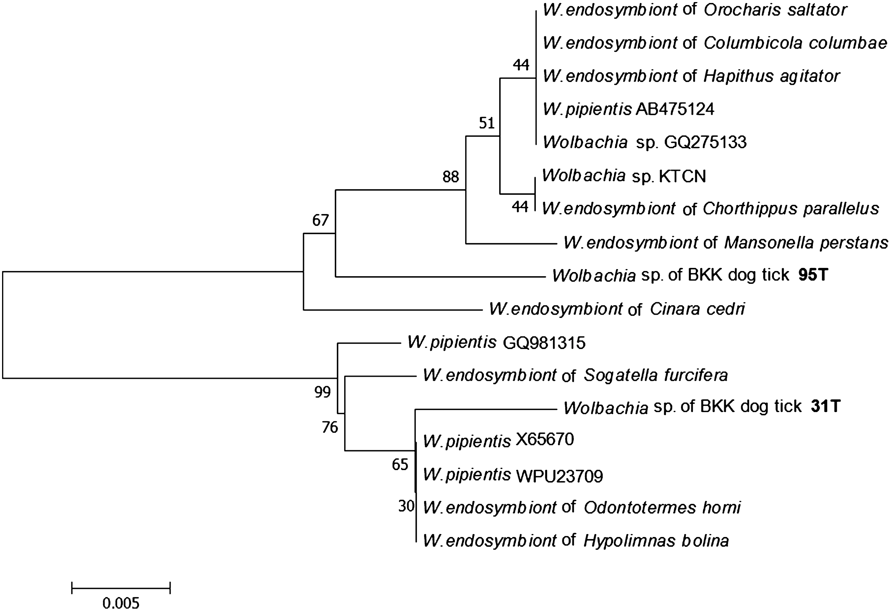
Phylogenetic reconstruction based on analyses of 279 bases of 16S ribosomal RNA gene showing position of Wolbachia sp. BKK dog tick 31T and 95T (deposited in GenBank under the accession numbers JF511466 and JF511465, respectively) detected in this work and 15 representatives of Wolbachia deposited in GenBank under the following accession numbers: Wolbachia pipientis WPU23709, W. pipientis X65670, W. pipientis GQ981315, W. pipientis AB475124, Wolbachia sp. GQ275133, Wolbachia endosymbiont of Cinara cedri AY620430, W. endosymbiont of Mansonella perstans AY278355, W. endosymbiont of Chorthippus parallelus FJ438538, W. endosymbiont of Odontotermes horni GQ422896, W. endosymbiont of Sogatella furcifera GQ206310, Wolbachia sp. KTCN AB508951, W. endosymbiont of Hapithus agitator DQ536098, W. endosymbiont of Hypolimnas bolina GU091499, W. endosymbiont of Orocharis saltator DQ536097, and W. endosymbiont of Columbicola columbae DQ498981. Phylogenetic analysis was conducted in MEGA4 (Tamura et al. 2007). Bootstrap values of 1000 resamplings are indicated at the branch nodes.
Discussion
This is the first report of Rickettsia sp., B. henselae, and B. clarridgeiae in fleas from this area. DNA of a single Rickettsia sp. and two Bartonella species were identified in these fleas using PCR with primers specific for different species-specific genes, followed by DNA sequencing to confirm the results. No co-infection was observed in this study. No PCR products were amplified in our negative controls, and all fleas that tested positive were also positive when other PCR amplifications using primers for different target genes were tested. Contamination of the samples was unlikely because fleas from different collections were tested on separate dates, and the negative controls were never contaminated.
In this work, the genotypic species of the Rickettsia sp. BKK2007 were detected in fleas found on dogs in Bangkok. This rickettsia has been detected in fleas at the Thai-Myanmar border, Rickettsia sp. RF2125 (Parola et al. 2003c), and at the United States, Rickettsia sp. cf1and5 (Reeves et al. 2005). In a phylogenetic analysis based on the alignment of the gltA gene, Rickettsia sp. RF2125 clustered together with R. felis (Parola et al. 2003c). Recently, the same rickettsia genotype in fleas from rats in Egypt was named Rickettsia sp. SE313 (Loftis et al. 2006). This genotype was also demonstrated in fleas in Uruguay and in Hungary (Venzal et al. 2006, Hornok et al. 2010). Although R. felis has been reported worldwide, this study did not demonstrate the presence of R. felis in the fleas and ticks collected.
Therefore, our findings support the notion that these bacteria might be carried by cat and dog fleas and that they are distributed worldwide. This rickettsia is not known to be a pathogen in humans or dogs. The public health implications of this new Rickettsia sp. are unknown. Consequently, the pathogenicity and biology of this rickettsia should be studied. In addition, the Rickettsia detected in C. felis might be antigenically similar to R. felis and cross react when analyzed using an indirect fluorescent antibody test. Further research should clarify the roles of fleas in the natural history of rickettsial pathogens in Thailand.
This is the first time that dog fleas in Bangkok were found to be associated with Rickettsia sp. Interestingly, it was found in 43.42% flea specimens collected from dogs but none from cats. Although fleas from which it was detected included both C. canis and C. felis, the percentage of positive specimens in C. canis (62.82%) was higher than in C. felis (22.97%) (p<0.0001). These data suggest that dog may be a main reservoir of this rickettsia.
Although, Rh. sanguineus is known to be the main vector of Rickettsia conorii, an agent of rickettsiosis in humans (Raoult and Roux 1997), no rickettsial DNA was detected in any of the brown dog tick specimens collected from the domestic dogs included in the study. This is of importance for epidemiologic research.
We show the evidence of B. henselae and B. clarridgeiae in fleas from Bangkok, and not only C. felis but also C. canis is a vector for B. henselae as reported in Spain (Blanco et al. 2006, Márquez et al. 2009). Of 53 flea specimens collected from cats in Bangkok, B. henselae was present in 15.1% and B. clarridgeiae was present in 3.8%. The cat is the main reservoir for B. henselae and B. clarridgeiae, whether or not the cat has symptoms. The number of fleas from which Bartonella DNA was detected in cats with a history of fever (rectal temperature: >102.5°F) (4/19, 21.05%) was not different from cats without a history of fever (6/35, 17.14%). Therefore, the fever in these cats was unlikely to have been caused by Bartonella.
Anaplasmataceae DNA was detected in 19 of the 304 (6.25%) tick specimens. In this study, E. canis, A. platys, and Wolbachia spp. were confirmed in ticks from dogs, with a rate of detection of 3.3%, 2.3%, and 0.66%, respectively. E. canis and A. platys are important veterinary etiologic agents in dogs. In Thailand, Suksawat et al. (2001a) reported a dog that was co-infected with E. canis and A. platys. They also reported that of 49 dogs 10 were infected with E. canis and 5 were infected with A. platys (Suksawat et al. 2001b).
Wolbachia spp. are Anaplasmataceae members related to rickettsial pathogens. They are obligate intracellular bacterial parasites that infect a wide range of invertebrate hosts including insects, crustaceans, arachnids, and nematodes (Fenn and Blaxter 2006). In Thailand, there are many reports of Wolbachia in mosquitoes, but few studies of this organism in fleas and ticks (Noda et al. 1997). In 2003, Hirunkanokpun et al. (2003) studied tick-associated bacteria, including Wolbachia, in 334 ticks collected from 10 locations in Thailand. Wolbachia DNA was not found in any ticks in that study.
Our results may be the first report of the presence of E. canis, A. platys, and Wolbachia spp. DNA in Rhipicephalus ticks in Thailand. It is possible that the ticks that yielded the E. canis and A. platys DNA could have been feeding on bacteremic dogs. This indicates that the Rhipicephalus ticks collected from dogs in Bangkok may harbor both canine pathogens, including E. canis and A. platys, and the endosymbiotic bacteria of insects. A. platys and Wolbachia spp. are not known to be pathogens in humans. E. chaffeensis DNA was not detected in either this tick study or that of Parola in 2003 (Parola et al. 2003a, 2003b).
In summary, these results provide evidence that, in Bangkok, fleas from dogs are commonly infected with Rickettsia sp. related to R. felis and fleas from cats are often infected with Bartonella spp., whereas ticks can harbor E. canis, A. platys, and Wolbachia spp. The rickettsia organisms are widespread around Bangkok, but the pathogenicity of this rickettsia has not been established in humans or dogs. Thus, further study of this rickettsia should be of concern in Thailand.
Footnotes
Acknowledgments
This research was supported by Siiraj Grant Research and Development and Medical Education, Faculty of Medicine Siriraj Hospital, Mahidol University.
Disclosure Statement
No competing financial interests exist.