
Abstract
We conducted a serosurvey for West Nile virus (WNV) infection in equines in Costa Rica in 2004. Antibodies to WNV were detected in 28% of the horses using an epitope blocking ELISA that is specific for WNV. WNV infection was confirmed for a subset of these sera by plaque reduction neutralization tests and Western blot. This is the first evidence of WNV activity in Costa Rica.
Introduction
Materials and Methods
Sera were obtained from horses in the north-western region of Costa Rica (Fig. 1, Liberia, Filadelfia, and Bolson) on December 8–15, 2004. The age of the horses ranged from 6 months to 18 years (Table 1), although the age of some horses was not known. The horses were asymptomatic at the time of sera collection, were not known to have traveled away from the area, and had not been vaccinated against WNV. Sera were lyophilized and sent to The University of Queensland, Australia, for assessment. Upon receipt, the sera were gamma irradiated, re-hydrated, and tested for antibodies to WNV. All sera were initially screened by blocking ELISA using the WNV-specific monoclonal antibody 3.1112G (Hall et al. 1995, Blitvich et al. 2003a). Recombinant WNV NS1 antigen produced in a baculovirus system was used as coating antigen. The ability of the test sera to block the binding of monoclonal antibody 3.1112G to the recombinant WNV antigen was compared with the blocking ability of control horse serum without antibody to WNV. Data were expressed as relative percentages and inhibition values ≥30% indicated the presence of viral antibodies (Blitvich et al. 2003a).
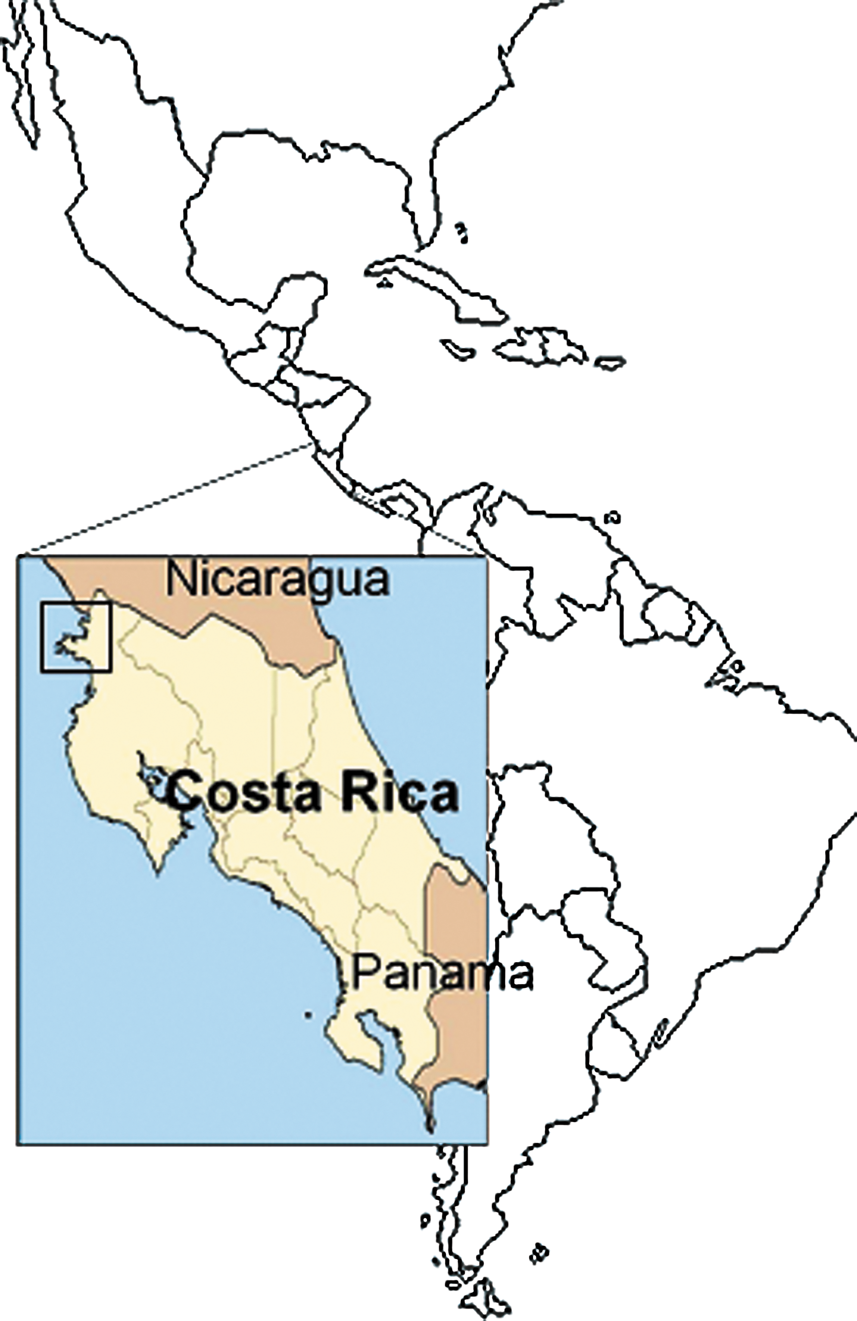
Map showing the approximate location of Liberia, Filadelfia, and Bolson in Costa Rica (boxed). Color images available online at
Age at time of serum collection (December 2004).
Only samples for which age of horse was known are included in this table.
Blocking ELISA performed using WNV-specific mAb 3.1112G. Inhibition values ≥30% are considered significant.
mAb, monoclonal antibody; WNV, West Nile virus.
Plaque reduction neutralization tests (PRNT) were performed using WNV (strain NY99-35261-11) and St. Louis encephalitis virus (SLEV; strain TBH-28) and African Green Monkey kidney (Vero) cells following standard protocols (Beaty et al. 1995). Results were expressed as the reciprocal titer of the serum dilution that reduced the number of plaques by ≥90% (PRNT90). Samples that returned a WNV PRNT90 titer that was at least fourfold greater than the corresponding SLEV PRNT90 titer were considered to have antibodies to WNV.
Sera were further assessed by Western blot using a peptide from domain I of the WNVNY99 envelope protein (WN19), using previously described procedures (Hobson-Peters et al. 2008).
Results
Of 101 serum samples tested, 28 (27.7%) were positive by blocking ELISA. A breakdown of the age distribution of ELISA-positive horses is shown in Table 1. In summary, 24% (6/25) of horses up to 2 years of age were positive for WNV-reactive antibodies in the blocking ELISA, compared to 35% of the serum samples taken from horses of 3–18 years of age. To validate the ELISA results, a subset of sera were further tested by PRNT using WNV and SLEV (Table 2). SLEV was included in these experiments because it is the most closely related flavivirus found in Central America (Reisen 2003) and may occasionally cause infections in horses (Farfan-Ale et al. 2006). The PRNT was performed on eight serum samples that were positive by blocking ELISA: five samples that were negative by blocking ELISA and three samples that were from horses that had been experimentally infected with WNVNY99. Of the eight sera that were positive by blocking ELISA, five were also positive for WNV by PRNT and three had antibodies to an undetermined flavivirus (Table 2). Hence, three of these horses had seroconverted to a flavivirus that could not be identified using this particular assay. As expected, the sera from the horses that were experimentally infected with WNVNY99 were positive for WNV by PRNT.
Blocking ELISA performed using WNV-specific mAb 3.1112G. Inhibition values ≥30% are considered significant.
Undetermined flavivirus.
< 20.
Reactivity of these sera to peptide WN19 has been published previously (Hobson-Peters et al. 2008).
Sera from horses experimentally infected with WNVNY99.
Reactivity could not be determined in Western blot.
PRNT, plaque reduction neutralization tests; SLEV, St. Louis encephalitis virus.
In addition to assessment by PRNT and blocking ELISA, this subset of sera were further assessed by Western blot using a peptide from domain I of the WNVNY99 envelope protein (WN19) that was previously shown to have diagnostic potential in WNV-specific serological assays (Hobson-Peters et al. 2008). The sera from 4 of the 5 Costa Rican horses that had antibodies to WNV by blocking ELISA and PRNT (Table 2, samples A9, A11, B5, and B10) reacted to peptide WN19 by Western blot. Consistent with these findings, the sera from all 5 of the Costa Rican horses that were negative for antibodies to WNV by blocking ELISA and PRNT (A2, A3, A4, B21, and C21) did not recognize peptide WN19.
Four sera (A8, A18, D7, and D11) did not fit this pattern. Serum A8 gave a positive result in the blocking ELISA and WNV PRNT, but did not recognize peptide WN19. Serum A18 contained antibodies that reacted to peptide WN19 and gave a positive result in the blocking ELISA, indicating the presence of WNV-specific antibodies, but the PRNT could not differentiate the response to WNV and SLEV due to similar titers to both viruses. Sera D7 and D11 were negative for antibodies to WNV by PRNT and did not react to peptide WN19 by Western blot, but were positive by blocking ELISA. Nevertheless, there was good overall correlation between recognition of peptide WN19 and WNV PRNT diagnosis.
Discussion
These data provide the first evidence of WNV activity in Costa Rica and support the gradual movement of the virus south from North America. In the subset of sera assessed by PRNT, those from horses confirmed as seropositive for WNV also gave inhibition values >30% in the blocking ELISA. Three sera (A18, D7, and D11) had neutralizing antibodies to both WNV and SLEV to a similar titer but also gave an inhibition value >30% in the blocking ELISA, indicating seroconversion to WNV. A number of sera with a similar profile of reactivity were collected in southern Mexico (Farfan-Ale et al. 2006, Morales-Betoulle et al. 2006). It is possible that these horses may have been exposed to both WNV and SLEV, or to another unidentified flavivirus. In a previous equine serosurvey conducted in Mexico, one serum with WNV-neutralizing antibodies also cross-neutralized the South American flavivirus, Ilheus virus, although to a lower titre (Farfan-Ale et al. 2006). No reports were found in which equines neutralized Bussuquara virus (Loroño-Pino et al. 2003, Farfan-Ale et al. 2006).
Our serosurvey indicated that between 18% and 28% of the horses living in the north-western areas of Costa Rica had been exposed to WNV, depending on the diagnostic assay used. This is lower than the seroprevalence for WNV in horses in northern Mexico in 2002 (62%) (Blitvich et al. 2003b) and southern Mexico in 2003/2004 (52%) (Farfane-Ale et al. 2006). However, the seroprevalence is likely to be indicative of how long the virus was circulating in the area before the samples were taken, as equine sera collected between 2001 and 2003 in El Salvador revealed only 14% of the horses had been exposed to WNV (Cruz et al. 2005) and only 13% of horse sera collected from Guatemala in late 2003–early 2004 were confirmed to have neutralizing antibodies to WNV by PRNT (Morales-Betoulle et al. 2006). Further, 9% of the horses sampled in Columbia in September and October of 2004 had antibodies to WNV (Mattar et al. 2005). Considering this serosurvey was conducted in only a small region near the border of Nicaragua, it would be of interest to conduct a more extensive survey of other areas of Costa Rica.
We took this opportunity to perform a small field evaluation of peptide WN19 as a diagnostic antigen (Hobson-Peters et al. 2008). The data indicated that there was good correlation between the PRNT results and those obtained with peptide WN19 in Western blot. Since all the WNV isolates from Mexico and Argentina tested to date belong to the WN02 genotype (reviewed in Blitvich et al. 2008, Kramer et al. 2008), as do the isolates from Puerto Rico (Barrera et al. 2008), it is likely that the horses assessed in the report were also exposed to this WNV genotype. This is significant when assessing the peptide WN19 results, as the WN02 strains harbor an envelope protein V159A substitution, compared to the WNVNY99 strain and this substitution lies within the sequence constituting peptide WN19. If these horses were in fact exposed to WNV of the WN02 genotype, it confers further diagnostic utility of this peptide and provides evidence that a mutation within this region has not affected its diagnostic sensitivity. Of additional importance is the specificity peptide WN19 appears to confer to the diagnosis of the sera assessed herein. Cross-reactivity problems in flaviviral serological assays have been well documented (Kuno 2003). In the subset of sera that were assessed by PRNT and Western blot, all samples that were confirmed as positive to WNV by PRNT were also positive by Western blot. With the exception of one sample (A18), all sera for which a definitive diagnosis could not be made by PRNT, were negative to WN19 in Western blot. Whether this peptide can be exploited to differentiate between WNV and SLEV infections requires further investigation.
Footnotes
Acknowledgments
The authors wish to thank Richard Bowen, Colorado State University, Fort Collins, CO, for generously supplying WNV-positive horse serum, and David Clark, The University of Queensland, Brisbane, Australia, for providing the recombinant NS1 antigen. This research was supported by the Australian Biosecurity CRC for Emerging Infectious Disease.
Disclosure Statement
No competing financial interests exist.