
Abstract
A 1-year-old castrated male Saanen goat was observed to have drooping and edema of the left ear consistent with published accounts of gotch ear in cattle associated with a tick bite. The goat's left ear was edematous from the tip of the pinna to the base of the ear. No signs of trauma or infectious processes were observed. Three engorged Gulf Coast ticks (Amblyomma maculatum) were observed attached inside the ear. Ticks were removed and the ear biopsied at tick attachment sites. The affected ear was treated topically with betadine after removal of the ticks. No other treatment was administered. The goat remained free of clinical signs and the edema of the ear resolved within 3 days after tick removal. No clinical adverse effects of the condition were evident. All three ticks were positive for spotted fever group rickettsia by polymerase chain reaction analysis and showed 100% similarity with the homologous sequence of Rickettsia parkeri. There was no immunohistochemical evidence of spotted fever group rickettsia in the ear samples, supporting the hypothesis that gotch ear is not due to rickettsial infection. This report represents the first apparent case of gotch ear in a goat.
Introduction
A yearling, castrated, male Saanen goat weighing 36.4 kg (80 lb) was evaluated on June 7, 2010 by the owner/veterinarian for drooping of the left ear of 1 day duration. Three partially engorged adult GCTs (two female and one male), A. maculatum, were removed from inside the left ear. No other tick species were observed on the animal. An 8-mm punch was used for biopsy of the tick attachment site on the affected ear. The sample was transferred to neutral-buffered 10% formalin and evaluated using hematoxylin and eosin stain and an immunohistochemical assay for spotted fever group Rickettsia spp. (Paddock et al. 1999). DNA extracts for each of the three attached ticks were evaluated using a nested polymerase chain reaction (PCR) assay designed to amplify a segment of the ompA gene based on a published protocol (Sumner et al. 2007). Extracts were also evaluated using a nested PCR assay designed to amplify a segment of the 17-kDa antigen gene as previously described (Paddock et al. 2004).
The wether's ear was treated topically at the tick attachment and biopsy sites with betadine solution. No further treatment was administered. At the time of evaluation, the wether was afebrile. This particular animal is housed with a herd of ∼40 Saanen dairy goats including kids, bucks, wethers, and does in east central Mississippi. There were no signs of trauma to the wether, either to his ear or to any other part of his body, and no abnormalities other than drooping of the left ear and significant edema in the full extent of the ear (Fig. 1). The wether was observed for a 1-month period after removal of ticks and no other clinical signs of disease were observed. None of the other members of the herd displayed similar signs or was observed to have any ticks attached.
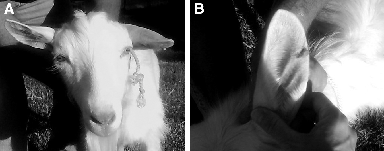
Saanen wether with edematous, drooping ear
Results and Discussion
Two of the three ticks were positive for spotted fever group rickettsiae (SFGR) by PCR of the ompA gene and all three were positive for SFGR by PCR of the 17-kDa antigen gene. An amplicon for the 17-kDa antigen gene from all three ticks was successfully sequenced and showed 100% similarity with the homologous sequence of R. parkeri. Hematoxylin and eosin–stained sections of the ear biopsy specimen showed ulceration and necrosis of the epidermis and diffuse and extensive neutrophilic inflammatory cell infiltrates involving the full thickness of the dermis. These infiltrates were accompanied by necrosis, collections of serum, and occasional extravasated erythrocytes. Some foci of epithelial hyperplasia were also identified at the margins of these infiltrates. No conspicuous bacteria were seen using Warthin-Starry and Lillie-Twort stains. There was no immunohistochemical evidence of SFGR, supporting the hypothesis that gotch ear is not due to rickettsial infection. One month after biopsy, no damage to the cartilage or signs of permanent disfiguration of the ear were appreciated.
Documentation of GCT infestation in cattle causing a condition consistent with the description of gotch ear has been attributed to the presence of large numbers of ticks (Williams et al. 1978), but there has been no description of gotch ear in a goat in the veterinary literature prior to this report. The wether described in this case had only three partially engorged ticks, but none fits the case description of gotch ear (Edwards et al. 2010). Even though the ticks were only partially engorged and lesions at the attachment sites were minimal, the ear was extremely edematous and drooping. Our observations are consistent with Ewing's observation in a dog (Ewing et al. 1997). That animal was part of a human granulocytic ehrlichiosis transmission study in which one of the dogs suffered a severe reaction in the ear described as an inflammatory response and severe edema, which the authors attributed to a reaction from A. maculatum (Ewing et al. 1997). The reaction subsided a few days after tick removal, which is the same pattern observed in the goat in this study. This suggests that gotch ear may occur in both ruminants and nonruminants.
Footnotes
Acknowledgments
The authors gratefully acknowledge Erle Chenney, Department of Basic Sciences, Mississippi State University, for technical assistance. This article has been approved for publication as Journal Article No. J-11882 of the Mississippi Agricultural and Forestry Experiment Station, Mississippi State University.
Disclaimer
The findings and conclusions described in this manuscript are those of the authors and do not necessarily represent the opinions of the U.S. Department of Health and Human Services.
Disclosure Statement
None of the authors has any commercial associations that might create a conflict of interest in connection with the submitted manuscript. No competing financial interests exist.