
Abstract
Human granulocytic anaplasmosis is increasingly being recognized in the United States and Europe. Ixodes ricinus tick is considered the primary vector of the causative pathogen, Anaplasma phagocytophilum, in Europe. Although previous studies in humans support the presence of A. phagocytophilum in Greece, detection of A. phagocytophilum DNA in I. ricinus ticks has been not yet reported. A total of 405 I. ricinus ticks (45 pools) collected from goats in eight prefectures of northern Greece were tested for the presence of A. phagocytophilum DNA. Four pools of ticks collected in three prefectures revealed a sequence with 99%–100% similarity to known A. phagocytophilum sequences, providing the first molecular evidence for the circulation of A. phagocytophilum in I. ricinus ticks in Greece.
Introduction
Although the clinical manifestations of HGA vary, the majority of patients have a moderately severe febrile illness accompanied by headache, myalgia, and malaise. The first case of HGA was described in 1994 in the United States, and the first description of the disease in Europe was in 1997 (Chen et al. 1994, Petrovec et al. 1997). Since then, HGA has become increasingly recognized in the United States and several European countries.
There is serological evidence in humans suggesting that the pathogen is present in Greece (Daniel et al. 2002, Chochlakis et al. 2008). Moreover, A. phagocytophilum has been recently detected in six human HGA cases in southern Greece (Chochlakis et al. 2009, Giadinis et al. 2011).
However, detection of A. phagocytophilum DNA in I. ricinus has been not yet reported. The purpose of the present study was to test Greek I. ricinus ticks for the presence of A. phagocytophilum.
Materials and Methods
I. ricinus, collected from goats in June 2006 from eight prefectures of northern Greece (Serres, Drama, Chalkidiki, Kavala, Kastoria, Imathia, Pella, Kilkis), were tested in this study; they had been previously tested for tick-borne encephalitis virus (Pavlidou et al. 2008). Ticks were pooled according to species, date, and collection site. Each pool contained 3–10 ticks. DNA was extracted using the QIAampTissue Kit (Qiagen, Hilden, Germany), and the extracts were stored at −20°C.
PCR amplifications were performed using the primer pair GE9f-GE10r, amplifying a 919-bp fragment of the 16S rRNA gene of A. phagocytophilum (Chen et al. 1994). PCR products were purified using the QIAquick PCR purification spin kit (Qiagen) and sequenced. All sequences were processed using nucleotide Blast (National Center for Biotechnology Information;
Data analysis
The rate of infection in tick pools was estimated using the following formula: maximum likelihood estimation=1 − (1 − Y/X)1/m , where Y=number of positive pools, X=number of pools, and m=number of organisms per pool. This formula assumes that when a PCR product is positive from a pool of five ticks, only one tick in the pool is considered to be infected (Walter et al. 1980).
Results
A total of 405 I. ricinus were tested. Of the 45 pools tested, 4 were PCR positive. The maximum likelihood estimation for A. phagocytophilum was calculated at 0.01. The positive ticks were collected from Kavala, Kastoria, and Chalkidiki prefectures (Fig. 1). Sequence analysis of the obtained 16S rRNA gene amplicons (841 bp) revealed 99%–100% similarity to various A. phagocytophilum sequences already deposited in GenBank: 100% to A. phagocytophilum sequence detected in I. ricinus in Germany (FN390892) and 99% to A. phagocytophilum sequences detected in I. ricinus in Turkey (FJ172530) and Ixodes persulcatus in Russia (HM366586). Based on A. phagocytophilum sequences detected mainly in European countries, as well as on sequences corresponding to distinct Anaplasma species (A. bovis, Ehrlichia chaffeensis, A. marginale), a phylogenetic tree was constructed (Fig. 2). The evolutionary history was inferred using the Neighbor-Joining method. The bootstrap consensus tree inferred from 500 replicates was taken to represent the evolutionary history of the taxa analyzed. The percentage of replicate trees in which the associated taxa clustered together in the bootstrap test (500 replicates) are shown next to the branches. The evolutionary distances were computed using the Kimura two-parameter method.
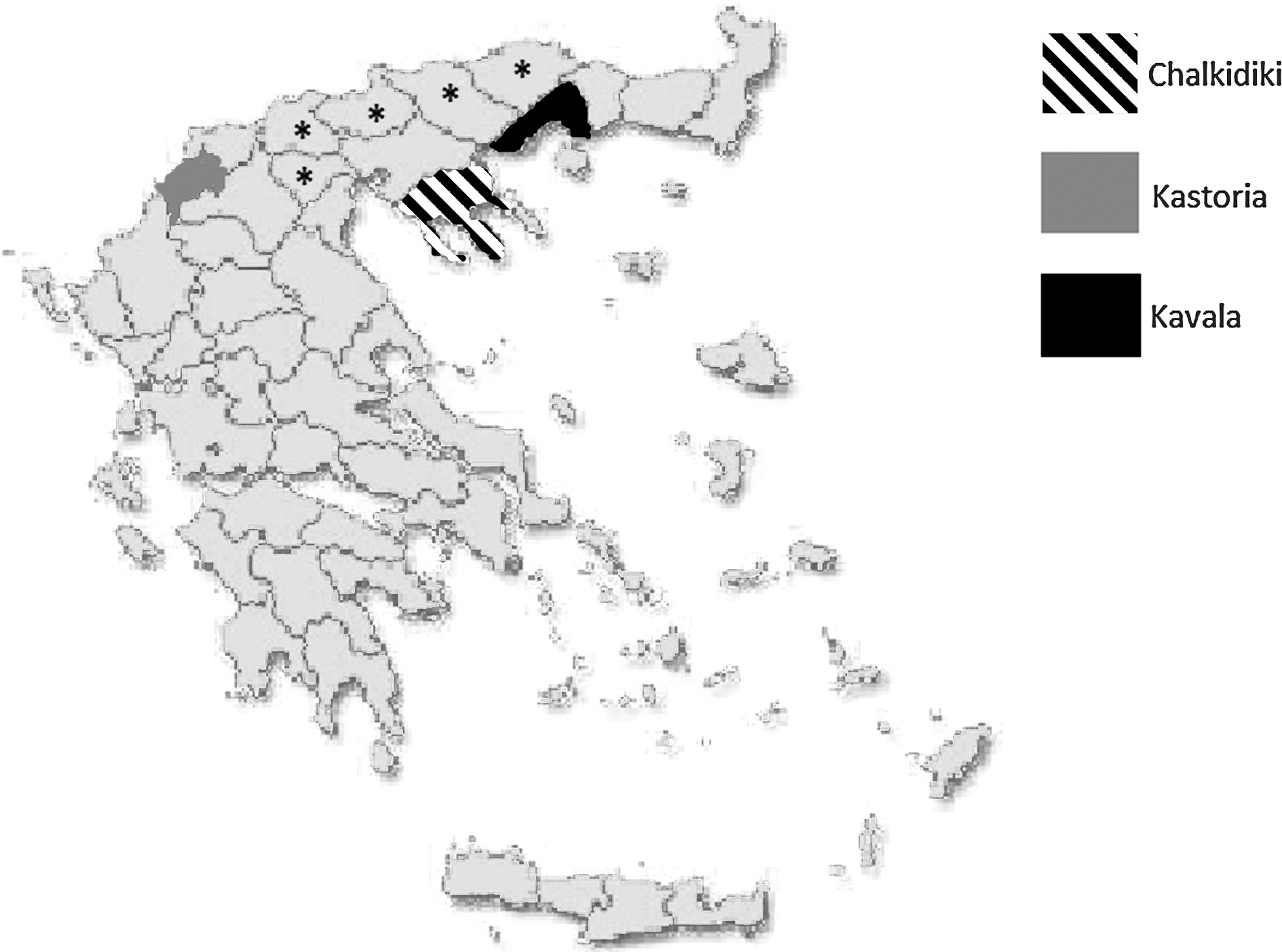
Map of Greece showing Kavala, Kastoria, and Chalkidiki prefectures, where Anaplasma phagocytophilum was detected. The black asterisks show the other prefectures from where Ixodes ricinus were also collected.
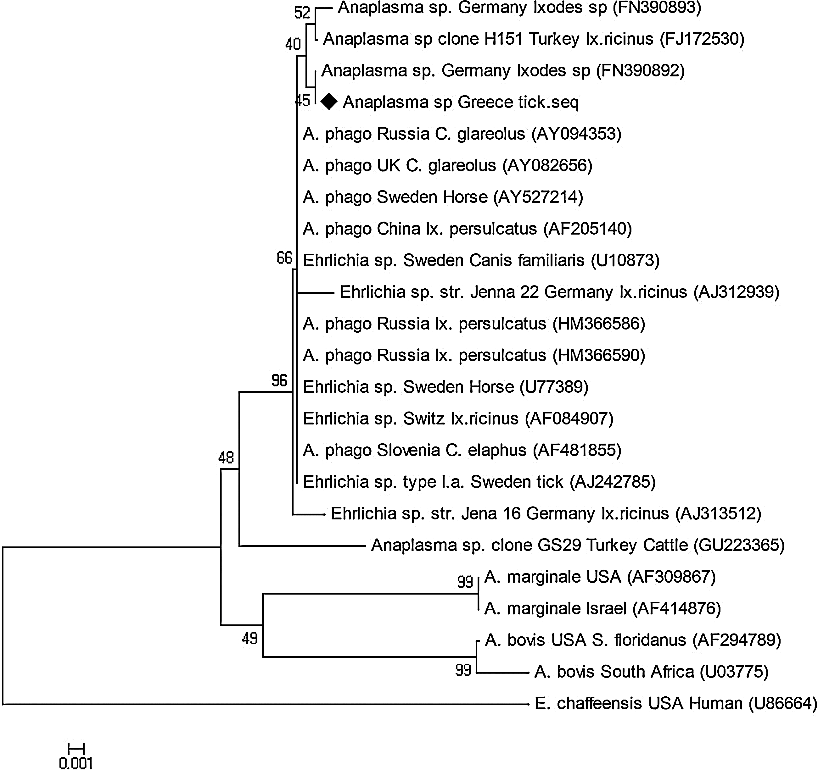
Phylogenetic tree demonstrating the correlation of the strain detected in our study with already published A. phagocytophilum and Anaplasma spp. sequences.
Discussion
A. phagocytophilum is considered an emerging human pathogen. Ixodes ticks play an essential role in the maintenance of A. phagocytophilum in nature, with I. ricinus being the primary vector of this organism in Europe.
The presence of A. phagocytophilum in I. ricinus ticks was reported for the first time in Europe in 1997 (von Stedingk et al. 1997), and since then the pathogen has been found in most parts of the European continent (Blanco and Oteo 2002), with prevalence in I. ricinus substantially varying from one geographical region to another. It can be as low as 1% (United Kingdom, Switzerland, France, Sweden) or between 24% and 34% (Bulgaria, Poland, Italy, Germany, Spain) (Oteo et al. 2001, Lillini et al. 2006).
Previous studies have indicated the presence of I. ricinus in northern Greece (Papadopoulos et al. 1996, Pavlidou et al. 2008) and have demonstrated the occurrence of A. phagocytophilum in both humans and sheep (Daniel et al. 2002, Giadinis et al. 2011). More recent studies in southern Greece (Crete) showed a high seroprevalence of A. phagocytophilum, and (Chochlakis et al. 2008) six human cases have been also recorded (Chochlakis et al. 2009). Comparing the strain detected in the present study with the strains detected in human cases in Crete, a 95% similarity was revealed. The difference among the strains may be attributed to a number of factors including the long distance between the two regions, the fact that Crete is a close microenvironment, the climate (dry in Crete, but wet in northern Greece), and the presence of different density of vectors.
In the present study, the infection rate for A. phagocytophilum in I. ricinus was found to be rather low. This might be due to the small sample size or the fact that ticks were collected only during a specific time point (summer time) and not throughout the year.
The results of our study provide the first molecular evidence for the presence of A. phagocytophilum in I. ricinus in Greece and suggest that HGA should be included in the differential diagnosis of febrile cases, especially when a tick bite is reported.
Footnotes
Disclosure Statement
No conflicts of interest were declared.