
Abstract
There are no comprehensive studies on the performance of commonly used point-of-care diagnostic enzyme immunoassay for common arthropod-borne canine pathogens. A comparative evaluation of an immunochromatographic test for these infections with a comprehensive polymerase chain reaction (PCR) test panel was performed on 100 pet dogs and 100 stray dogs without obvious clinical symptoms. Of the 162 positive test results from both immunochromatographic test and PCR, there was 85.2% concordance. The 24 discordant results between serology and PCR occurred in tests involving Ehrlichia canis (14) and Anaplasma platys (10), which may be related to the time of infection. No positive cases of borreliosis or rickettsiosis were detected. One important limitation of the immunochromatographic test was its lack of testing for babesiosis and hepatozoonosis. The former is the most prevalent arthropod-borne canine infection in our cohort (41%). Coinfections were found in 19% stray dogs and 6% of pet dogs with both tests (p<0.01). Seventeen and 8 samples from stray and pet dogs, respectively, were initially positive in the PCR test for Ehrlichia. However, on sequencing of the PCR amplicon, 10 from stray and 2 from pet dogs were found to be Wolbachia sequences instead, with 100% nucleotide identity to the 16S rRNA sequence of Wolbachia endosymbiont of Dirofilaria immitis. The presence of Wolbachia DNAemia (6%) correlated well with the molecular test and immunochromatographic antigen test for D. immitis.
Introduction
Materials and Methods
Animals
Two groups of dogs were included in the study. The first group were pet dogs (n=100) brought to the care of veterinarians for routine health checking from March to July 2010. After clinical evaluation, relevant specimens were sent to a veterinary pathologist for laboratory investigations. Peripheral EDTA blood was taken, and the plasma was tested by the SNAP 4Dx for antibodies against Anaplasma phagocytophilum, Ehrlichia canis, Borrelia burgdorferi, and D. immitis antigen. The second group were stray dogs (n=100) captured by the government Agriculture, Fisheries, and Conservation Department from June 2009 to January 2010. EDTA blood samples were collected by veterinarians in the kennels during euthanasia for polymerase chain reaction (PCR) and SNAP 4Dx testing.
Immunochromatographic assay
SNAP 4Dx test was performed according to the manufacturer′s instructions by the same investigator.
PCR and DNA sequencing for blood pathogens
DNA was extracted from EDTA whole blood samples using EZ1 mini kit (QIAgen, Hilden, Germany) according to the manufacturer′s instructions. The DNA was eluted in elution buffer and was used as the template for PCR. The primer sequences are listed in Table 1. The sequences of the PCR products were compared with known sequences by BLAST analysis against the NCBI database (Yuen et al. 2001).
Quantitative PCR
Quantitative PCR was performed on the PCR-positive samples (14 for E. canis and 81 for Babesia) using TaqMan Universal PCR Master Mix with StepOnePlus Real-Time PCR System (Applied Biosystems, Foster City, CA) (Lau et al. 2009). The primers and probes are listed in Table 2. This set of quantitative PCR primers and probe for Ehrlichia does not cross amplify Wolbachia. Two plasmids containing the target sequences were used for generating the standard curves. The limit of detection for either species is 10 plasmid copies per reaction.
Phylogenetic characterization
Phylogenetic tree was constructed by the neighbor-joining method using Kimura′s two-parameter correction with ClustalX 1.83. The 316, 292, 338, 150, 264, and 171 bp of amplicons from the 16S rRNA of E. canis, 16S rRNA of Wolbachia endosymbioint of D. immitis, 18S rRNA of Babesia, 18S rRNA of Hepatozoon canis, groEL of Anaplasma platys, and ITS of D. immitis, respectively, from all positive samples were included in the analysis.
Data analysis
The chi-square test and Student′s t-test were used where appropriate to assess whether data obtained in the two groups of dogs significantly differed. A p-value of <0.05 is considered statistically significant.
Nucleotide sequence accession numbers
Partial nucleotide sequences of 16S rRNA gene (E. canis and Wolbachia endosymbiont of D. immitis), 18S rRNA gene (Babesia gibsoni, Babesia canis, and H. canis), groEL gene (A. platys), and ITS (D. immitis) obtained in this study have been lodged within the GenBank sequence database under accession numbers HQ718601 to HQ718730.
Results
Comparison between SNAP 4Dx and PCR
There were 162 positive test results from both SNAP 4Dx and PCR with 85.2% concordance between the two tests. The results were summarized in Table 3. Concordance between SNAP 4Dx and PCR was 100% for D. immitis. Discrepancies between serologic and PCR results were observed for 24 test results (Table 4) involving E. canis and A. platys. Thirteen samples had positive serology but negative PCR for the respective pathogens, whereas 3 A. platys and 8 E. canis samples had positive PCR but negative serology results.
Based on both serology and PCR studied. Co-detection of Wolbachia and D. immitis were excluded.
PCR, polymerase chain reaction.
Prevalence of individual pathogens
We did not find any B. burgdorferi, Rickettsia, and O. tsutsugamushi infection in our samples. The commonest pathogen detected is Babesia, which is present in 48% and 33% of stray and pet dogs, respectively (p<0.05). Quantitative PCR test showed that there is a significantly higher Babesia DNA copy number in pet versus stray dogs (4.8×10⁷ vs. 2.26×10⁶ copies/mL) (p<0.005). Sequencing of the 18S rRNA gene showed that most of the Babesia positive cases are B. gibsoni (accounting for 91.6% [stray] and 93.9% [pet]) rather than B. canis, all of the latter are B. canis subspecies vogeli.
The prevalence of E. canis was of 8% and 6% in stray and pet dogs, respectively. There are no significant differences in the prevalence and DNA copy number between the two groups of dogs (p>0.5). E. canis antibodies were found in 2 stray and 10 pet dogs; 1 and 3 dogs, respectively, from the two groups were also positive for Anaplasma antibodies. In the two E. canis antibody-positive stray dogs, the one that was positive for both Ehrlichia and Anaplasma antibodies was PCR-positive for E. canis (1.07×10⁵ copies/mL) but PCR-negative for Anaplasma. The one that was positive for E. canis antibody alone had a very weak color change on SNAP 4Dx, and the sample was PCR-negative for E. canis. In the 10 pet dogs with positive E. canis antibodies, 5 of them were PCR-negative, suggesting that the antibodies represented a past infection. On the other hand, 1 pet and 2 stray dogs had positive E. canis PCR (ranging from 1.13×10⁵ to 1.43×10⁶ copies/mL) but negative E. canis antibodies on SNAP 4Dx. We suspect that these may represent a hyperacute ehrlichial infection before detectable antibodies were developed in the infected dogs.
The prevalence of anaplasmosis was 8% and 0% in stray and pet dogs, respectively. Anaplasma antibodies were present in 12 dogs (9 stray and 3 pet), either alone (6 stray) or with heartworm (2 stray) or E. canis (1 stray and 3 pet) antibodies. All the pet dogs were PCR-negative for Anaplasma, signifying possible past infections. Anaplasma PCR was positive in 8 stray dogs, 6 of which were also PCR positive for E. canis. Sequencing of the groEL amplicon showed that there was 1 (0.2%) base difference between our samples and that of A. platys (AF4781291) but >16 (3.4%) base difference with that of A. phagocytophilum (HM7520981), indicating that all the PCR-positive cases were A. platys (Fig. 1A).
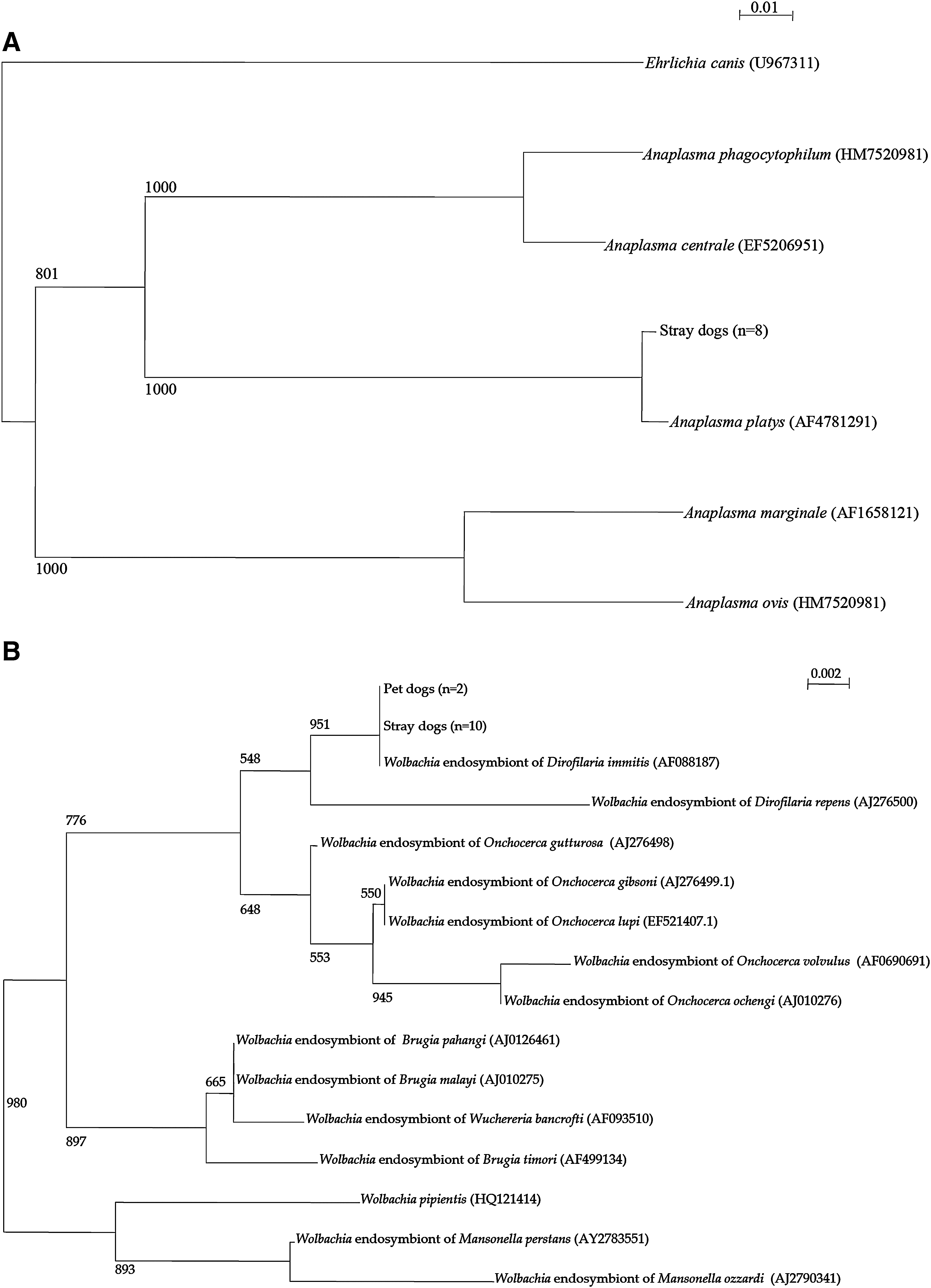
Phylogenetic relationships based on sequences identified in this study.
D. immitis DNAemia was found in 12 dogs (10 stray and 2 pet, p<0.025); all of them were positive for circulating heartworm antigen. Sequencing of the ITS showed that all cases have 100% nucleotide identity to those of the reported strains.
Hepatozoonosis is uncommon in our sample with only 2% and 1% prevalence in stray and pet dogs, respectively. Sequencing of the 18S rRNA amplicon showed that all belonged to H. canis.
Co-infection by 2 or more pathogens was observed in 19% of stray dogs and 6% of pet dogs (p<0.01). The combinations of pathogens in dogs with coinfection are shown in Table 3.
Incidental finding of Wolbachia DNAemia
Seventeen and 8 samples from stray and pet dogs, respectively, were initially positive in the Ehrlichia PCR reaction. However, on sequencing of their PCR amplicons, 10 from stray and 2 from pet dogs were found to be Wolbachia sequences instead, with 100% nucleotide identity to the 16S rRNA sequence of Wolbachia endosymbiont of D. immitis (AF088187) (Fig. 1B). This can be explained by the significant homology between the Ehrlichia and Wolbachia 16S rRNA sequences (Fig. 2). Therefore, we designed another Wolbachia-specific 16S rRNA primer pair and found that all cases with D. immitis DNAemia also had Wolbachia DNAemia when tested again by Wolbachia-specific PCR test. Sequencing of the 16S rRNA gene of these amplicons showed that they are truly Wolbachia endosymbiont of D. immitis but not other dog arthropod-related Wolbachia species (Fig. 1B). False-positive Ehrlichia PCR results were eliminated by the Tagman quantitative PCR for Ehrlichia using a specific probe or PCR primers for E. canis that will not cross react with Wolbachia.
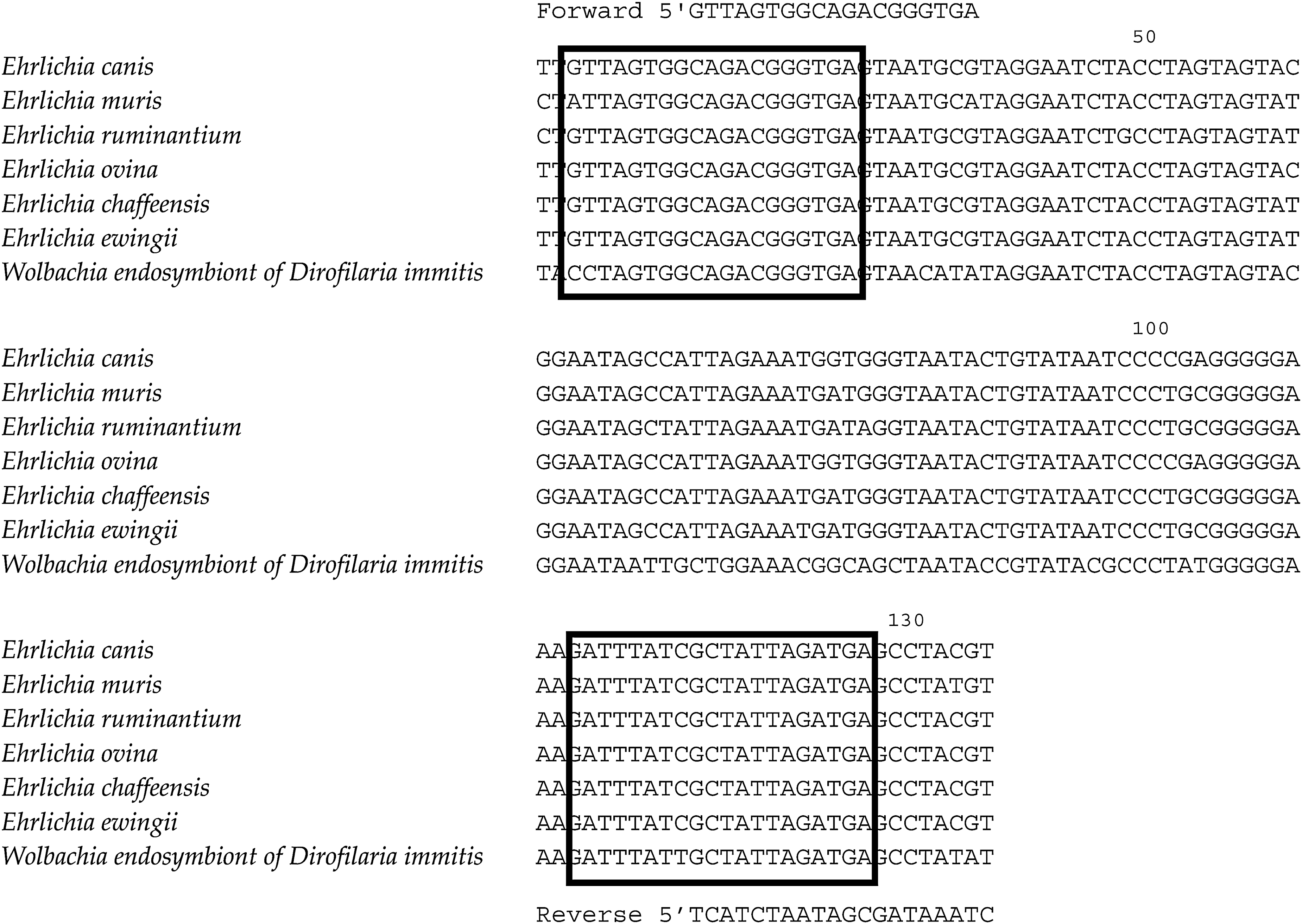
Nucleotide sequence alignment of 16S rRNA gene of Ehrlichia spp. and Wolbachia endosymbiont of D. immitis, showing regions containing the 5′ and 3′ primers used for Ehrlichia polymerase chain reaction.
Discussion
Rapid and accurate detection of veterinary pathogens is important for clinical management of sick animals because of the limited sensitivity and specificity of clinical examination and problems associated with empirical treatment. This is the first systematic study to compare SNAP 4Dx against PCR and determine the prevalence of eight canine vector-borne pathogens in Hong Kong. It provides a better understanding of the strengths and limitations of serologic tests and the local prevalence of these pathogens.
The overall concordance between SNAP 4Dx and PCR is 85.2%. Discrepancies between the commercial immunochromatographic test and our PCR tests involved E. canis and A. platys (Table 4). We suspect that these PCR-positive and antibody-negative cases represented early infections before the development of antibody responses. For example, an E. canis-seronegative dog had one of the highest bacterial DNA load in blood (4.03×10⁵ copies/mL). It was presumably diagnosed during acute infection before seroconversion, which shows that molecular tests do have an important role in diagnosis at the hyperacute stage of the disease and are not confounded by positive antibody response due to past exposures as in immunochromatographic tests. On the other hand, the antibody-positive but PCR-negative cases may represent past infections that may have been treated, spontaneously resolved, and progressed to the subclinical chronic stage with low levels of bacteremia, or due to cross-reacting antibodies.
Around 10% of dogs in our study had evidence of Ehrlichia and/or Anaplasma infection by serology or PCR. E. canis is the only Ehrlichia species found in Hong Kong, presumably due to the absence of Amblyomma americanum tick, which is the vector for Ehrlichia ewingii and Ehrlichia chaffeensis.
All the Anaplasma amplicons in our series belonged to A. platys, the vector of which is the brown dog tick Rhipicephalus sanguineus. A. phagocytophilum but not A. platys can cause human granulocytic anaplasmosis. The absence of A. phagocytophilum in our study suggests that the risk of autochthonous human granulocytic anaplasmosis is low in Hong Kong. In contrast to A. phagocytophilum, canine infection due to A. platys is often asymptomatic despite the presence of recurrent thrombocytopenia. This may explain the 8% prevalence among apparently healthy stray dogs, which is higher than the prevalence in pet dogs, presumably due to more intense exposure to tick vectors. E. ewingii, E. chaffeensis, Neorickettsia risticii, and Neorickettsia helminthoeca are other canine Anaplasmataceae pathogens, whereas E. chaffeensis and A. phagocytophilum also cause human infections.
Although we initially did not specifically look for Wolbachia DNA in our samples, the E. canis primers unexpectedly picked up Wolbachia, which can only be differentiated from E. canis by sequencing the PCR product. This cross reaction could be due to significant homology between their 16S rRNA sequences (Fig. 2). The 16S rRNA primers for Ehrlichia and Anaplasma are known to amplify Wolbachia, and sequencing of the PCR products is essential for differentiating the 3 genera of Anaplasmataceae (Unver et al. 2003, Little 2010). Similar findings have also been previously reported when H. canis was unexpectedly detected using Babesia primers due to a high degree of homology between the 18S rRNA sequences of the 2 organisms (Spolidorio et al. 2009). Wolbachia are endosymbionts of arthropods and filarial nematodes. Humans and animals with filariasis develop immune responses to Wolbachia antigens, and the pro-inflammatory antigens from Wolbachia participates in the pathogenesis of filariasis and affects the clinical outcome of Dirofilaria infections (Bandi et al. 2001, Bazzocchi et al. 2003, Kramer et al. 2008, Dingman et al. 2010). Elimination of Wolbachia with tetracyclines is clinically beneficial in the treatment of filariasis and can possibly reduce transmission (Rossi et al. 2010).
The prevalence of D. immitis is highly variable, ranging from 0.24% to over 50% in different countries (Lee et al. 2010). The prevalence of canine heartworm infection is 6% in Hong Kong, with a significantly higher prevalence among stray than pet dogs, possibly related to the intensity of exposure to mosquito vectors. Accurate diagnosis is important, because the infection can result in severe cardiopulmonary disease. Diagnosis is commonly made by either blood smear examination by the modified Knott′s method or antigen detection. In recent years, molecular diagnosis by using PCR to detect D. immitis DNA in canine blood is also possible. We found 100% concordance between SNAP 4Dx and PCR for the diagnosis of D. immitis infection, and all dogs with D. immitis infection also had positive Wolbachia PCR. Unfortunately, since the detection of Wolbachia DNAemia was not initially planned as part of the study, we did not perform blood film examination for microfilaremia and, hence, could not correlate the presence of Wolbachia DNAemia with microfilaremia. Although the use of antigen detection kits provides rapid results for immediate management decisions and most of the antigen detection tests are highly sensitive and specific (Nelson et al. 2005), PCR for D. immitis DNA is a potentially useful adjunct in cases with low levels of microfilaremia, as the sensitivity of antigen detection tests may be lowered in such cases (Vezzani et al. 2008). However, the added benefits of PCR over serology need to be demonstrated by further studies. On the other hand, in certain parts of the world where other filarial parasites are endemic, these point-of-care tests (primarily targeted against D. immitis) may not be clinically useful. With careful selection of primers and/or the use of sequencing, molecular testing could be a better diagnostic test in these areas where non-D. immitis filariases are common. Wolbachia PCR may provide additional information, because the sensitivity of D. immitis antigen detection tests could be limited in dogs with only male or young female worms, as the antigens are derived from the genital organs of adult female worms.
We found no evidence of active canine infection due to Rickettisa, O. tsutsugamushi, and B. burgdorferi in our cohorts. Although the role of dogs as a reservoir host for these bacteria in Hong Kong is probably minor, we cannot completely exclude their existence, because past infections may not be detectable by PCR and serologic tests are required for confirmation. Rickettsia felis is an emerging zoonotic pathogen in many parts of the world, with reservoirs in both the cats and dogs. B. burgdorferi sensu sticto is primarily found in North America and Europe, and the main vertebrate reservoir hosts are small mammals (especially rodents). However, both B. burgdorferi sensu sticto and sensu lato have been isolated from animals in mainland China and Taiwan (Chao et al. 2002, Zhang et al. 1997, Shih et al. 1998a, 1998b). Dogs and humans are accidentally infected by hard tick bites, though neither are important reservoir hosts.
In previous studies from Spain, France, Italy, Czech, the United Kingdom, and the United States, coinfection of vector-borne pathogens in dogs ranged from 0% to over 50%, depending on the location, pathogens, and methods of detection (Shaw et al. 2005, Solano-Gallego et al. 2006, Amusategui et al. 2008, Beall et al. 2008, Kybicová et al. 2009, Pantchev et al. 2009, Couto et al. 2010, Otranto et al. 2010, Tzipory et al. 2010). Evidence of co-infection was found in 19% of stray and 6% pet dogs in our study (p<0.01). The higher prevalence in stray dogs can be explained by the higher risk of exposure to arthropod vectors in the environment.
One of the most important limitations of the point-of-care test is the lack of testing for babesiosis, which is the commonest arthropod-borne infection in both pet and stray dogs in Hong Kong. Most of these are caused by B. gibsoni with only 7.3% due to B. canis. Although serologic diagnosis of babesiosis using immunofluorescent antibody testing is commercially available, it is rather cumbersome for most veterinarians and requires separate laboratory support. Canine babesiosis is a tick-borne infection with a global distribution caused by B. canis (subsp. canis, vogeli, and rossi), B. gibsoni, B. microti, B. equi, and B. conradae. B. gibsoni generally causes hemolytic anemia, fever, lethargy, hepatosplenomegaly, hemoglobinuria, and icterus. The other less important tick-borne infection is hepatozoonosis. As expected, the Old World species H. canis (transmitted by R. sanguineus) is the only species found in our study, though the pathogen is not common in our dog population. The absence of American canine hepatozoonosis (H. americanum) in Hong Kong is probably related to the absence of its tick vector Amblyomma maculatum. The American form of the disease is generally more severe, and infected animals are more debilitated and often fatal.
Compared with conventional blood film examination, PCR offers a highly sensitive means for detecting blood-borne pathogens. A broad range of pathogens can be detected by PCR studies. Sequencing studies also allow differentiation of species or subspecies that may have similar morphological appearances. The accurate speciation of the pathogens carries epidemiologic or prognostic significance. Molecular studies allow the detection of newly described pathogens for which serologic tests are generally not available. However, at the moment, the turnaround time for molecular testing is still longer than blood film examination and point-of-care testing, and the availability of molecular diagnostics for veterinary service is still limited in many countries. A better organization and delivery of such techniques is highly desirable not just for better care of the sick animals but also to allow epidemiologic study of these pathogens and their zoonotic potentials.
Footnotes
Acknowledgments
This work is partly supported by the Consultancy Service for Enhancing Laboratory Surveillance of Emerging Infectious Disease for the Department of Health, Hong Kong; Tung Wah Group of Hospitals Fund for Research in Infectious Diseases; the Research Fund for the Control of Infectious Diseases of the Health, Welfare, and Food Bureau, Hong Kong; and the Providence Foundation Limited in memory of the late Dr. Lui Hac Minh. We are grateful for the generous support of Mrs. Carol Yu, Professor Richard Yu, The Hong Kong Sanatorium and Hospital, Mr. Hui Hoy, and Mr. Hui Ming in the genomic sequencing platform. The support of Mr. Alan C. K. Wong, Director of Agriculture, Fisheries, and Conservation, is indispensable to the project; and all the work from the staff of the Agriculture, Fisheries, and Conservation Department, Hong Kong, including Dr. Thomas Sit and Dr. Howard K. H. Wong, is gratefully acknowledged.
Disclosure Statement
No competing financial interests exist.