
Abstract
A biological method was used to synthesize stable silver nanoparticles that were tested as mosquito larvicides against Aedes aegypti, Anopheles stephensi, and Culex quinquefasciatus. Annona squamosa leaf broth (5%) reduced aqueous 1 mM AgNO3 to stable silver nanoparticles with an average size of 450 nm. The structure and percentage of synthesized nanoparticles was characterized by using ultraviolet spectrophotometry, X-Ray diffraction, Fourier transform infrared spectroscopy, and scanning electron microscopy methods. The median lethal concentrations (LC50) of silver nanoparticles that killed fourth instars of Ae. aegypti, Cx. quinquefasciatus, and An. stephensi were 0.30, 0.41, and 2.12 ppm, respectively. Adult longevity (days) in male and female mosquitoes exposed as larvae to 0.1 ppm silver nanoparticles was reduced by ∼30% (p<0.05), whereas the number of eggs laid by females exposed as larvae to 0.1 ppm silver nanoparticles decreased by 36% (p<0.05).
Introduction
Noble metal nanoparticles have been studied because of their unique optical properties. Gold and silver have a broad absorption band in the visible region of the electromagnetic spectrum (Kreibig and Vollmer 1995, Mulvaney 1996). The properties of these metals change depending on their shape, size, and the surrounding medium, and they have been used in advanced technologies in medicine, opto-electronics, and chemical catalysis, in sensors, for drug delivery, and for etching and cutting (Che and Bennett 1989, Elghanian et al. 1997, Haruta 1997, Valden et al. 1998, Fujimoto 2003, Kruusing 2004, El
Silver (Ag) is a versatile element with applications in the clothing, appliance, and semiconductor industries. Nanoparticles of this metal manifest antibiotic activity against bacteria, viruses, and eukaryotes (Shirkhanzadeh et al. 1995, Kim et al. 2007,Magana et al. 2008, Rai et al. 2009), and nanosized silver has been used to treat immunologic and inflammatory disease (Shin et al. 2007).
Noble-metal nanomaterials have been synthesized by using hard-template (Zhou et al. 1999), bioreduction (Canizal et al. 2001), and solution phase syntheses (Sun et al. 2003). Conventional redox synthesis methods use hazardous chemicals as reducing agents or require significant energy input (Cushing et al. 2004). However, there is growing interest in environmentally safe reducing agents. These so-called “green” reducing agents include microorganisms, such as Pseudomonas stutzeri (Klaus-Joerger et al. 2001), Verticilium spp. (Mukherjee et al. 2001), Fusarium oxysporum (Sastry et al. 2003), Fusarium semitactum (Basavaraja et al. 2008), and Thermomonospora spp. (Ahmad et al. 2003), as well as vascular plants such as alfalfa (Gardea Torresdey et al. 2003), neem (Sastry et al. 2004), and geranium (Shankar et al. 2003).
Annona squamosa L. (Family: Annonaceae), commonly known as custard apple, is cultivated throughout India as an edible fruit. The phytochemical constituents of An. squamosa include alkaloids (Mukhlesur et al. 2005), terpinoids, glycosides (O'Farrell 1975), flavanoids (Kotkar et al. 2002), corticosteroids, and essential oils. Some of these constituents exhibit anticytotoxic (Mukhlesur et al. 2005), antioxidant (Shirwaikar et al. 2004), and antipesticidal properties.
In the study reported here, we describe the use of An. squamosa leaf extract as a 'green' reducing and stabilizing agent for the synthesis of silver nanoparticles. We also report the toxicity of silver nanoparticles to mosquito larvae and pupae and the effect of exposure of immature mosquitoes to silver nanoparticles on survival and reproduction in the adult mosquitoes.
Materials and Methods
Collection of plant materials and preparation of leaf broth
An. squamosa leaves were collected from the Maruthamalai Hills, near the Bharathiar University campus in Coimbatore. Leaves were washed with distilled water and dried for 2 days at room temperature. A plant leaf broth was prepared by placing 5 g of the leaves (finely cut) in a 300 mL flask with 100 mL of sterile distilled water. This mixture was boiled for 5 min, decanted, stored at −4°C, and used in our tests within 1 week.
Mosquito rearing
The eggs of Aedes aegypti, Anopheles stephensi, and Culex quinquefasciatus were collected from different locations (cisterns, cattle tanks, temporary pools, impoundments, overhead tanks, ditches, etc.) in Coimbatore District, Tamil Nadu, India. These were returned to the laboratory and transferred (in approximately the same aliquot numbers of eggs) to 18 cm L×13 cm W×4 cm D enamel trays containing 500 mL of water where they were allowed to hatch.
Mosquito larvae were reared (and adult mosquitoes held) at 27°C±2°C and 75%–85% relative humidity (RH) in a 14:10 (L:D) photoperiod. Larvae were fed 5 g ground dog biscuit and brewers yeast daily in a 3:1 ratio. Pupae were collected and transferred to plastic containers with 500 mL of water. The container was placed inside a screened cage (90 cm L×90 cm H×90 W) to retain emerging adults, for which 10% sucrose in water solution (v/v) was available ad libitum. On day 5 postemergence, the mosquitoes were provided access to a rabbit host for blood feeding. The shaved dorsal side of the rabbit was positioned on the top of the mosquito cage in contact with the cage screen (using a cloth sling to hold the rabbit) and held in this position overnight. Glass Petri dishes lined with filter paper and containing 50 mL of water were subsequently placed inside the cage for oviposition by female mosquitoes.
Synthesis, characterization, and formulation of silver nanoparticles
Ten milliliters of leaf broth was added to 190 mL of 1 mM aqueous AgNO3 solution for reduction of Ag+ ions. The effect of reaction time on synthesis rate and particle size of the prepared silver nanoparticles was determined by allowing the reaction to proceed in a water bath at 95°C with reflux (5 refluxes) for between 10 min and 4 h. Silver nanoparticle solutions obtained in this manner were purified by centrifugation at 15,000 rpm for 20 min followed by redispersion of the pellet in deionized water. Repeated centrifugation and re-dispersion methods (Tsaur and Fitch 1987) were used to remove impurities.
Formation of silver nanoparticles from 5% An. squamosa leaf broth with 1 mM AgNO3 solution at 95°C was indicated by the appearance of a yellowish brown color in the reaction vessel (Shankar et al. 2004). The intensity of change was recorded as a function of reaction time (10, 30, 60, 120, and 240 min) by using a UV-3600 Shimadzu spectrophotometer at 1 nm resolution. The resulting density of synthesized nanoparticles was 89% silver, 8% carbon, and 3% oxygen.
The structure and composition of freeze-dried purified silver particles was analyzed by using a 10 kV ultra high resolution scanning electron microscope (FEI QUANTA-200 SEM) and energy dispersive X-ray spectroscopy. The surface groups of the nanoparticles were qualitatively confirmed by using Fourier transform infrared (FTIR) spectroscopy (Stuart 2002), with spectra recorded by a Perkin-Elmer Spectrum 2000 FTIR spectrophotometer. X-ray diffraction (XRD) using Cukα radiation (PANanlytical X'pert Pro MPD diffractometer) allowed us to determine the crystalline structure of the silver nanoparticles. Powder X-ray analysis was made by using a Philips PW 1050/37 diffractometer, operating at 40 kV and 30 mA, with a step size of 0.02° (2θ).
Ten milliliters of the synthesized silver nanoparticles was diluted in 90 mL of distilled water for the preparation of a 10% stock solution. Each of the five different concentrations of nanoparticles (0.001, 0.01, 0.1, 1.0, and 10 ppm) was prepared by dilution of the stock solution in distilled water. All stock solutions and dilutions thereof were refrigerated at −4°C and used within 4 months of synthesis.
Larval/pupal toxicity test
Twenty-five larvae (instars 1–4) or pupae were placed in 249 mL of de-chlorinated water in a 500 mL glass beaker, and 1 mL of the desired concentration of silver nanoparticles was added. In fact, 0.5 mg larval food was provided for each test concentration. Tests of each concentration against each instar and the pupae were replicated thrice. In each case, the control comprised 25 larvae or pupae in 250 mL of distilled water. Control mortality was corrected by using Abbott's formula (Abbot 1925), and percentage mortality was calculated as follows:
Oviposition and adult longevity studies
The effect of larval exposure to silver nanoparticles on oviposition by female mosquitoes was determined by placing 20 male and female mosquitoes of similar size and age from each treatment group and the respective control into separate screened cages (30 cm L×30 cm W×30 cm H). Sucrose solution was available to adults ad libitum. Commencing 3 d after blood feeding and until oviposition ceased, eggs or egg rafts were collected daily from a plastic bowl (12 cm H×12 cm dia) containing 500 mL water that was placed in each cage. The average number of eggs laid by each female in each cage was calculated as the total number of eggs collected in each cage divided by the number of females originally placed in that cage.
To characterize adult longevity, the number of live male and female mosquitoes in each cage was recorded at the same time each day until all mosquitoes had died. This value was divided by the number of males or females originally placed in to the cage to obtain percent survival according to the number of days since emergence.
Statistical analysis
The median Lethal Concentration (LC50) and the respective 95% fiducial limits observed for the larvae of each mosquito species were estimated by using probit analysis. Mean adult longevity and mean fecundity data were analyzed by using analysis of variance methods, where the life span of a mosquito (days) and the number of eggs oviposited were dependent variables, and the concentrations used were independent variables. The level of significance used in all tests was 5%. Statistical significance of mean differences was assessed by Tukey's honestly significant difference (HSD) test. Analyses were made by using SPSS Software version 14.0.
Results and Discussion
Ultra-violet visible absorption spectroscopy studies
The UV absorption spectra of silver nanoparticles as a function of reaction time is shown in Figure 1. There was maximum absorption between 438 and 442 nm with average maximum absorption at 440±1.6 nm. The surface plasmon peak of silver nanoparticles at 440 nm steadily increased with reaction time and became saturated at 120 min, indicating complete reduction of the silver nitrate. Final absorption intensities at 440 nm were 1.3 absorbance units (a.u.) in 10 min, 1.8 a.u. in 30 min, 1.9 a.u. in 60 min, 2.4 a.u. in 120 min, and 2.0 a.u. in 240 min.

Ultra-violet light absorbance of synthesized silver nanoparticles in 1 mM AgNO3 in 5% Annona squamosa leaf broth at different reaction times.
The UV absorption spectra of silver nanoparticles as a function of AgNO3 concentrations are shown in Figure 2. The effect of AgNO3 concentration (1, 2, and 4 mM) on silver nanoparticle formation (5% An. squamosa leaf broth, 60 min, 95°C) was that particle size increased with AgNO3 concentration. Maximum absorption peaks were at 438 nm in 1 mM AgNO3, 458 nm in 2 mM AgNO3, and 461 nm in 4 mM AgNO3. Final absorption intensities were 1.9 a.u. in 1 mM AgNO3, 3.8 a.u. in 2 mM AgNO3, and 4.4 a.u. in 4 mM AgNO3.

Ultra-violet light absorbance of synthesized silver nanoparticles in three different concentrations of AgNO3 in 5% An. squamosa leaf broth.
The absorption peak varied as a function of reaction time and silver nitrate concentration. As the size of the particles decreased, the energy gap increased, and the absorption peak shifted to a higher energy level. These observations are consistent with the results of other studies (gold nanotriangles synthesized using lemongrass extract) (Rai et al. 2006) that have shown the reduction rate to increase with increased reaction temperature. In addition, it is known that the absorbance of silver nanoparticles depends on the size and shape of the particles (Sosa et al. 2003). The surface plasmon peak we observed (440 nm) is consistent with the results of other studies that show peaks at 490 nm (Raghunandan et al. 2010), 430 nm (Song and Kim 2009), 400–420 nm (Pyne et al. 2010), 410 nm (Das et al. 2009), and 425 nm (Maliszewska et al. 2009).
XRD studies
To verify the result of the UV–vis analysis, the sample of the Ag+ exposed to the leaf broth of An. squamosa was examined by XRD (Fig. 3). There were intense silver nanoparticle (AgNP) diffraction peaks at 38.10°, 44.44°, 64.47°, 77.53°, and 81.62° 2θ, corresponding to facets 111, 200, 220, 311, and 222 of the face-centered cubic crystal structure. Sathyavathi et al. (2010) reported diffraction peaks at 44.50°, 52.20°, and 76.7° 2θ, which correspond to the (111), (200), and (220) facets of the face-centered cubic crystal structure. However, other XRD reports of silver nanoparticles (Bar et al. 2009, Das et al. 2009, Nirmala et al. 2010, Raghunandan et al. 2010) are in general agreement with the just-cited results.
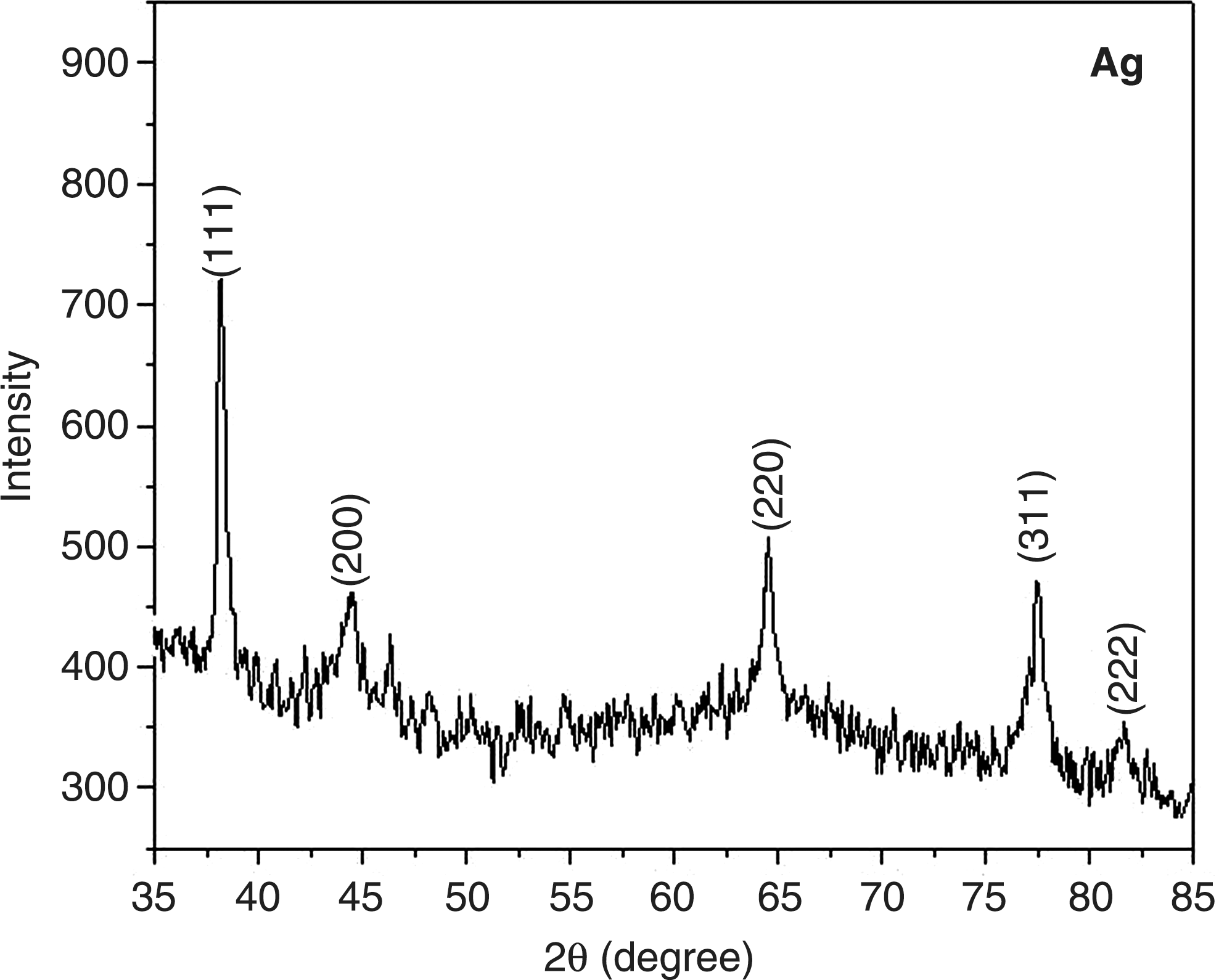
X-ray diffraction pattern of synthesized silver nanoparticles in 1 mM AgNO3 in 5% An. squamosa leaf broth.
FTIR spectroscopy studies
The FTIR spectra of aqueous silver nanoparticles prepared from the An. squamosa leaf extract (Fig. 4) show transmittance peaks at 1262.1, 979.4, 847.0, 712.4, 660.7, 501.6, and 434.5/cm. These peaks indicate that the carbonyl group formed amino acid residues and that these residues “capped” the silver nanoparticles to prevent agglomeration, thereby stabilizing the medium (Sathyavathi et al. 2010). The disappearance of secondary metabolites after the bioreduction of silver nanoparticles was also confirmed by FTIR spectroscopy and is thought to result from the reduction of Ag ions by the polyols, which themselves are oxidized to unsaturated carbonyl groups with a broad peak at 1650 cm−1 (Jain et al. 2009).

Infra-red light transmittance of synthesized silver nanoparticles in 1 mM AgNO3 in 5% An. squamosa leaf broth.
Scanning electron microscopy studies
Scanning electron micrographs enabled visualization of the size and shape of the silver nanoparticles (Fig. 5). Particle shape was highly irregular with diameters that ranged from 200 to 500 nm. The scanning electron microscopy (SEM) results indicate that the silver powder consists of a nanometrical conglomerate which contains ∼89% silver, 8% carbon, and 3% oxygen. In addition, the SEM image shows that the “capped” silver particles were stable in solution for at least 8 weeks.

Scanning electron micrograph of synthesized silver nanoparticles.
Bioassay
For Ae. aegypti at silver nanoparticle concentrations >1 ppm, >76% mortality was observed, whereas at the 0.001 ppm concentration acute mortality was low and some third or fourth instars failed to pupate and/or died (Table 1). The LC50 was 0.02, 0.07, 0.19, 0.30, and 0.56 ppm for first through fourth instars and for pupae, respectively. For Cx. quinquefasciatus, 100% mortality was observed at the 10 ppm concentration for all instars and the pupae (Table 2). The LC50 values of instars 1–4 and pupae were 0.04, 0.13, 0.29, 0.41, and 0.79 ppm, respectively.
Significant at p=0.005 (heterogeneity factor used in calculation of confidence limits).
SE, standard error.
Significant at p=0.005 (heterogeneity factor used in calculation of confidence limits).
At the 10 ppm silver nanoparticle concentration, the mortality rate for An. stephensi was 100% for instars 1–3 and 98% and 89%, respectively, for fourth instars and pupae (Table 3). The LC50 for instars 1–4 and pupae were 0.16, 0.29, 0.48, 2.12, and 3.74 ppm, respectively.
Significant at p=0.005 (heterogeneity factor used in calculation of confidence limits).
A comparison of the silver nanoparticle LC50 values for Ae. aegypti, Cx. quinquefasciatus, and An. stephensi is shown in Figure 6. The values are consistently higher for An. stephensi, particularly for instar 4 and the pupa, compared with Cx. quinquefasciatus or Ae. aegypti. The figure also indicates that LC50 values increased as larvae aged.

Median lethal concentrations (LC50) of synthesized silver nanoparticles to larvae and pupae of three mosquito species.
The results of this study show that silver nanoparticles can be used to kill larvae of Ae. aegypti, Cx. Quinquefasciatus, and An. stephensi. Silver nanoparticles are most effective for this purpose against Ae. aegypti followed by Cx. quinquefasciatus and An. stephensi. The mortality effect of silver nanoparticles on mosquito larvae may be enabled by the small size of the particles, which allows passage through the insect cuticle and into individual cells where they interfere with molting and other physiological processes. Safaepour et al. (2009) reported that a 1 μg/mL concentration of silver nanoparticles inhibited cell growth by <30%, whereas at 5 μg/mL, cell growth was inhibited by >60%.
High concentrations of silver nanoparticles (0.1, 1.0, and 10 ppm) significantly reduced adult longevity (p<0.05, HSD). In addition, at these concentrations, many emerging adults had defective wings and/or legs and abnormal morphologies of the abdomen and laid relatively few eggs, many of which failed to hatch. The effect of silver nanoparticles on mean adult longevity and mean numbers of eggs per female in An. stephensi is shown in Table 4. At the 0.001 ppm nanoparticle concentration, mean longevity in males was reduced from 20 to 18.9 days and in females from 35 to 33.3 days. At the 0.01 ppm concentration, mean longevity in males/females was reduced to 16.7/30.1, to 14.1/25.4 days at the 0.1 ppm concentration, to 9.5/18.9 days at the 1.0 ppm concentration, and to 4.2/11.7 days at the 10.0 ppm concentration. In terms of fecundity, the average number of eggs laid per female was inversely proportional to the concentration of silver nanoparticles. Thus, an average of 140 eggs were recorded for the mosquitoes in the control group, whereas in the treatments, the number of eggs was 122, 105, 89, 66, and 37 at the 0.001, 0.01, 0.1, 1.0, and 10 ppm, concentrations, respectively.
Means within each columns followed by the same letters are not significantly different (p<0.05, HSD).
HSD, honestly significant difference; SD, standard deviation.
Conclusion
From the present study we conclude that the leaf broth of An. squamosa can be used as an effective capping as well as reducing agent for the synthesis of silver nanoparticles. Silver nanoparticles synthesized by the method just mentioned are stable for at least 8 weeks. Additional research is needed to improve the efficiency of production of silver nanoparticles using An. squamosa leaf extract as a reducing and stabilizing agent and to increase the biological activity of the product against mosquito larvae to levels commensurate with commercially available microbial larvicides. We also seek to develop the methods and techniques necessary for green synthesis of silver nanoparticles by using microorganisms, such as bacteria, for mosquito control.
Footnotes
Disclosure Statement
No competing financial interests exist.