
Abstract
Zoonotic cutaneous leishmaniasis, caused by Leishmania major (L. major), is endemic in Tunisia. Several rodents have been identified as reservoir hosts of parasites. This study reports, for the first time, the natural infection with L. major zymodeme MON-25 in a specimen of least weasel: Mustela nivalis Linnaeus, 1776 (M. nivalis) collected in Sidi Bouzid. This finding justifies further research on larger samples of this animal to verify its role as a potential reservoir host for cutaneous leishmaniasis in Tunisia.
Introduction
The present study demonstrated, for the first time in the endemic area of central Tunisia, that least weasel: Mustela nivalis Linnaeus, 1776 (M. nivalis) can be naturally infected by L. major zymodeme MON-25.
Materials and methods
Study site
The specimen of M. nivalis was captured in the endemic area of zoonotic cutaneous leishmaniasis at the village of Ouled Mhamed (average Altitude 310 m; N35 52 E9 30), which is a part of Sidi Bouzid in central Tunisia. The weasel was trapped using an unabated pincer trap placed in the mouth of a burrow of Psammomys obesus in a shoal where plants of the family Chenopodiaceae (Salsola, Suaeda, and Arthrocnemum spp., with occasional Atriplex sp.) represented the much-disturbed remnants of the edge of the area.
Body measurements and age determination
The specimen was anesthetized, weighted to the nearest 0.1 g on a pan balance, and then sacrificed by complete cardiac exsanguination. Phenotypic characteristics and external measurements (the head, the body, the tail, the hind feet, and the ear) were recorded. Samples of heart, liver, kidney, spleen, lung, brain, and homogenate of ears were taken, then frozen, and stored in liquid nitrogen for further analysis.
Detection of Leishmania Infection
The weasel was sexed and searched for cutaneous lesions throughout the body. Biopsies from a cutaneous lesion and from both ears were macerated in physiological saline. A part of the suspension obtained from the lesion was smeared on a slide, stained by May-Grunwald-Giemsa, and observed at 1,000×. Another sub sample of the suspension was placed in coagulated rabbit serum for in vitro culture (Ben Ismail et al. 1989). The culture of parasite was identified by the World Health Organization (WHO) Leishmania Reference Center in Montpellier.
Isoenzyme characterization
Starch gel electrophoresis was performed according to Rioux et al. (1990), using the following 15 enzyme systems: malate dehydrogenase, MDH, EC 1.1.1.37; malic enzyme, ME, EC 1.1.1.40; isocitrate dehydrogenase, ICD, EC 1.1.1.42; 6-phosphogluconate dehydrogenase, PGD, EC 1.1.1.44; glucose-6-phosphate dehydrogenase, G6PD, EC 1.1.1.49; glutamate dehydrogenase, GLUD, EC 1.4.1.3; NADH diaphorase, DIA, EC 1.6.2.2; purine nucleoside phosphorylase, NP1, EC 2.4.2.1; purine nucleoside phosphorylase, NP2, EC 2.4.2.*; glutamate-oxaloacetate transaminases, GOT1 and GOT2, EC 2.6.1.1; phosphoglucomutase, PGM, EC 5.4.2.2; fumarate hydratase, FH, EC 4.2.1.2; mannose phosphate isomerase, MPI, EC 5.3.1.8; and glucose phosphate isomerase, GPI, EC 5.3.1.9. WHO referenced strains of L. tropica, L. donovani, L. major, and L. infantum were used as references.
Results
A small carnivore was captured in November 2009 in the endemic area of leishmaniasis in Central Tunisia, at the Governorate of Sidi Bouzid. Using morphological criteria, this animal was identified as a least weasel, M. nivalis, and confirmed at the Center for Biology and Management of Populations in Montpellier, France. The least weasel has a cylindrical body and is active diurnally. Its pelage is mostly brown-reddish on the dorsum and whitish on the ventrum. The tail is wholly brown and terminates with a hair brush (Fig. 1). The specimen was a male weighing 212 g; the head and body, tail, hind feet, and ear measured 242, 113, 45, and 18 mm, respectively. Clinical examination of the body showed that M. nivalis had a hairless inflammatory lesion at the back with a diameter of 4 mm and an infiltrated nodule covered with scab (Fig. 1).
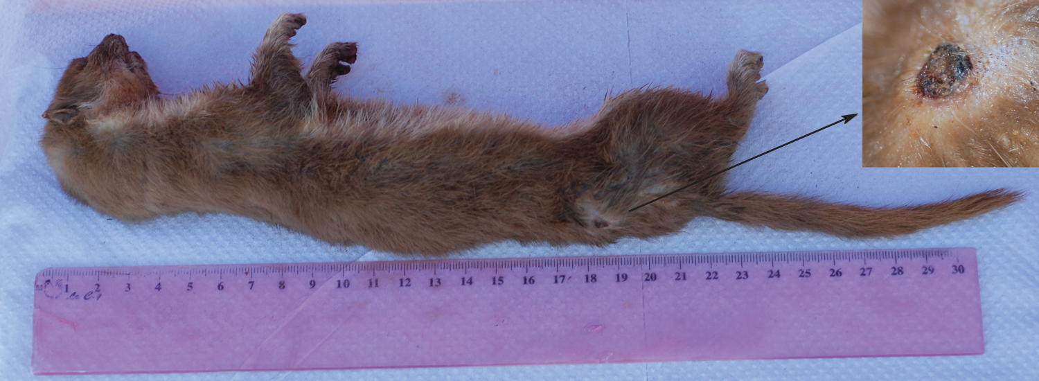
Tunisian Mustela nivalis with an ulcerated lesion caused by Leishmania major in the back as shown in the top right of the figure. Color images available online at
Parasite identification
The smear taken from the lesion, stained with Giemsa, showed amastigote forms of Leishmania parasites. Parasites in culture were transferred to the WHO Leishmania Reference Center in Montpellier and referenced as LEM 6057. They were confirmed as L. major zymodeme MON-25 and coded as L. major MMST/TN/2009/NEMS.
Discussion
In the North Africa region, the M. nivalis is encountered in Morocco, Algeria, and Tunisia. Three species of mustelid are known in Tunisia: the otter (Lutra lutra), the weasel, and the striped weasel (Poecilogale albinucha) (Gharaibeh 1997). M. nivalis is a specialized predator of small mammals that is capable of entering the underground burrows and nests of their prey. This can explain the capture of this animal in a Psammomys obesus biotope. Previously, this animal was only known in the north of Tunisia (Gharaibeh 1997), whereas our capture was made in the Governorate of Sidi Bouzid. This finding would suggest that the distribution of M. nivalis in Tunisia is poorly known. The least weasel has been found to be infected with macro-parasites using small mammals as intermediate hosts and might also encounter micro-parasites that infect their prey animals or that are present in the soil or in the air of the runways (Jennings et al. 1982). However, despite several previous attempts, Leishmania parasites have never been isolated from the least weasel (Dubrovsky 1966, Bettini 1980). In the present article, we confirmed the infection of the M. nivalis by L. major MON-25, the causal agent of zoonotic cutaneous leishmaniasis in North Africa. This finding might imply just an incidental infection of the M. nivalis by the Leishmania parasite. However, further research on larger samples of this animal is needed to verify its role as a potential reservoir host for cutaneous leishmaniasis caused by L. major Mon-25.
Footnotes
Acknowledgments
The authors extend their sincere thanks to Dr. Jean Marc Duplantier (Center for Biology and Management of Populations in Montpellier, France) for his help in identifying the weasel. They thank Francine Pratlong for technical contribution in isoenzyme typing of Leishmania parasites in the WHO Leishmania Reference Center of Montpellier. The authors are also indebted to Dr. Dhafer Laouini (Pasteur Institute of Tunis) for reviewing the article. They are grateful to Hichem Dridi, who facilitated access to the field.
Disclosure Statement
The present study is a part of a research project supported by US-NIAID-NIH, Grant number 1P50AI074178-01, and approved by the Institutional Review Board of Pasteur Institute of Tunis. All animal experimentations were in agreement with the guidelines of International Guiding Principles for Biomedical Research Involving Animals.