
Abstract
Orf virus is the etiological agent of contagious ecthyma, a severe exanthematic disease that affects small ruminants. Orf virus is zoonosis that is associated with occupational contact with infected animals in human disease. Clinically, contagious ecthyma is characterized by the appearance of vesicles, pustules, ulcers, and papillomatous proliferative lesions on the skin of the lips and nostrils. Here we describe a case of lethal cutaneous multifocal Orf virus infection in goats in the Amazon region of Brazil. Exanthematic lesions were collected and epidemiological and clinical data were obtained. Orf virus was detected using PCR amplification of the whole B2L, VIR, and VEGF open reading frame. Phylogenetic analysis revealed that this virus clustered together with the Orf virus samples isolated during classical contagious ecthyma. The present work is the first to report a severe proliferative Orf virus case in South America.
Introduction
In small ruminants, CE is usually characterized by highly infectious lesions on the lips, tongue, and around the mouth (Mazur et al. 2000; Abrahão et al. 2009). Clinically, CE is characterized by the appearance of pustules and papillomatous proliferative lesions on the skin of the lips and nostrils (Mazur et al. 2000). In more severe cases, the skin of the eyes, feet, vulva, or udder also may be affected. Although less common, lesions can be found on the navel, esophagus, intestines, and respiratory tract (Leite-Browning et al. 2008). The lesions are initially characterized by macules, which progress to papules, vesicles, and pustules, until scab formation after 3–4 weeks. Depending on the location, infected animals may be unwilling to nurse, eat, or walk (Mazur et al. 2000; Abrahão et al. 2009). Although ORFV is epitheliotropic, viral infections can be associated with systemic symptoms. Live vaccines reduce the duration and severity of clinical signs, but do not prevent subsequent infection. In humans, lesions are usually observed on the hands, face, and arms, and last for 6–7 weeks. Although severe cases of CE have been described around the world (Abu and Housawi 2010), there are no reports of severe proliferative ORFV cases in South America. Here we describe a case of lethal cutaneous multifocal ORFV infection in goats in the Amazon region of Brazil.
Materials and Methods
During a technical visit to a rural property located in the northeastern state of Pará in the Brazilian Amazon, a case of caprine exanthematic disease was reported. Two goats showing proliferative lesions on the lips and other areas of the body were examined by veterinarians, who revealed a case of exceptionally severe CE (Fig. 1). One of the goats presented weight loss, anorexia, and proliferative papillomatous nodules ranging in size from 3–8 cm, keratinized with irregular edges and located on several regions of the body. The most affected sites were the inner thigh, abdomen, axilla, the base of the tail, and the distal part of the limbs and lips (Fig. 1). This goat died 20 days after our initial visit. The other sick animal recovered, with its lesions healing a few weeks later. Both were 2-month-old females from an anglo-nubian breed.
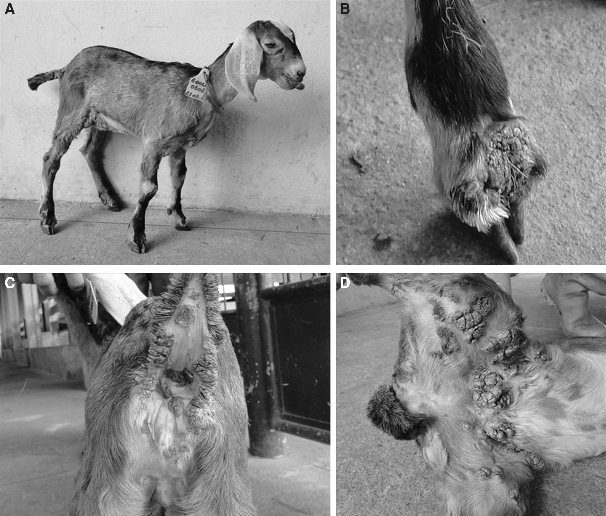
Cutaneous multifocal Orf virus infection in a goat. Severe exanthematic lesions on the mouth (
Epidemiological data were obtained from a questionnaire given to the farm manager and by the observations of our research group. Information related to the disease, age, sex, and breed of affected animals, a history of similar cases, property management, herd size, and animal origins were investigated. The animals were fed with grass and a commercial chow for sheep and received water and mineral salt ad libitum. The property houses a total of 2000 sheep and 100 goats living in intensive or semi-intensive systems, divided into small flocks. All the sick animals belonged to the same flock of 30 goats living in concrete bays. The property manager reported that the localized lesions had been previously observed in the mouth of young lambs; however, this was the first time that a lethal cutaneous multifocal CE case was observed on the property. No animal had been vaccinated against CE.
Scabs were collected from the lesions in the inner thigh of the animal that presented with acute clinical symptoms. The material was packed in plastic bottles, frozen at −70°C, and sent to the laboratory for analysis. The samples (20 mg) were mechanically homogenized in 200 μL of phosphate-buffered saline (PBS) in a tube using a pellet pestle device (Abrahão et al. 2009). The homogenates were centrifuged at 2000g for 3 min, and the supernatant was collected (Abrahão et al. 2009). Since attempts at viral isolation failed in many cell lines and primary cell culture (data not shown), we decided to use molecular methods of viral detection. The virus was detected using PCR amplification of the whole B2L open reading frame as previously described (Hosamani et al. 2006), using 2 μL of the supernatants as a template. In addition, to improve our analysis, the virus interferon resistance gene (VIR) and the vascular endothelial growth factor gene (VEGF) were also amplified, as described previously (Inoshima et al. 2010). A Brazilian sheep ORFV scab, MT05 (Abrahão et al. 2009), was used as PCR positive control. All of the experimental and control samples were screened for orthopoxviruses (OPV) using PCR (Abrahão et al. 2010).
The PCR B2L product was purified using the QIAquick Gel Extraction Kit (Qiagen, Valencia, CA), and cloned into the pGEMT-easy vector (Promega, Madison, WI). Four clones were sequenced in both orientations using M13 universal primers (Mega-BACE sequencer; GE Healthcare, Buckinghamshire, U.K.). The sequences were aligned with previously published PPV sequences from GenBank by using the ClustalW method, manually aligned using MEGA software version 3.1 (Arizona State University, Phoenix, AZ), and adjusted to equal lengths of 1137 bp. Multiple alignments of deduced amino acid sequences were generated. Phylogenetic trees were constructed by the neighbor-joining method with 1000 bootstrap replicates using the Tamura 3 parameters model implemented by MEGA 3.1. The ORFV full-length B2L, VIR, and VEGF sequences were deposited in GenBank and named ORFV-PA11.
Results and Discussion
An expected the PPV B2L gene fragment (1200 bp) was amplified from the caprine samples and from the MT05 positive control. OPV PCR did not generate any specific amplicons. The comparison of the PPV B2L sequences demonstrated a high degree of identity among ORFV-PA11 and other ORFV strains (Fig. 2). The nucleotide and amino acid sequences demonstrated that the ORFV-PA11 was closer to the ORFV vaccine strain isolate (AY278209). The sequences of ORFV-PA11 and ORFV vaccine strain showed 99.7% and 99.5% similarity at the nucleotide and amino acid level, respectively. Among the Brazilian ORFV isolates, ORFV-PA11 was closer to ORFV-MT05 (FJ665818) (Abrahão et al. 2009) (Fig. 2), isolated from a sheep from the state of Mato Grosso that shares a long border region with Para. Both VIR and VEGF phylogenetic analyses revealed the similarity between ORF-PA11 and ORFV NZ2, showing 100% identity between these viruses in both genes (Fig. 3), similar to that observed in B2L trees (Fig. 2B and C).

Molecular analysis of ORFV-PA. (
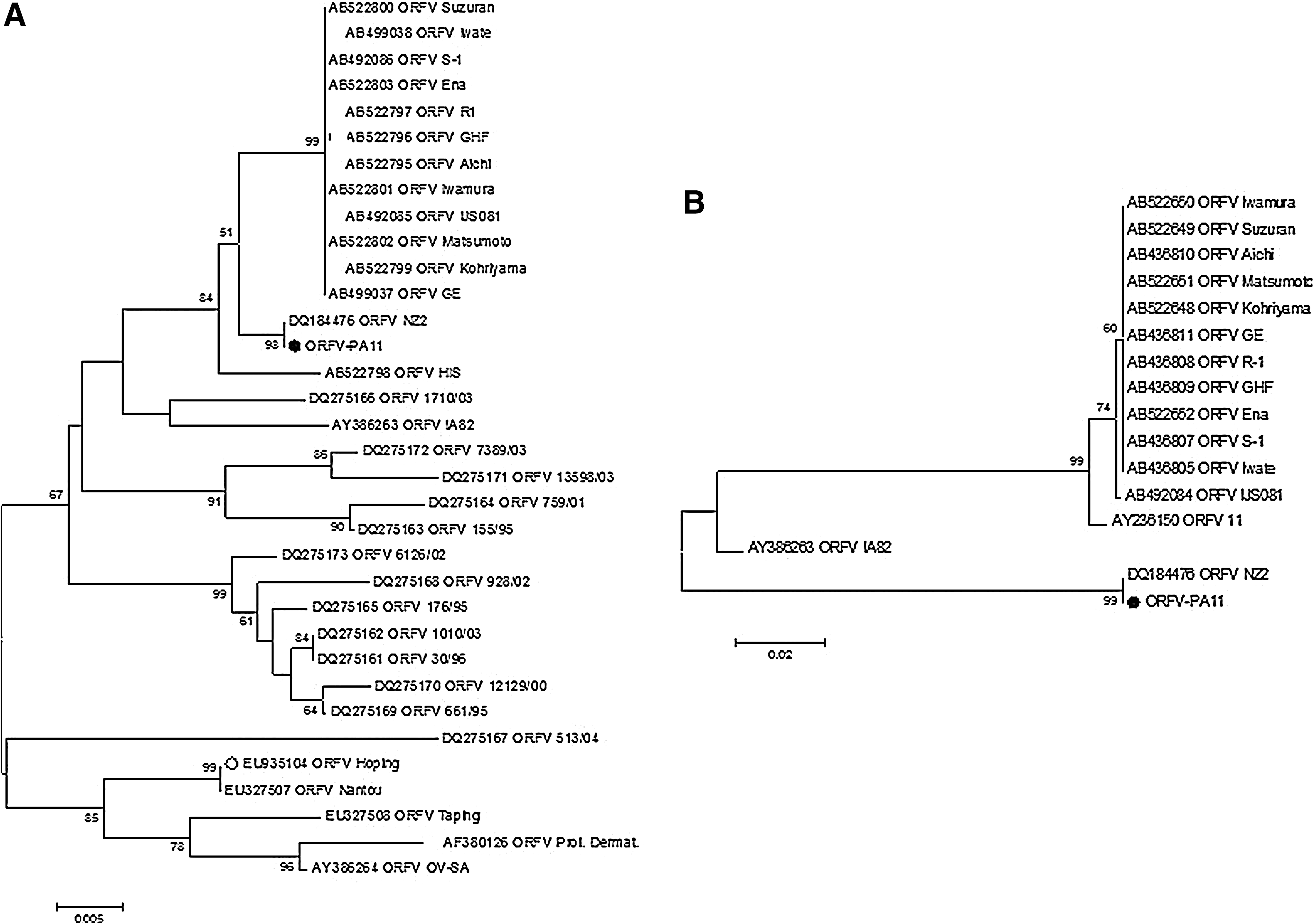
ORFV-PA11 phylogenetic tree based on the VIR (
ORFV is endemic in Brazil (Mazur et al. 2000; Abrahão et al. 2009). Vaccination has been implemented in some regions, yet veterinary and economic losses remain significant. The clinical picture of the disease and the molecular findings confirmed that an ORFV was the etiologic agent of the CE outbreak in Pará State of the Brazilian Amazon region. To establish new pasture areas, the emancipation of caprine and ovine herds in northern Brazil occurs concomitantly with the degradation of the Amazon rainforest. This anthropogenic disturbance causes profound changes in the ecological structure of the forest and may favor the occurrence of new or more severe cases of viral infections. Although our data demonstrated that ORFV-PA11 was the etiological agent of a lethal cutaneous multifocal CE case, the phylogenetic analysis revealed that this virus clustered together with the ORFV samples isolated during classical CE. Two hypotheses could explain these results: (1) the immunological status of the goat was a determinant in the severe clinical manifestations, and thus disease morbidity was not associated with the ORFV sample, or (2) B2L, VIR, and VEGF are not good molecular markers for genetic discrimination of ORFV isolates with different virulence profiles. Both hypotheses have been investigated by our group in the context of other Brazilian CE outbreaks. Epidemiological surveillance can reduce the frequency of ORFV outbreaks, and when combined with genetic studies, fill important gaps in Brazilian ORFV biology.
Footnotes
Acknowledgments
We thank MSc. João R. dos Santos, Angela S. Lopes, Ilda M.V. Gama, Andreza de Carvalho, and colleagues from Laboratório de Vírus (ICB-UFMG). Financial support was provided by Conselho Nacional de Desenvolvimento Cientifico e Technologico (CNPq), Coordenaçáo de Aperfeiçoamento de Pessoal de Nível Superior (CAPES), Fundagáo de Amparo a Pesquisa de Minas Gerais (FAPEMIG), and Ministério da Agricultura Pecuária e Abastecimento (MAPA).
Author Disclosure Statement
No competing financial interests exist, however, E.G.K. has a CNPq fellowship.