




Abstract
Coxiella burnetii, the agent of Q fever, is an intracellular bacterial pathogen. It has a nearly cosmopolitan distribution. We conducted a serological survey of domestic sheep herds for infections with C. burnetii in Wyoming following reports of abortion and open ewes. Based on the serologic evidence, there was no link between reproductive problems and exposure to C. burnetii. However, the overall prevalence of C. burnetii in WY sheep was 7%, which indicates that the agent is present in the environment and could pose a threat to public health.
Introduction
Wyoming is ranked third in the United States for sheep production (National Agricultural Statistics Service 2008), but little work has been done on the prevalence of C. burnetii infection in this state. In the 2007 Census of Agriculture-Wyoming (USDA 2009), there were 902 farms raising 412,000 sheep with a range of flock sizes from 1–24 to >5,000. The flock size for this study ranged from 300 to 400 (2), 700 to 800 (1), >1,000 (2), >2,000 (2), and >3,000 (1). In general, the smaller flocks are in more urban areas with closer contact with other domestic ruminants, and this was the case for the flocks sampled for this study. This compares to the 2002 Census which reported 966 farms raising 459,662 sheep. C. burnetii was first reported in Wyoming in 1939, when the causative agent was isolated from ticks in the southern part of the state (Davis 1939). Two human cases linked to goat herds were reported in Wyoming in 2001 (Schara and Cornish 2001). Human cases of Q fever, from California, were linked to the presence of sheep from Idaho and Wyoming that graze on winter pastures in California (Cone et al. 2006).
Following an epizootic of bluetongue virus in sheep in northern Wyoming, during the late summer and fall of 2007, sheep ranchers reported reproductive problems and open animals. Open animals were those that should have been pregnant but were not. Sera were collected from seven different ranches, and serologic surveys conducted in our laboratory of the open and aborting ewes indicated low levels of exposure to bluetongue virus (Miller et al. 2010). We further tested these samples for antibodies against C. burnetii to evaluate the possible role of C. burnetii in ovine abortions in Wyoming and to assess the potential exposure of sheep ranchers in this region.
Materials and Methods
All animal handling and serum collection was approved by the University of Wyoming Animal Care and Use Committee, and access to sheep was in accordance to approved USDA permits and agreements. We tested sera from 1120 sheep out of 8 flocks: three flocks that range in the Big Horn Basin (flock A, 211 animals; flock B, 452 animals; and flock C, 30 animals sampled), one from the Black Hills (flock D, 74 animals), one from the western slopes of the Wind River Mountains (flock E, 227 animals), one from the eastern slope of the Big Horn Mountains (flock F, 110 animals), and one from southeastern Wyoming (flock G, 16 animals). All of the ewes tested from Flock D had aborted, were excessively thin, or were late to lamb. Flock F had problems in the year. Flock E was a control flock that had not reported reproductive diseases.
IFA was performed using gamma-irradiated, acetone-fixed C. burnetii Phase II antigen. The IFA buffer (phosphate-buffered saline+1% normal rabbit serum+0.02% thimerosol) was prepared without bovine serum albumin to prevent cross reactions between contaminating bovine gamma-globulins and the FITC-labeled rabbit anti-sheep IgG(γ) conjugate (KPL, Gaithersburg, MD). All sheep sera were initially screened at a 1:64 dilution, positive samples were confirmed by duplicate screening, and then two-fold serial dilutions were tested to determine the endpoint titer of all positive samples. Pearson's chi-square statistical analysis was used to compare results between ranches and between ewes, lambs, and rams or wethers.
Results
Sera were collected from seven ranches from different sheep-producing areas of Wyoming (Figs. 1 and 2). Overall, 83/1120 (7.4%) of the sheep tested positive at a dilution of 1:64 for antibodies reactive against C. burnetii; details for individual flocks are shown in Table 1. Sheep from all but one flock (C) were seropositive for C. burnetii; the sample size from flock C was smaller (n=30), and the lack of seropositive sheep could be a result of this small sample size. The seroprevalence in individual positive flocks ranged from 1.4% to 12.5%. The seroprevalence in yearlings (8.52%) was similar to that in adult sheep (6.90%, p=0.34). Similar distributions of IFA titers were noted for seropositive lambs, ewes, and rams or wethers (p=0.94).
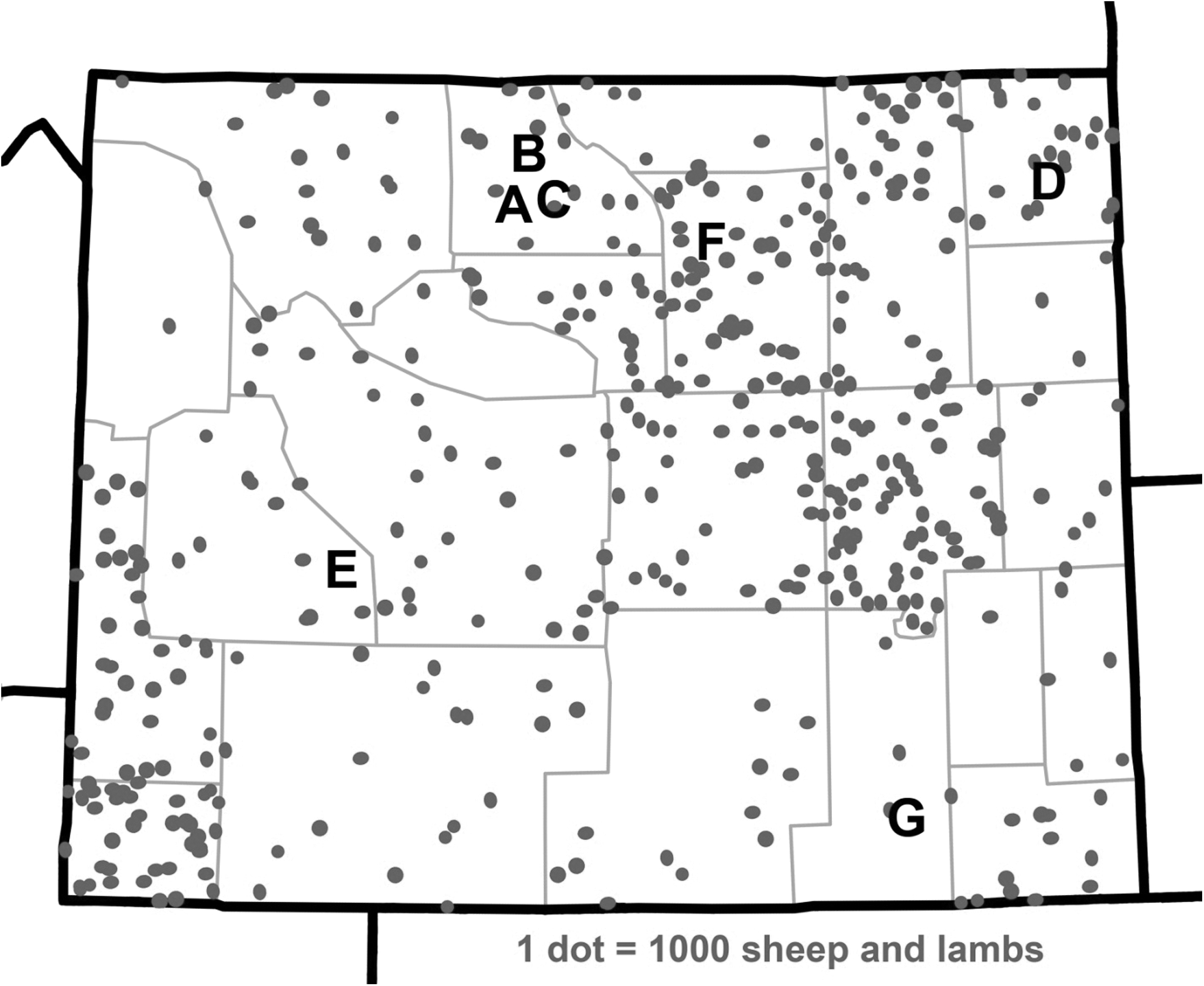
Map showing the approximate location of the seven flocks tested for antibodies against Coxiella burnetii, overlaid on the overall distribution of sheep and lambs in the state of Wyoming. Image adapted from the 2002 U.S. Census of Agriculture (2008).

The location of Wyoming relative to the United States.
Number positive/number tested are shown. The distribution of endpoint titers for seropositive animals is shown.
All of the tested ewes from flock D had suffered reproductive problems approximately 2–3 months prior to sera collection. However, the seroprevalence in flock D (1.4%) was low, with a single seropositive ram and no seropositive ewes; this seroprevalence was similar to that found in flock F (p=0.81) and in flock E (p=0.42), from which no reproductive problems were reported. In the two flocks with the lowest seroprevalence (D and F), the endpoint titers for positive sera were low (≤1:128), and serologic cross reaction with other organisms cannot be ruled out. Higher titers, up to 1:2048, were seen in flocks with higher seroprevalence rates. The highest seroprevalences were seen in flocks B (12.4%) and G (12.5%), located respectively in the Big Horn Basin and in southeastern Wyoming.
Discussion
Evidence of exposure to C. burnetii was found in sheep throughout Wyoming. However, the seroprevalence versus C. burnetii did not correlate with the intensity of reproductive problems reported in these flocks; thus, C. burnetii was unlikely to be the primary cause. The relatively small number of flock sample was a result of the reported problems and prevalence of Q fever in Wyoming might be significantly different. A larger study is needed to determine overall infection rates. Regardless of this the data show that C. burnetii is present in some flocks. Follow-up studies by USDA and Wyoming State Veterinary Laboratory demonstrated that some sheep were seropositive for ovine progressive pneumonia virus, Cache Valley virus, and other orthobunyaviruses (per. comm.). Other nonviral causes of abortion such as Toxoplasma were not examined. Our data are, however, compatible with a stable enzootic cycle of C. burnetii exposure among sheep flocks in Wyoming. Seroprevalence rates and titers were similar in lambs and in adult sheep. These ewe lambs were 10–12 months old at the time of testing, so no remaining maternal antibodies would be expected. Exposure to C. burnetii and seroconversion must occur very early in life in these flocks.
Although the cause of the ovine reproductive problems was not identified, this study revealed a significant, underrecognized public health risk of Q fever in Wyoming. An overall seroprevalence in sheep of 7%, up to 12.5% in some flocks, is not an insignificant public health threat. The seroprevalence in sheep from WY was comparable to that of sheep from Nova Scotia, which had a 6.7% overall seroprevalence rate (Marrie et al. 1985). Sheep in neighboring states played roles in outbreaks of Q fever in humans but serologic studies are generally lacking (Rauch et al. 1987). The presence of C. burnetii in enzootic cycles in these ranches could result in significant long-term environmental contamination with organisms from infected sheep, leading to human exposures via aerosol, blood contact, or handling of parturient animals. Although ranch workers, abattoir workers, and veterinarians are traditionally considered to be at risk for exposure to Q fever, recent experience in the Netherlands underscores the potential for C. burnetii organisms to disseminate to urban areas via windborne spread (Schimmer et al. 2008). C. burnetii should therefore be considered a possible cause of illness in humans or livestock throughout the state of Wyoming when patients present with compatible clinical signs.
Footnotes
Acknowledgments
We thank Larry DeBrey, Andy Fabian, and Bill Yarnell for their assistance with animal care and handling, and Bryce Reece and the Wyoming Wool Growers Association for providing sheep for this study. The use of trade names in this document does not constitute an official endorsement or approval of the use of such commercial hardware or software. Do not cite this document for advertisement. The views expressed in this article are those of the authors and do not reflect the official policy or position of the Department of the Air Force, the Department of Defense, or the Department of Health and Human Services.
Disclosure Statement
No competing financial interests exist.