
Abstract
There is a lack of knowledge regarding the prevalence of Dirofilaria immitis and Ehrlichia spp. in coyotes in Oklahoma and Texas. Documenting the prevalence of these vector-borne disease agents in coyotes from Oklahoma and Texas underscores the importance of wild canids as reservoir hosts that infect companion animals and humans. To learn more about the sylvatic cycle of D. immitis and Ehrlichia spp. in coyotes from Oklahoma and Texas, we tested for infection with and exposure to, respectively, these disease agents. Coyote carcasses were collected opportunistically from animal control experts and hunters in seven counties in Oklahoma and Texas from January to March, 2010. Serum samples from 77 coyotes were tested with a commercial ELISA test. Five (6.5%) coyotes had D. immitis antigens, and four (5.2%) had antibodies to Ehrlichia spp. The overall prevalence of D. immitis was low relative to studies from the eastern United States. Little is known about the prevalence of Ehrlichia spp. throughout the United States, but coyotes from rural Oklahoma in the current study had a higher exposure rate than those reported from California, and a lower rate than data from an earlier study from Oklahoma.
Introduction
Little is known about the occurrence of Ehrlichia spp. in coyotes. One study of 21 coyotes in Oklahoma found a high prevalence of infection with Ehrlichia chaffeensis, but did not detect infection with Ehrlichia canis (Kocan et al. 2000). Similarly, a study of coyotes in California did not detect antibodies to Ehrlichia spp., although exposure to Anaplasma phagocytophilum was prevalent (Pusterla et al. 2000). However, both studies were limited in the number and geographic extent of the coyotes examined.
Materials and Methods
Coyotes were collected opportunistically from the United States Department of Agriculture Animal and Plant Health Inspection Service (USDA-APHIS) wildlife service aerial hunting program, and the Oklahoma Predator Hunters' Association (OPHA) hunting contests, with one sample from Payne County, Oklahoma opportunistically collected as recent road kill. Serum samples were tested using a commercial ELISA (SNAP 4DX; D. immitis: sensitivity 99.2%, specificity 100%; Ehrlichia spp.: sensitivity 96.2%, specificity 100%, according to the manufacturer's literature; IDEXX Laboratories Inc., Westbrook, ME), according to the manufacturer's directions. This assay detects antigens of D. immitis and antibodies to A. phagocytophilum, Borrelia burgdorferi, and Ehrlichia spp. Samples were collected from 77 coyotes from six Oklahoma counties and one Texas county, including 29 from Craig County, Oklahoma (36°45′N, 95°08′W); six from Creek County, Oklahoma (35°50′N, 96°19′W); two from Logan County, Oklahoma (35°56′N, 97°31′W); 33 from Okmulgee County, Oklahoma (35°40′N, 95°58′W); two from Payne County, Oklahoma (36°08′N, 97°00′W); one from Roger Mills County, Oklahoma (35°37′N, 99°38′W); and four from Collingsworth County, Texas (35°02′N, 100°20′W).
Results and Discussion
Five (5/77 or 6.5%) coyotes infected with D. immitis were detected: two males in Creek, Oklahoma (2/6), and one female each in the counties of Okmulgee, Oklahoma (1/33), Craig, Oklahoma (1/29), and Collingsworth, Texas (1/4) (Fig. 1). None of the coyotes collected had antibodies to A. phagocytophilum or B. burgdorferi. Antibodies to Ehrlichia spp. were detected in one male and one female coyote (5.2%) from Craig and Creek counties, respectively (Fig. 1). One male from Creek County tested positive for infection with D. immitis and exposure to Ehrlichia spp.
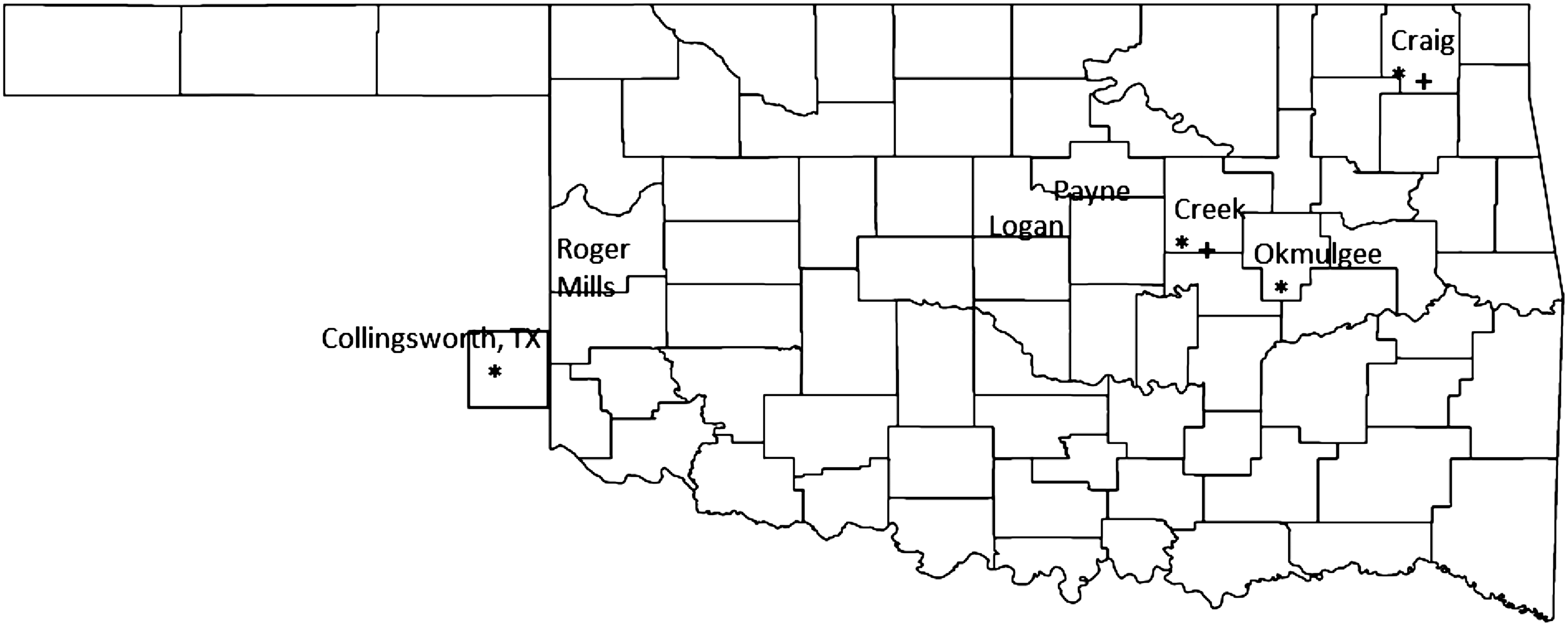
Map of Oklahoma counties indicating where coyotes were collected, and which counties had heartworm-positive (*) and Ehrlichia spp.-positive (+) samples. Labeled counties with no symbol indicate that all coyotes were negative for both diseases. Map modified from the U.S. Census Bureau:
The overall prevalence of D. immitis in coyotes in Oklahoma was low relative to data from eastern states, but similar to some studies in the western United States. Sacks and associates (2004) collected coyotes at the county level in California, and found prevalence estimates from 0–25% per county. On the other hand, studies in eastern states have reported high prevalences of D. immitis in coyotes, ranging from 16% to as high as 71% (Custer and Pence 1981; Nelson et al. 2003). Bowman and colleagues (2009) found D. immitis antigens to be prevalent in domestic dogs in Craig and Creek Counties at a rate of 2.1–4%; there were not enough data to report on dogs from Okmulgee and Collingsworth Counties. Although we were not able to sample coyotes from all counties in Oklahoma, we found D. immitis-infected coyotes only in the eastern half of the state. This suggests that the pattern of canine heartworm infection in coyotes in Oklahoma may reflect the larger, continental pattern observed in dog surveys, although one coyote from west Texas was also positive (Bowman et al. 2009). Our collection methodology was primarily through hunting (76/77 coyotes), which may bias the sample relative to the age or health of the complete coyote population. However, we do not know the direction of the bias, so we cannot speculate about the effects on our results. More extensive and intensive sampling would better elucidate the geographic and epizootiological patterns of coyote infection with dog heartworm within Oklahoma.
Coyotes in Oklahoma had a higher prevalence of exposure to Ehrlichia spp. than those collected in California, and lower than those collected from a limited area in Oklahoma (Kocan et al. 2000, Pusterla et al. 2000). The SNAP test cannot distinguish between antibodies to E. chaffensis and E. canis (O'Connor et al. 2006, Bowman et al. 2009). Due to the cross-reactivity of the SNAP test in detecting Ehrlichia spp. antibodies, the 5.2% seroprevalence of Ehrlichia spp. detected in the present study may be due to past or current infection with E. canis, E. chaffeensis, both organisms, or another unknown Ehrlichia spp. Domestic dogs in this region are also commonly exposed to Ehrlichia spp.; Creek County had a prevalence of 2.1–15% in domestic dogs, while not enough data were available in Craig County to calculate the prevalence (Bowman et al. 2009).
The occurrence of vector-borne diseases in coyotes can be dynamic, contributing to variations in observations. We only examined coyotes older than 1 year of age. It is possible that our samples were not representative of all coyotes in Oklahoma and Texas. An age or condition bias, if present, may skew our estimates of the prevalence of D. immitis and Ehrlichia spp. in coyotes (Nelson et al. 2003). All of the coyotes tested in the present survey were collected from rural areas. In a similar survey conducted in Illinois, coyotes from rural areas had a 16% prevalence of D. immitis infection, while those around Chicago had a prevalence of 41% (Nelson et al. 2003; Gehrt 2011). It has also been shown that the prevalence of D. immitis infection was higher in mosquitoes collected from urban areas in Oklahoma compared to those from rural areas (Paras 2011). It is possible that the prevalence of D. immitis infection may be higher in coyotes living in urban areas of Oklahoma and Texas.
Footnotes
Acknowledgments
The authors wish to thank IDEXX for donation of the SNAP tests, the USDA-APHIS and OPHA for donation of the carcasses, and two anonymous reviewers for editorial assistance. This work was supported by the Oklahoma Agricultural Research Station under Hatch Project 2702 (to M.H.R.).
Author Disclosure Statement
No competing financial interests exist.