
Abstract
Head and body lice are strict, obligate human ectoparasites with three mitochondrial clades (A, B, and C). Body lice have been implicated as vectors of human diseases, and as the principal vectors of epidemic typhus, relapsing fever, and Bartonella quintata-associated diseases (trench fever, bacillary angiomatosis, endocarditis, chronic bacteremia, and chronic lymphadenopathy). Using molecular methods (real-time and traditional PCR), we assessed the presence of Bartonella quintana DNA in black head lice collected from three locations in Sénégal. DNA from B. quintana was identified in 19 lice (6.93%) collected from 7 patients (7%) in Dakar. B. quintana-positive lice collected from three subjects were identified as clades C and A.
Introduction
Materials and Methods
The study was conducted during October and December 2010 and January 2011. After ethical approval for the search for pathogens responsible for non-malarial fever, and after informed consent was obtained from parents for minors, we collected head lice from different locations in Sénégal: Kaolack City (14°14’N,16°08’W), Dakar City (14°34’N,17°29’W) and its suburbs, and two villages in the Fatick region, Dielmo and Ndiop (14°20’N,16°25’W). All lice were preserved dry in sterile conditions, except two that were kept in ethanol at room temperature and then sent to our reference center in Marseille in January 2011. Pictures of the ventral and dorsal sides of each louse were taken in the laboratory (Fig. 1). Before DNA isolation, each louse was rinsed twice in sterile water for 15 min. Then total genomic DNA was extracted from each louse using a QIAamp Tissue kit (Qiagen, Hilden, Germany) per the manufacturer's instructions. The extracted genomic DNA was stored at −20°C under sterile conditions to avoid cross-contamination until the PCR assays were performed. DNA was used as a template in a previous study that utilized an RT-PCR assay targeting a portion of the Bartonella 16S-23S intergenic spacer region (ITS) (Angelakis et al. 2009), and a specific B. quintana gene, fabF3, encoding 3-oxoacyl-(acyl-carrier-protein) synthase II (Angelakis et al. 2011a). Negative controls (DNA from uninfected lice and sterile water) and positive controls (DNA from Bartonella elizabethae) were included in each assay. Nine head lice infected with B. quintana DNA collected from three subjects (three lice per person), and two non-infected lice collected from two other subjects, were randomly selected for amplification and sequencing of the mitochondrial gene cytochrome b (cytB), as previously described (Li et al. 2010). Lice with B. quintana DNA were collected from two subjects from Keur Massar and one from Diamaguene (Dakar). The non-infected lice were collected from one subject in Yeumbeul and one in Dielmo. For phylogenetic analysis, we used MEGA 4.1 (Molecular Evolution Genetic Analysis; The Biodesign Institute, Tempe, AZ), and for data comparison, we used Epi Info version 6.0 (Centers for Disease Control and Prevention, Atlanta, GA). A p value<0.05 was considered significant.
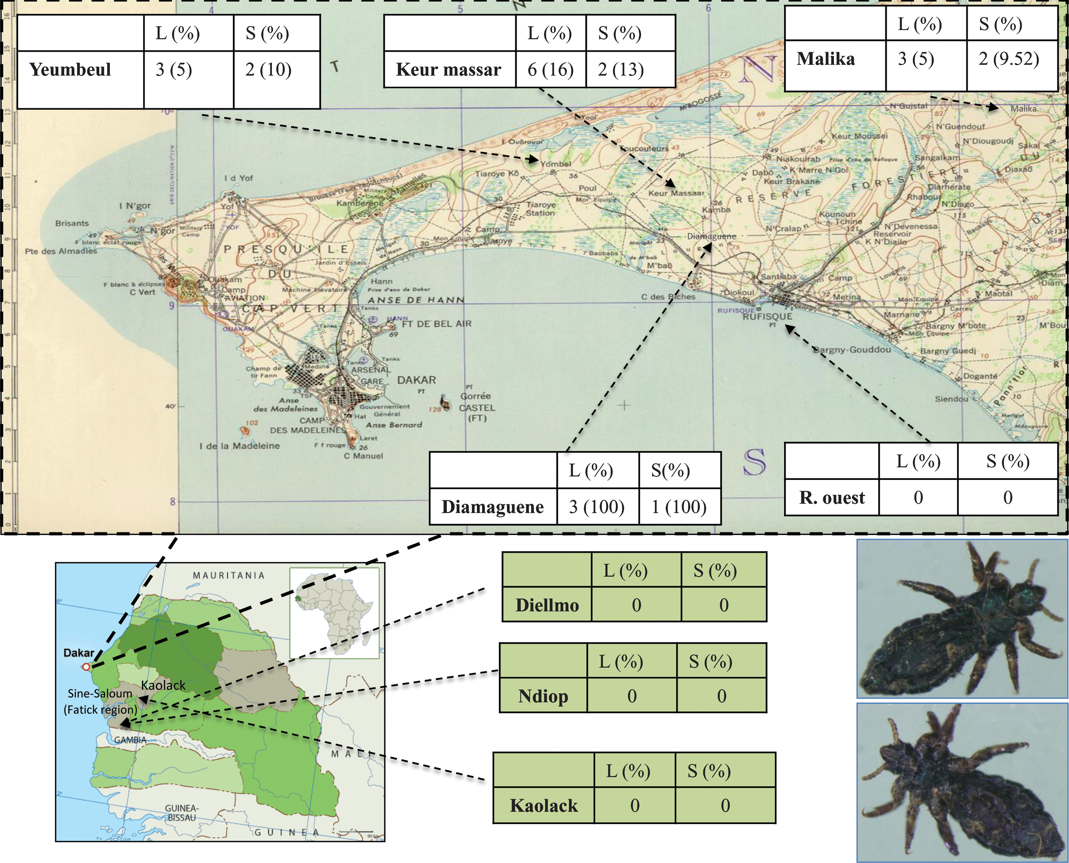
Lice infected with DNA from Bartonella quintana and subjects infested with these lice in Sénégal [2010; L (%), number and percentage of infected lice; S (%), number and percentage of subjects infested with infected lice].
Results
Overall, we tested 274 adult head lice collected from 100 females (78 children, 14 youths, and 8 adults). From each person, we collected 1 to 10 adult head lice. B. quintana was identified in 19 lice (6.93%) collected from 7 patients (7%). Four children (5.12%), one youth (7.14%), and 2 adults (25%) were infested with head lice infected with B. quintana (Table 1). No significant difference was found according to age (p=0.52 by the Kruskal-Wallis test). Positive and negative controls yielded the expected results in all tests. All the B. quintana-positive lice were found in Dakar and in its suburb. No positive lice were found in the villages Dielmo and Ndiop. Among B. quintana-positive lice from Dakar, 7 were from the Yeumbeul district, 6 were from the Keur Massar district, 3 were from the Diamaguene district, and 3 were from the Malika district (Fig. 1). All these positive lice were kept dry without ethanol.
A 316-bp fragment was obtained from each of 11 lice (GenBank numbers JN400090, JN400091, JN40009, JN969581, JN969582, JN969583, JN969584, JN969585, JN969586, JN969587, and JN969588). We found that 4 B. quintana-positive head lice belonged to clade A and 5 belonged to clade C (Fig. 2). The non-infected lice belonged to clade A.
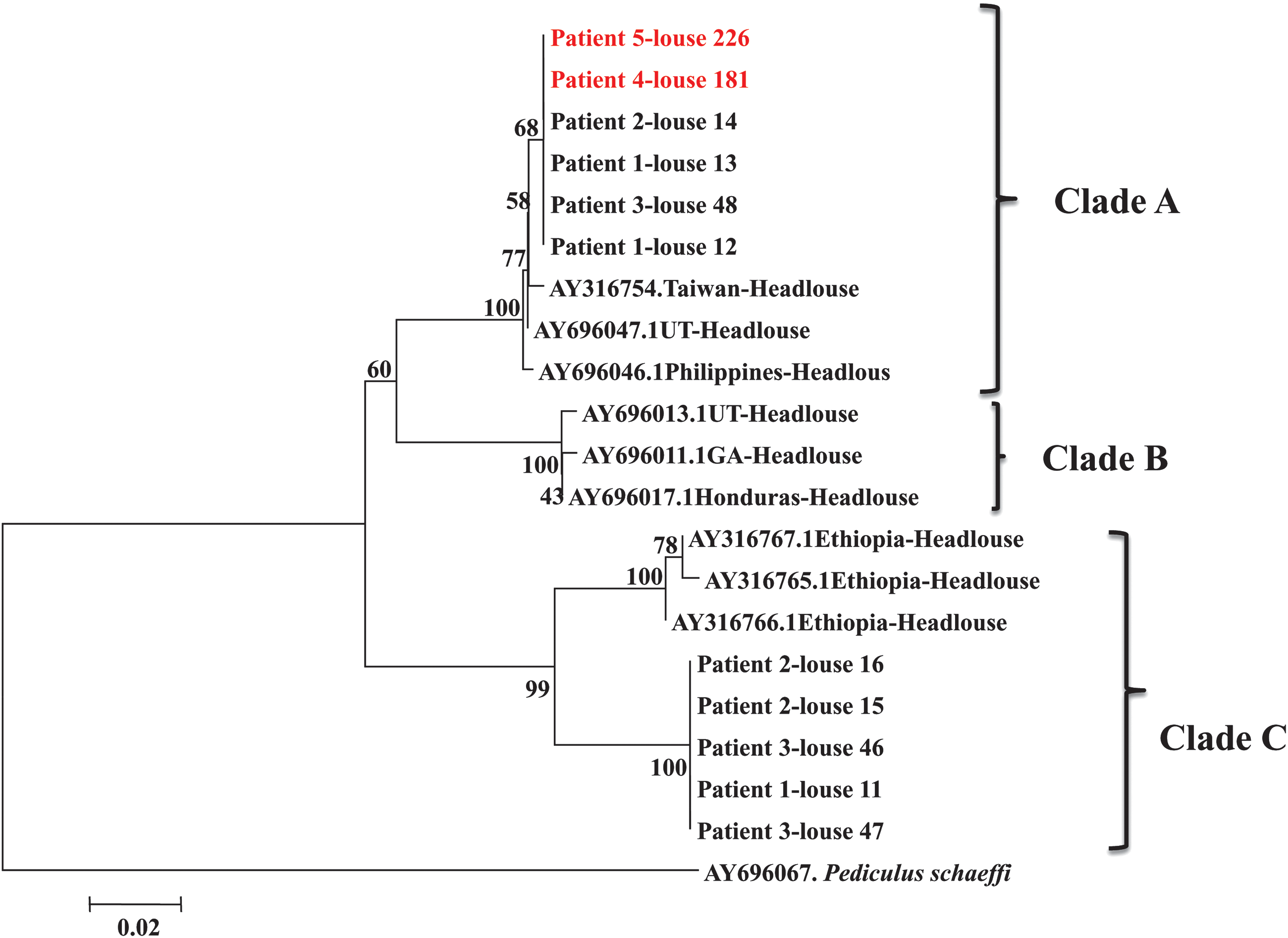
Neighbor-joining tree of head lice collected from five subjects in Sénégal (2010) based on cytochrome b gene sequencing (black type, lice with DNA from B. quintana; red type, non-infected lice).
Discussion
In this study, we confirmed the presence of DNA from B. quintana in head lice collected from Dakar (Sénégal). Clade C head lice are known to be prevalent in Ethiopia (Kittler et al. 2003; Reed et al. 2004; Raoult et al. 2008), and in Nepal (Sasaki et al. 2006), and here we found that they are also present in Sénégal. Co-infestation of the same subject with different louse clades has been described, but only between body and head lice; co-infestation with different head louse clades has never before been reported (Angelakis et al. 2011b). In addition, in Ethiopia, type C head lice were found on a subject with type A body lice, but we did not observe co-infestation by head lice of clades A and C on the same subject (Angelakis et al. 2011b). It appears that dual transmission cycles of lice are occurring, and that type C head lice do not inhibit the development of type A head louse outbreaks.
Poor living conditions and crowded shelters provide ideal conditions for the spread of lice. The role of the head louse in the maintenance and transmission of B. quintana remains to be determined because all attempts to cultivate B. quintana from nits or larvae have failed (Angelakis et al. 2011a). DNA from B. quintana has been detected exclusively in head lice collected from people living in poverty, and Sasaki and associates were the first to identify B. quintana in head lice collected from two heavily infested homeless children in Nepal (Sasaki et al. 2006).
In addition, DNA from B. quintana was detected in head lice collected from 3 alcoholic homeless adults in San Francisco (Bonilla et al. 2009; Schroff 2010), and in head louse nits collected from a homeless man in Marseille (Angelakis et al. 2011a). People living in more hygienic conditions were not infested with lice infected with B. quintana, and in a previous study all attempts to detect B. quintana DNA in head lice collected from schoolchildren failed (Fournier et al. 2002).
In this study, no homeless persons were included, but all subjects lived in poor conditions. Recently, DNA from B. quintana was also found in head lice and body lice collected from subjects in Ethiopia living in conditions of poverty (Angelakis et al. 2011b).
In conclusion, we identified the presence of B. quintana DNA in clade A and C head lice from Dakar, Sénégal. In Dielmo and Ndiop we tested a small number of lice and we did not find any lice with B. quintana DNA. As a result, we believe that a larger number of lice should be tested in the future in these areas. Moreover, further studies using head lice collected from more subjects, and from subjects in different countries, are needed to determine if head lice infected with B. quintana having phylotype C exist in African countries other than Ethiopia and Sénégal.
Footnotes
Author Disclosure Statement
No competing financial interests exist.