
Abstract
Bovine-associated zoonotic infectious diseases pose a significant threat to human health in the Lao People's Democratic Republic (Lao PDR). In all, 905 cattle and buffalo serum samples collected in northern Lao PDR in 2006 were used to determine seroprevalence of five major bovine zoonotic infectious diseases that included Taenia saginata cysticercosis, bovine tuberculosis, Q-fever, bovine brucellosis, and bovine leptospirosis. Five enzyme-linked immunosorbent assays (ELISAs) were used to test for the presence of antibodies to the diseases, except Taenia saginata, for which we tested for the presence of Taenia metacestode circulating antigens. The overall highest prevalence was for T. saginata (46.4%), with lower prevalence for Q-fever (4%), leptospirosis (3%), tuberculosis (1%), and brucellosis (0.2%). Although there were no significant differences in the proportion of seroprevalence between sex and age of the animals sampled, there were significant differences between the provincial distributions. Further studies are required to determine the seroprevalence of these infections in other locations in Lao PDR, as well as other animal species including humans, in order to develop effective prevention and control strategies. This is the first study to investigate the prevalence of bovine zoonotic infectious agents in the Lao PDR. Positivity was demonstrated for all diseases investigated, with the highest prevalence for T. saginata antigen and Coxiella burnetti antibodies. For T. saginata, there were significant differences in the provincial distribution. Approximately 16% seroprevalence of Coxiella burnetti was noted in Xayabuly Province; however, there are no clear reasons why this was the case, and further studies are required to determine risk factors associated with this observation.
Introduction
The Lao PDR is one of the poorest countries in the Southeast Asian region, and it has a predominantly rural-based agricultural economy. Cattle and buffalo production makes a significant contribution to the Lao PDR's rural economy, with approximately 31% and 48% of households raising cattle and buffalo, respectively (Stür et al. 2002). Cattle and buffalo native to the Southeast Asia region are the predominant breeds and are raised in extensive production systems with limited health inputs (Stür et al. 2002). Approximately 27%, 48%, and 25% of all production is accounted for in the northern, central, and southern regions of the Lao PDR, respectively (Department of Livestock and Fisheries 2008). Households raising bovines tend to vary according to ethnicity, with some of the most poor and vulnerable ethnic groups dependent on cattle and buffalo for their livelihood, but they are also potentially exposed to debilitating zoonotic diseases.
Taenia saginata is endemic in the Lao PDR, and is considered one of the NZDs (World Health Organization 2009, 2010). Humans are the only definitive host acquiring the adult tapeworm (taeniasis) following ingestion of beef contaminated with the larval metacestode stage of the parasite (cysticercus). Tapeworm carriers act as a source of infection for cattle and buffalo, who become infected with cysticerci after ingesting eggs on fecally-contaminated pasture. Only T. saginata has been reported in the southern and central Lao PDR, and both T. solium and T. saginata have been reported from the northern Lao PDR (Conlan et al. 2011a). In previous studies, the prevalence of taeniasis varies in different areas, and estimates range from 0–14% (Conlan et al. 2008). In the northern Lao PDR, T. saginata is the predominant species infecting humans (unpublished data). Brucellosis is also considered a NZD (World Health Organization 2009, 2010) with worldwide distribution, and is caused by bacteria of the genus Brucella. Bovine-associated brucellosis is normally caused by serovars of B. abortus, but may be caused by B. melitensis in areas where cattle and buffalo are raised in close proximity to goats and sheep. The bacterium is readily transmitted to humans by occupational exposure via the oral, respiratory, and conjunctival routes, or by ingestion of dairy products. Human infection causes acute febrile illness and undulant fever, which may progress to the chronic form, resulting in musculoskeletal, cardiovascular, and central nervous system complications. Leptospirosis and Q-fever (Coxiella burnetti) are responsible for undifferentiated febrile illnesses in many rural communities in Southeast Asia, including the Lao PDR (Laras et al. 2002), and Thailand (Suttinont et al. 2006), and if left untreated may lead to complications. However, little is known about the role of bovines in the epidemiology of these diseases in the Lao PDR. Zoonotic tuberculosis caused by Mycobacterium bovis, like brucellosis has worldwide distribution and is considered an NZD (World Health Organization 2009, 2010) but the threat to human populations in the Lao PDR is unknown. The principal mode of transmission of M. bovis is via the consumption of unpasteurized dairy products, but it can also be transmitted through close contact with infected animals, which is the most common route of transmission in the Lao PDR.
Like many developing countries, the Lao PDR has a poor veterinary infrastructure and the detection capability for zoonotic diseases is limited. The evidence base for the presence of bovine-associated zoonoses has not previously been investigated in the Lao PDR, therefore veterinary and public health stakeholders are not in a position to assess the burden and impact of disease on society. This retrospective study describes the serological prevalence of five major bovine-associated zoonoses: bovine cysticercosis (T. saginata), zoonotic tuberculosis (Mycobacterium bovis), Q fever (Coxiella burnetti), brucellosis (Brucella abortus), and leptospirosis (Leptospira icterohaemorrhagiae serovar hardjo) in the year 2006.
Materials and Methods
Inventory of cattle and buffalo serum samples and sample selection
A total of 905 samples from 67 villages in 33 districts of 5 northern provinces (Fig. 1), collected between April and May 2006, were stored at −80°C according to Office International des Épizooties (OIE) guidelines and biosafety precautions. The geographical distribution of the samples tested in this study are described in Table 1.
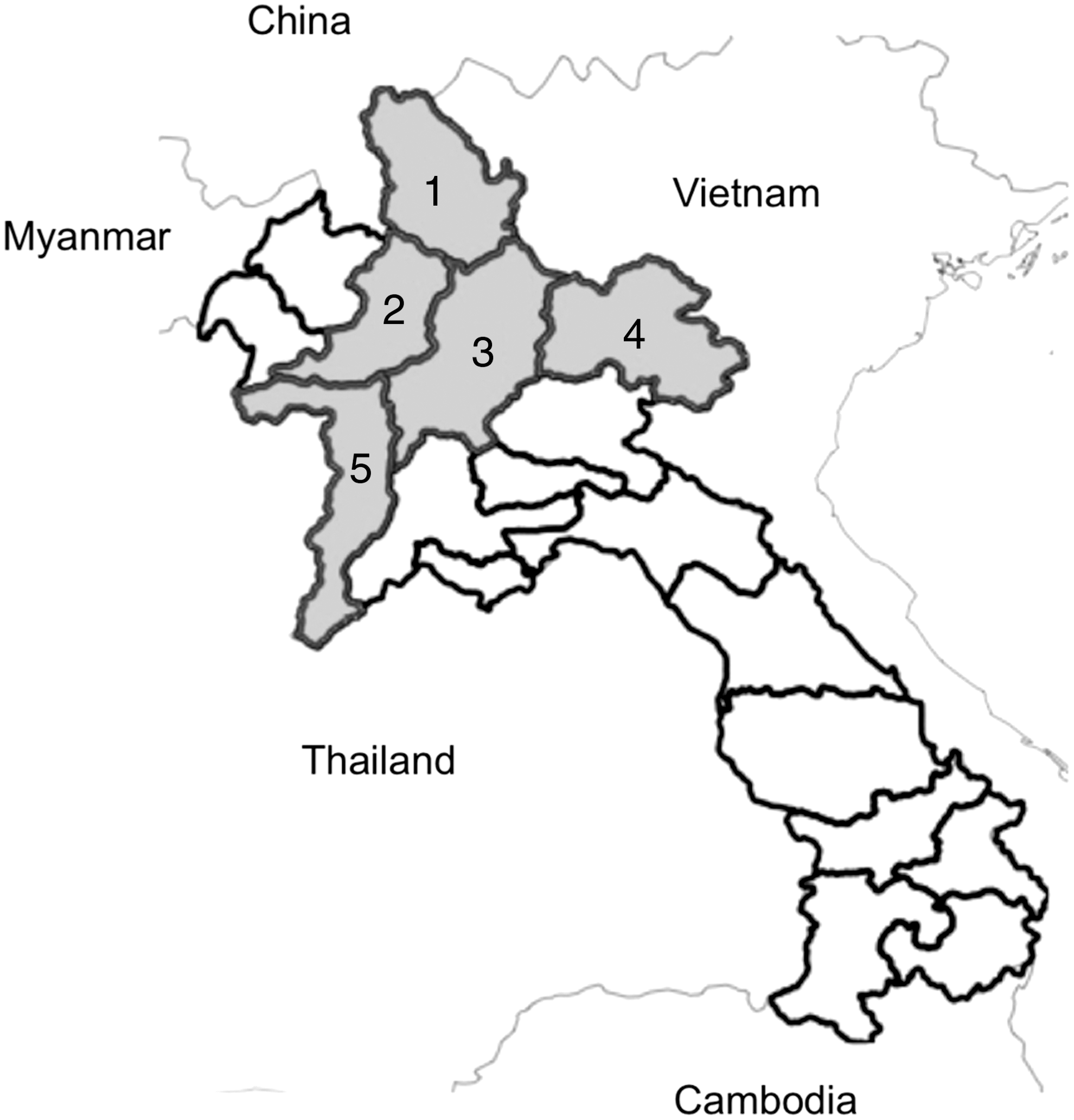
Study sites in northern Laos PDR (1, Phongsaly province; 2, Oudomxay province; 3, Luangprabang province; 4, Huaphan province; 5, Xayabuly province).
Serological ELISA assays
The following enzyme-linked immunosorbent assays (ELISAs) were used for the detection of antibodies: CHECKIT* Q-Fever Antibody Test Kit (cat. no. QFT1135T; IDEXX Labs, Westbrook, ME); Bovine Brucellosis Ab ELISA (cat. no. EB43-01; BioNote, Gyeonggi-do, Korea); Bovine Tuberculosis Ab ELISA (cat. no. EB43-03; BioNote); and Leptospira IgM ELISA (BioNote). All assays were used following the manufacturer's recommended practices and methodologies. The bovine serum samples were tested by ELISA for the presence of Taenia metacestode circulating antigens (Brandt et al. 1992; Dorny et al. 2000, 2002), using modifications introduced by Dorny and associates (2004).
Calculation of ELISA results and determination of diagnostic cutoffs
Diagnostic cutoffs for the CHECKIT* Q-Fever Antibody Test Kit, Bovine Brucellosis Ab ELISA, and Bovine Tuberculosis Ab ELISA were determined using the manufacturer's recommended controls and calculations. The Leptospira IgM ELISA (BioNote) is an assay in development, and the manufacturer did not suggest a fixed diagnostic cutoff. The diagnostic cutoff was determined by plotting the optical densities (ODs) (lowest to highest) to determine the distribution of positive and negative Leptospira antibody populations using the results of the cattle and buffalo sera from the Lao PDR. A conservative cutoff value of 0.25 was selected (see the legend to Fig. 2 for details). The diagnostic cutoff for the cysticercosis antigen ELISA was calculated as described by Dorny and associates (2000), using a panel of eight negative sera representative of the Lao bovine population. A ratio for each test was calculated by dividing the OD of the test sample by the cutoff value; a ratio >1 was considered positive. Samples were retested if the coefficient of variation was more than 50%, or if the OD of the test sample approached the cutoff.

Distribution of optical densities (ODs) for the BioNote Leptospira IgM ELISA using cattle and buffalo sera from the Lao PDR. The diagnostic cutoff (0.25 OD) was determined by sorting the ODs from lowest to highest, and plotting to determine the distribution of positive (slope) and negative (lower plateau region) populations.
Data analysis
Data analysis was performed using Stata version 10 (StataCorp LP, College Station, TX). Serological prevalence was calculated as the proportion of animals that had detectable antibodies or antigen in the sample population. The Fisher's exact test or Pearson's chi-square test (depending on the positivity rate) was used to explore associations between species, age, sex, and province. Associations were considered significant if p≤0.05.
Results
Prevalence of bovine Taenia saginata cysticercosis
In total, 46.4% (422/905) of the animals tested had evidence of bovine cysticercus antigen (Table 2). There was no significant difference in prevalence by sex and age, although there was a declining prevalence with increasing age. Significant differences were observed between species and province (both p<0.001). Cattle demonstrated substantially higher prevalence compared to buffalo. The highest provincial prevalence was in Xayabuly, and the lowest was in Huapanh.
95% confidence interval.
Pearson's chi-square test.
ELISA, enzyme-linked immunosorbent assay.
Seroprevalence of bovine brucellosis
Overall, only 0.2% (2/905) of the animals sampled were seropositive for Brucella abortus antibodies (Table 3). There were no significant differences in the proportion of seropositive animals between species, sex, age, and province. Seropositivity was noted only in Luang Prabang (0.6%) and Phongsaly (0.6%). Older animals, especially adult breeders (3–10 years; 0.2%), and older animals (>10 years; 1.4%), demonstrated the only seropositivity.
95% confidence interval.
Pearson's chi-square test.
Fishers Exact test.
Seroprevalence of bovine tuberculosis (Mycobacterium bovis)
Overall, only 1.0% (9/905) were seropositive for Mycobacterium bovis antibodies (Table 3). There were no significant differences in the proportion of seropositive animals between species, sex, and age. Significant differences were noted between provinces (p=0.001), with high positivity noted in Xayabuly (3.2%), and Luang Prabang (2.5%) provinces. Adult breeders (3–10 years) demonstrated the only seropositivity.
Seroprevalence of Q-fever
Overall, 4.0% (36/905) were seropositive for Coxiella burnetti antibodies (Table 3). There were no significant differences in the proportion of seropositive animals for species and sex. However, significant differences were noted between provinces (p<0.001), with very high seroprevalence noted in Xayabuly (15.9%), with substantially lower results in Luang Prabang (3.8%), Huapahn (1.3%), and Phongsaly (0.6%). There was no seropositivity recorded in Oudomxay. Significant differences in seropositivity between age groups were noted (p=0.008), with 5.4% in adult breeders (3–10 years), and only 1.1% for juveniles (1–3 years).
Seroprevalence of bovine leptospirosis
Estimation of the diagnostic cutoff of the BioNote Leptospira IgM ELISA of OD 0.25 was determined by plotting the distribution of positive and negative sample populations (Fig. 2). Using this cutoff, overall, 3.1% (28/905) of the animals sampled were seropositive for Leptospira icterohaemorrhagiae serovar hardjo bovis antibodies (Table 3). There were no significant differences in the proportion of seropositive animals for sex and age. Significant differences were noted between species (p=0.001), with seroprevalence of 6.1% in cattle, while it was only 1.7% in buffalo. Significant differences were also noted between provinces (p=0.036), with seroprevalence noted in Luang Prabang (5.7%), Xayabuly (3.2%), Huapanh (3.8%), and Phongsaly (1.2%). There were no seropositive animals recorded in Oudomxay.
Discussion
This is the first study to investigate the prevalence of bovine zoonotic infectious agents in the Lao PDR. Positivity was demonstrated for all diseases investigated, with the highest prevalence seen for T. saginata antigen and Coxiella burnetti antibodies. There were significant differences in the provincial distribution of all infectious agents, with the exception of bovine brucellosis. Oudomxay Province was seronegative for all bovine zoonoses tested except for T. saginata.
Assay sensitivity and specificity can have a potentially large effect on the accuracy of the seroprevalence estimates. In this study, we used four commercial ELISAs and one “in house” test for the qualitative assessment of seropositivity, as the assays were: (1) reportedly demonstrated to have high levels of accuracy for sero-surveillence compared to gold standard or reference assays; (2) simple and amenable to the testing of large numbers of samples; and (3) affordable and applicable to a non-reference laboratory environment. The IDEXX Q Fever ELISA had a reported sensitivity of 95% and specificity of 100% compared to the reference complement fixation test (Kittelberger 2009). The bovine tuberculosis ELISA used recombinant antigens, and the manufacturer reports sensitivity and specificity of 90.0% and 98.4% (isolation confirmed), and 86.9% and 99.0% (intradermal skin test confirmed), respectively, with antibodies detected 4–8.5 weeks post-infection (BioNote 2011a). The Brucella ELISA used a lipopolysaccharide Brucella abortus antigen, and the manufacturer claims that the assay has been standardized using OIE standard sera with a sensitivity of 100% and specificity of 97%, and is reported to be 2048 times more sensitive than the Rose-Bengal test (BioNote 2011b). The Leptospira IgM ELISA is currently under development, and can be used with cattle, pig, and dog sera, and the manufacturer claims sensitivity of 97.2% (102/105) and specificity of 99.1% (344/347), compared to the serological gold standard microscopic agglutination test (MAT) (personal communication, BioNote).
While the cysticercosis Ag-ELISA has high sensitivity (92.3%) for animals with more than 50 cysticerci present (Van Kerckhoven et al. 1998), the test is genus-specific and cannot differentiate between closely related taeniid species, meaning that cross-reactions may have occurred if cattle and buffalo were infected with cysticerci of T. hydatigena. This dog tapeworm is typically associated with sheep and goats, but is known to be prevalent in the dog and pig populations of Southeast Asia (Conlan et al. 2011a, 2011b). There are no reports of bovine T. hydatigena infections in Asia; however, the possibility that some of the sampled cattle and buffalo were infected with T. hydatigena cysticerci cannot be ruled out. Nevertheless, this study has clearly demonstrated that T. saginata cysticercosis is hyperendemic in the bovine population of the northern Lao PDR. This observation corresponds to the high prevalence of taeniasis in humans, for whom estimates were 13.1%, 9.2%, and 5.4% for Oudomxay, Luang Prabang, and Huapanh provinces, respectively (Conlan et al. 2011b). The Taenia species causing human infections in the northern Lao PDR are often not determined, but recent data on worms recovered from 35 taeniasis cases demonstrated that 94.3% (33/35) were T. saginata (Conlan et al. 2011b). The prevalence of having consumed uncooked beef in the northern Lao PDR peaks at greater than 55% in the 40- to 54-year-old age group and open defecation is very common (Conlan et al. 2011b), providing the parasite with suitable conditions to thrive. Furthermore, 90% of taeniasis cases in the northern Lao PDR reported having had a history of taeniasis, indicating a high re-infection rate, and that tapeworm infections do not act as a deterrent to consuming uncooked beef (Conlan et al. 2011b).
This is the first study that has determined bovine-associated seropositivity for Coxiella burnetti in the Lao PDR. High seroprevalence (nearly 16%) of Coxiella burnetti was noted in Xayabuly province; however, there are no clear reasons why this should be the case, and further studies are required to determine risk factors associated with this observation, in addition to human serological surveys in at-risk populations. Reports of Q-fever in the human and animal populations of Southeast Asia are scarce. In Thailand, studies from more than 40 years ago in domestic animals found seroprevalence in dogs of 28% and seroprevalence in ruminants of 2–6% (Suputtamongkol et al. 2003). The reservoir host range and the specific contact patterns in Xayabouly province that have caused this apparent high prevalence in cattle and buffalo remain to be determined, as does the assessment of Q-fever in undifferentiated fever patients in this region of the Lao PDR.
Brucella abortus antibodies were detected at very low levels (0.2%; 2/905), with the two positives found in older animals. However, the specificity of the assay was 97%, which means in a population of 905 we may expect approximately 33 false-positive samples, and hence it is possible that these are false-positive results. The low level of Brucella abortus antibodies contrasts with recent surveillance in Chiang Rai province, northern Thailand, which borders the Lao PDR, where bovine brucellosis is an emerging problem with 24% of dairy herds infected (Jittapalapong et al. 2008), and human brucellosis is a potential threat in livestock-producing areas of Thailand (Manosuthi et al. 2004). The difference between the Lao and Thai seroprevalence rates may be due to the extensive farming practices employed in the Lao PDR, and the low reliance on dairy products in Lao in contrast to those in Thailand.
Leptospira icterohaemorrhagiae serovar hardjo is normally associated with cattle and buffalo and is a known pathogen for humans, causing severe disease. In the Lao PDR, leptospirosis is a known cause of acute disease (Laras et al. 2002), particularly in flood-prone areas (Kawaguchi et al. 2008). In this study, using the BioNote Leptospira IgM ELISA, which did not have a fixed diagnostic cutoff due to its developmental nature, Leptospira antibodies were clearly demonstrated in cattle and buffalo, even when using a conservative cutoff. Nevertheless, it is clear that assessment of the samples by MAT would have been preferable. However, the MAT is best suited to reference laboratory settings, and commercial MAT testing is prohibitively expensive (approximately US$10 per sample) and was beyond the budget of this study. Importantly, the assay will only detect IgM antibodies that are present in the early phase of the infection, and we recommend that the Leptospira IgM ELISA used in this study be evaluated with samples from various species and geographical locations to determine the true sensitivity and specificity of the assay.
In conclusion, our results have shown that there is serological evidence for the five major bovine zoonotic diseases in the Lao PDR, with the highest prevalence of T. saginata antigen and Coxiella burnetti antibodies. Although there were no significant differences in the proportion of seroprevalence for sex and age, there were significant differences between the provincial distributions, with the exception of bovine brucellosis. Further studies should focus on the detection of Coxiella burnetti, Mycobacterium bovis, and leptospirosis in other animal species, and may help to develop an effective prevention and control strategy for those zoonotic diseases to minimize risks to humans.
Footnotes
Acknowledgments
The authors wish to thank Ms. Manivanh Phruaravanh and Ms. Vilaywan Soukvilai for excellent technical assistance in performing the ELISAs. Thanks to Dr. Fidel Malbas for critical comments on this manuscript. This study was funded by the UNDP/WB/World Health Organization Special Programme for Research and Training in Tropical Diseases Joint Small Grants Programme for Operational Research in Communicable Diseases. The authors are grateful to BioNote, Korea for the donation of leptospirosis, bovine tuberculosis, and Brucella abortus ELISAs. S.D.B. is supported by the Wellcome Trust of Great Britain.
Author Disclosure Statement
No competing financial interests exist.