
Abstract
In this study we endeavored to determine the seroprevalence of tick-borne infections in the military working dog (MWD) population in the Republic of Korea (ROK). Our sample population consisted of 182 serum samples from MWDs for 3 different years (1996, 2002, and 2007). In addition, 63 whole blood samples from 2007 were available for polymerase chain reaction (PCR). Serum samples were evaluated by a commercially available enzyme-linked immunosorbent assay (ELISA) and immunofluorescence assay (IFA) for Ehrlichia canis and Anaplasma phagocytophilum, and by ELISA only for Borrelia burgdorferi. PCR amplification of DNA was performed to screen for Ehrlichia canis, E. chaffeensis, E. ewingii, Anaplasma phagocytophilum, A. platys, Borrelia burgdorferi, and Rickettsia rickettsii, as well as Babesia and Theileria species using previously published primers and probes. A total of 56 (30.8%) MWDs were positive by at least one serologic test. Seroprevalences for Anaplasma and Ehrlichia were 4.4% and 0.6% based on the ELISA, and 24.7% and 22.5% based on the IFA, respectively. ELISA testing for Borrelia yielded 2 (1.1%) positive results. In parallel testing using both the ELISA and IFA tests, the percentages of dogs with one or more positive results were 34.1%, 25.9%, and 28.4%, for 1996, 2002, and 2007, respectively. There was no significant differences in seroprevalence based on location, year, breed, or sex of the MWD. There was poor agreement between IFA and ELISA test results. No MWD sample had a positive PCR result. MWDs stationed in Korea had serologic evidence of exposure to several tick-borne pathogens, but PCR testing did not identify any active infections.
Introduction
MWDs are important for all branches of the U.S. military. These highly-trained animals provide such services as explosive, mine, and drug detection; security and patrol; search and rescue; and guard duty. The tasks these dogs perform are critical to the fight in the global war against terrorism, as demonstrated by their presence in both Operation Iraqi Freedom and Operation Enduring Freedom (Department of Defense Military Working Dog Veterinary Services, 2004). Therefore, the health and well-being of these indispensable animals is of the upmost importance, which prompted the development of strict guidelines for their care and management. Proper procedures for preventive care and record-keeping are documented in The Handbook of Veterinary Care and Management of the Military Working Dog. To help control tick-borne infections, MWDs are required to have monthly topical treatment with a commercially available flea- and tick-prevention product (Department of Defense Military Working Dog Veterinary Services, 2004). Many studies examining the efficacy of fipronil (Frontline Plus®; Merial Limited, Duluth, GA), along with other commonly used preventive medications (i.e., imidacloprid and permethrin [Advantix®; Bayer HealthCare, Animal Health Division, Shawnee, KS]), and amitraz collars (Preventic Collars®; Virbac Animal Health, Inc., Fort Worth, TX), have shown that these products, which are marketed to offer protection against tick infestation for up to 30 days, are not 100% effective (Young et al. 2003; Hagimori et al. 2005; Estrada-Peña and Venzal Bianchi 2006; Otranto et al. 2005, 2008). Prior to the development of these residual products, which began in 1997, organophosphate dips were recommended for flea and tick control for MWDs.
There are several published reports from the Republic of Korea (ROK) of ticks, small mammals, and rodents harboring organisms that are considered tick-borne pathogens (Chae et al. 2003; Lee et al. 2003, 2005; Kim et al. 2003, 2006, 2007) These studies have demonstrated the presence of Ehrlichia canis, E. chaffeensis, E. ewingii, Anaplasma phagocytophilum, A. platys, Borrelia burgdorferi, Rickettsia rickettsii, and R. japonica, as well as several others. By far, the most common tick identified in these studies is Haemaphysalis longicornis, but other Haemaphysalis species and several Ixodes species of ticks are also present. There have been several case reports of tick-borne infections in wild animals, dogs, and humans in the ROK (Heo et al. 2002; Park et al. 2003; Jang et al. 2004; Song et al. 2004; Choi et al. 2005, 2009; Chung et al. 2006; Yu et al. 2008; Han et al. 2009; Camer and Lim 2009). Given the fact that tick-preventative measures may not be 100% effective, the possibility of dogs serving as a potential reservoir for tick-borne infections for humans, and the risk of illness in the MWD population, are valid concerns. These facts raised questions concerning the prevalence of tick-borne infections in the MWD population in the ROK. The MWDs are sent to the ROK immediately after initial training at Lackland Air Force Base in San Antonio, Texas, and these animals rarely if ever leave the Korean peninsula. All MWDs are screened for tick-borne infections by immunofluorescence assay (IFA) prior to departure from Lackland Air Force Base, ensuring that any tick-borne infection detected in the animals stationed in the ROK would have been acquired after arrival on the peninsula.
To date, there have been no published prevalence studies regarding tick-borne infections in dogs in the ROK. The primary objective of this retrospective study was to determine the seroprevalence of tick-borne infections in the MWD population in the ROK. We also wanted to examine whether there was a breed, sex, or age predisposition for these various tick-borne infections, and if location of the MWDs on the peninsula was correlated with seroprevalence. We also sought to compare the seroprevalence of tick-borne infections in recent years, when fipronil has been the primary flea and tick preventive medication used in MWDs, to the seroprevalence in a previous year when monthly organophosphate dips were used.
Materials and Methods
Sample selection
The analysis included serum samples from three separate years: 1996, the year prior to the use of fipronil; and 2002 and 2007, when fipronil was used. All MWDs are required to have blood drawn on an annual basis for banking purposes. These samples are submitted to the Food and Animal Diagnostic Laboratory (FADL), Fort Sam Houston, Texas, for storage. In June 2008, all samples from MWDs stationed in the ROK for the above-mentioned years were identified. Samples were excluded if there was insufficient serum available for analysis. When multiple submissions were available for an individual dog in the same year, only the first submitted sample was utilized for analysis. In the instance that an individual dog was present in multiple years, the only sample included for analysis was from the first year that serum was available for that animal. All samples from later years were excluded. Serum samples were drawn from the MWD at the duty location and shipped to FADL, where the samples were stored at −70°C. Samples from all dogs for the study were identified and shipped by overnight delivery to the Athens Veterinary Diagnostic Laboratory, where the samples were thawed, an aliquot of the serum was separated, and the remainder of the serum was returned to FADL for storage. The selected aliquots of serum were refrozen and stored at −70°C until needed.
The only year during our study when EDTA anticoagulated whole blood was banked from the MWDs in the ROK was 2007. As with the serum, samples were collected at the duty location of the MWD, shipped to FADL, and stored at −70°C. The samples were thawed, aliquots of the EDTA anticoagulated whole blood were collected, and the remainder of the sample was shipped back to FADL. If an animal had multiple banked EDTA anticoagulated whole-blood samples, only the first collected sample was utilized.
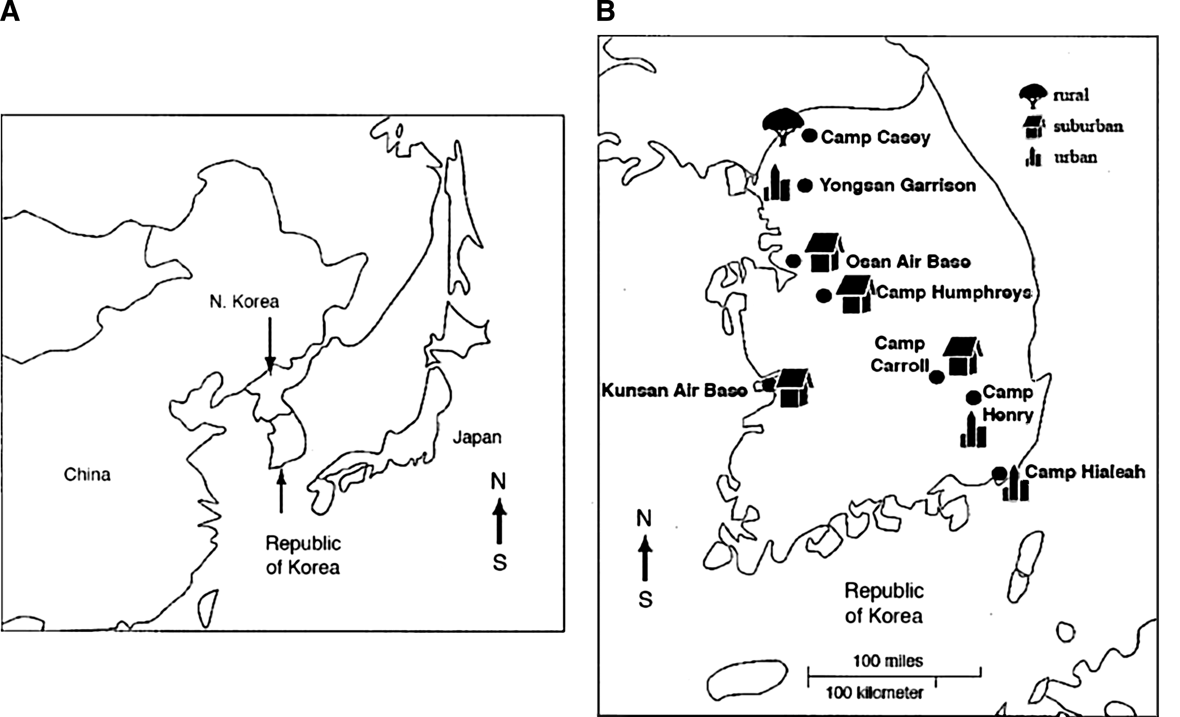
Serum and whole-blood samples were collected from MWDs at eight U.S. military locations throughout the ROK. Military installations that submitted samples during the study period included Camp Casey, located in a rural, mountainous area in the northwest; Yongsan Garrison, located in the city of Seoul; Osan Air Base, located in a suburban area south of the city of Osan; Camp Humphreys, located in a suburban area near the city of Pyeongtaek; Kunsan Air Base, located on the southwestern coastline near the city of Kunsan; Camp Henry, located in the city of Daegu; Camp Carroll, located in a semi-rural area west of Daegu; and Camp Hialeah, located on the southeastern coast in the city of Pusan (Fig. 1A and B). The location, age, breed, sex, and year of sample collection were recorded for each animal, when available.
(
Population characteristics
A total of 182 dogs were included in the study. Sixty-two dogs were from the northwestern region (Yongsan Garrison [55] and Camp Casey [7]), 71 were from the midwestern region (Camp Humphreys [11] and Osan Air Base [60]), 30 were from the southwestern region (Kunsan Air Base [30]), and 19 were from the southeastern region (Camp Henry [8], Camp Hialeah [10], and Camp Carroll [1]). There were 45 dogs in the 1- to 4-year-old age group, 74 were in the 5- to 8-year-old age group, and 56 were in the 9- to 12-year-old age group. Age could not be determined for 7 dogs. The breed distribution included 92 Belgian Malinois, 78 German shepherds, and 12 others, which consisted of Dutch shepherds (8), Belgian shepherds (2), and Labrador retrievers (2). The sex distribution included 31 female dogs and 119 males. Sex was not reported for 32 dogs. There were 88 dogs enrolled from 1996, 27 from 2002, and 67 from 2007.
Enzyme-linked immunosorbent assay
ELISA testing, using a commercially available assay, was performed according to the manufacturer's instructions (IDEXX SNAP® 4Dx®; IDEXX Laboratories, Inc., Westbrook, ME). This test identifies IgM and IgG antibodies to Ehrlichia canis, Anaplasma phagocytophilum, and Borrelia burgdorferi, as well as antigen of Dirofilaria immitis. Test results were read at 8 min. The samples were recorded as positive for a particular antibody if a color change was present at the appropriate sample spot. In addition a sample was considered positive for Dirofilaria immitis antigen if the appropriate sample spot showed a color change. No color change was considered as negative. Tests having equivocal results were repeated. Tests were also repeated if the positive control spot showed no color change.
Indirect immunofluorescence assay
A commercially available microimmunofluorescence (MIF) assay (Fuller Laboratories, Fullerton, CA) was used for the detection of E. canis and A. phagocytophilum antibodies. All testing materials and sera were allowed to equilibrate to room temperature prior to testing. Serum samples were diluted using phosphate-buffered saline (PBS) to a dilution of 1:80. Only samples demonstrating fluorescence at this dilution were considered positive. Tests were performed according to the manufacturer's instructions. The slides were read using a fluorescence microscope at 400× magnification. Sample wells were compared to the appearance of the positive and negative controls that were provided with the tests. End-point dilutions were not performed.
Polymerase chain reaction assay
DNA was extracted from 200 μL of EDTA anticoagulated whole blood using a commercially available product according to the manufacturer's instructions (UltraClean DNA BloodSpin Kit; MO BIO Laboratories, Inc., Carlsbad, CA). A housekeeping gene for canine glyceraldehyde-3-phosphate dehydrogenase (G3PDH) was used as an extraction positive control (Peters et al. 2003). All protocols used for PCR amplification were described elsewhere (Table 1). For detection of all Anaplasma and Ehrlichia species, real-time primers and a probe were utilized to amplify a portion of the 16S rRNA gene (Chae et al. 2003). Nested PCR amplification for a portion of the 16S rRNA gene for Anaplasma or Ehrlichia species was also used on all samples. Organisms detectable using this nested PCR protocol include Ehrlichia canis, E. chaffeensis, E. ewingii, and Anaplasma platys (Yabsley et al. 2008). Real-time primers and probes were used to amplify a portion of the msp2 gene of A. phagocytophilum, and a portion of the 23S rRNA gene of Borrelia burgdorferi (Courtney et al. 2004). For detection of Rickettsia rickettsii, real-time PCR was performed to amplify a portion of the gltA gene (Stenos et al. 2005). Conventional PCR was performed to amplify a fragment of the 18S rRNA gene common to Babesia and Theileria species (Yabsley et al. 2008). Master mix for PCR and the PCR set-up were performed in separate biosafety cabinets to prevent contamination. Specific pathogen-free water was used as a negative control for each PCR reaction. Positive control amplifications in the presence of canine G3PDH DNA were included with each PCR reaction to ensure that the procedure was performed appropriately. Sensitivities for PCR assays have been previously established (Chae et al. 2003; Peters et al. 2003; Courtney et al. 2004; Stenos et al. 2005; Yabsley et al. 2008).
Statistical analysis
The animals were grouped by age, sex, breed, duty location, and year of sampling. For statistical analysis, age was evaluated as a categorical variable, with groups consisting of dogs 1–4 years, 5–8 years, 9–12 years, and over 12 years of age. The MWDs were placed in 1 of 3 groups according to breed: Belgian Malinois, German shepherd, and others. Duty locations were grouped according to geographic location. The northwestern region consisted of Camp Casey and Yongsan Garrison, the midwestern region consisted of Osan Air Base and Camp Humphreys, the southwestern region contained only Kunsan Air Base, and the southeastern region consisted of Camp Henry, Camp Carroll, and Camp Hialeah. An exact test of homogeneity was used to evaluate the associations between prevalence and potential risk factors. McNemar's test was used to compare the proportions of positive results for ELISA and IFA tests performed on the same samples, and the kappa statistic was used to estimate agreement. Seroprevalence was calculated by taking the number of dogs testing positive in each year and dividing by the number of dogs tested that year. All statistical analyses were performed using commercially available software (Stata version 10; StataCorp LP, College Station, TX). A p value<0.05 was considered significant for all tests performed.
Results
Serologic testing
The results of serologic testing for A. phagocytophilum are summarized in Table 2. The ELISA test yielded significantly fewer positive results than did the IFA (p<0.001), and the agreement between the two tests was poor (kappa=0.12). When using the results of both tests in parallel, age was the only characteristic that was significantly associated with A. phagocytophilum prevalence, with dogs in the 1- to 4-year-old age group having the highest percentage of positive results.
Exact test of homogeneity for the proportion of dogs positive by IFA or ELISA.
Not recorded; dogs in these categories were excluded from group comparisons.
ELISA, enzyme-linked immunosorbent assay; IFA, immunofluorescence assay.
The results of serologic testing for E. canis are summarized in Table 3. As was the case for A. phagocytophilum, the ELISA test yielded significantly fewer positive results than did the IFA (p<0.001), and the agreement between the two tests was similarly poor (kappa=0.04). None of the evaluated characteristics was significantly associated with E. canis prevalence.
Exact test of homogeneity for the proportion of dogs positive by IFA or ELISA.
Not recorded; dogs in these categories were excluded from group comparisons.
ELISA, enzyme-linked immunosorbent assay; IFA, immunofluorescence assay.
Serologic testing for B. burgdorferi was performed using only the ELISA. Two (1.1%) dogs had a positive ELISA result for B. burgdorferi, with both being Belgian Malinois dogs sampled in 1996. One was a 10-year-old spayed female from Camp Hialeah, and the other was a 9-year-old dog of undetermined sex from Kunsan Air Base. All dogs were negative for D. immitis based on ELISA antigen testing.
No dog had a positive ELISA result for more than one organism, while 33 (18.1%) dogs had positive IFA results for both A. phagocytophilum and E. canis. When considering the results of all tests in parallel, the percentage of dogs having a positive result did not differ significantly between 1996 (34.1%), 2002 (25.9%), and 2007 (28.4%; p=0.655). Likewise, the percentage of dogs with a positive result during the year when organophosphate dips were used as the primary preventive measure (1996), did not differ significantly from the percentage of positives during the combined years when fipronil was used for tick prevention (2002 and 2007; 34.1% versus 27.7%; p=0.422).
Polymerase chain reaction assay
There were 63 EDTA-anticoagulated whole-blood samples available for PCR testing from 2007. All samples were negative for Anaplasma spp. or Ehrlichia spp. All positive controls displayed appropriate amplification. Furthermore, nested PCRs specific for A. platys, E. canis, E. chaffeensis, and E. ewingii were negative. In addition, PCR amplification for A. phagocytophilum, B. burgdorferi, R. rickettsii, and Babesia and Theileria spp. were negative. All samples tested displayed positive internal canine G3PDH DNA control amplification.
Discussion
This study utilized banked serum from 3 different years (1996, 2002, and 2007) from MWDs in the ROK for serologic testing for multiple tick-borne infections, as well as banked whole blood from 2007 for PCR analysis for the presence of DNA from multiple tick-borne organisms. Given the large number of dogs with a positive IFA result in 2007, it was surprising to find that there were no PCR-positive dogs. It is possible that the MWDs were exposed to ticks and tick-borne pathogens, but either did not have an active infection at the time of sampling, or had a low circulating quantity of DNA present, as is the case for chronic E. canis infections (Harrus et al. 1998; Eddlestone et al. 2004; Harrus and Warner 2011). The presence of DNA inhibitors, such as hemoglobin or immunoglobulins, may have prevented adequate amplification of DNA from any tick-borne organism. DNA could have been present in quantities that were below the detection limit of the assay; however, all internal controls performed as expected. Storage of the whole-blood samples may also have adversely affected DNA quality, but frozen EDTA-anticoagulated whole blood has previously been used successfully (Hobson-Peters and Toye 2005; Ally et al. 2009).
There was a large discrepancy between the results of the IFA and ELISA testing. Based on the ELISA testing, 8 (4.4%) dogs were seropositive for A. phagocytophilum, and 1 (0.6%) dog had a positive E. canis result. This is in contrast to the IFA testing, which identified 45 (24.7%) dogs that were seropositive for A. phagocytophilum, and 41 (22.5%) that had a positive E. canis result. These results are similar to those of a previous study that showed poor correlation of IFA and ELISA results when a single IFA E. canis titer of 1:80 to 1:160 was used (O'Connor et al. 2006). Another study compared IFA and ELISA results, finding poor agreement between the two testing modalities (Seaman et al. 2004). These results are not unexpected given the differences in the two testing methods. IFA uses a whole-cell preparation, which increases the number of antigens for antibodies to bind compared to the ELISA, which uses specific peptides of the outer membrane. In addition, IFA testing for E. canis is known to show cross-reactivity with antibodies for E. chaffeensis, E. ewingii, E. ruminantium, and A. phagocytophilum (Carrade et al. 2009; Harrus and Warner, 2011), while IFA testing for A. phagocytophilum has shown cross-reactivity with antibodies for A. platys and E. canis (Carrade et al. 2009). According to the manufacturer, the ELISA for E. canis will only show cross-reactivity with E. chaffeensis, while the ELISA for A. phagocytophilum will only show cross-reactivity with A. platys. Therefore it is expected that IFA would have a higher sensitivity than the ELISA, while the ELISA would have a higher specificity than IFA. It has been suggested that using an IFA titer of 1:320 as opposed to 1:80 may yield better agreement between ELISA and IFA results (Harrus et al. 2002); however, this would result in decreased sensitivity of the IFA, inappropriately labeling dogs with low antibody titers as negative. Also, multiple serum dilutions were not evaluated in the current study because sample quantities were limited.
There are several reports from the ROK that document tick-borne infections in dogs. One recent publication documented PCR evidence of B. gibsoni in dogs (Song et al. 2004). Still another report has documented E. chaffeensis infection in two dogs (Yu et al. 2008). Finally, a case of Lyme borreliosis was recently documented using a quantitative C6 assay (Choi et al. 2009). The dog with Lyme borreliosis in that report was found to be PCR-negative, but had appropriate clinical signs, responded to appropriate therapy, and the quantitative C6 assay documented a reducing titer, suggesting that this dog had active borreliosis. These reports document that tick-borne infections are a valid concern for dogs in the ROK. None of these reports document whether or not these animals were receiving adequate tick prevention. The most common tick in the ROK is H. longicornis. There has only been one report documenting efficacy of fipronil against this particular tick (Hagimori et al. 2005). This study documented that fipronil killed adult female H. longicornis ticks with an efficacy of 90% and 94.4% after 1 and 4 days, respectively. Given this result, it is plausible that the MWDs in this study could have become exposed to various tick-borne pathogens and become seropositive, even while receiving appropriate tick preventive treatment according to manufacturer instructions.
Of even greater concern than dogs developing tick-borne infections is the likelihood that humans may develop these same infections. It has been documented that dogs can serve as effective sentinels or sources of ticks for human infection with tick-borne organisms (Duncan et al. 2005; de Paiva Diniz et al. 2007; McQuistion et al. 2011; Smith et al. 2012), and there have been several case reports from the ROK of humans developing antibodies to E. chaffeensis, A. phagocytophilum, and spotted fever-group rickettsioses (Heo et al. 2002; Park et al. 2003; Jang et al. 2004; Choi et al. 2005; Chung et al. 2006; Han et al. 2009). Therefore, it is imperative that proper screening of dogs for ticks, as well as appropriate preventive medications, be utilized to prevent human cases of tick-borne infections. Although there was not a statistically significant difference between the seroprevalences observed in different years, the point estimates were numerically lower during the later years when fipronil was used as a tick preventive, compared to 1996, when organophosphate dips were used. Many factors other than tick prevention methods may influence seroprevalence, such as the effect of climatological changes on the tick and rodent populations. Changes in humidity as well as average rainfall have been shown to influence the rates that ticks become infected with A. phagocytophilum and E. chaffeensis (Chae et al. 2008).
A limitation of the current study is that EDTA-anticoagulated whole blood was only available for dogs sampled in 2007. Having whole blood available from dogs that were sampled in all years would have increased the sample size, and may have allowed the detection of DNA from tick-borne pathogens. Also, it would have been ideal to have a concurrent control population to allow for the comparison of dogs on different types of tick prevention programs, or to allow the comparison of MWDs to local civilian dogs that were not receiving tick prevention.
In conclusion, this study found serologic evidence of tick-borne infections in the MWD population in the ROK. However, there was no DNA evidence of active tick-borne infections, suggesting that the current tick-prevention treatment for MWDs seems to be appropriate. Further studies, including prospective studies examining the prevalence of these infections in the MWD population globally, could further advance our understanding of tick-borne infections, and would also enhance the overall health and well-being of MWDs.
Footnotes
Acknowledgments
The research was supported by IDEXX Laboratories, Inc., and Fuller Laboratories.
Author Disclosure Statement
No competing financial interests exist.