
Abstract
The enzootic cycle of Borrelia burgdorferi, the etiologic agent of Lyme disease, involves Ixodes spp. ticks and vertebrates. Resident tick Borrelia, harbored inside the midgut, are eventually expelled with the tick's saliva into the vertebrate host when a tick consumes a blood meal. During this 4- to 5-day feeding period I. scapularis will defecate onto the host's skin. Previously we detected borrelial DNA in tick feces throughout engorgement. In this study we report the microscopic examination for B. burgdorferi in nymphal excrement. Using immunofluorescence assays, we observed Borrelia in all mouse skin and capsule fecal swabs tested, although we could not culture the spirochetes. These results update our previous analysis by revealing that spirochetes can also be visualized in tick excrement. Furthermore, the results emphasize that borrelial contamination by defecation is a possibility, and that caution should be exercised by researchers investigating pathogen/host/vector interactions. The biological significance of the presence of non-culturable Borrelia in tick feces during engorgement is unclear.
Introduction
There is little information regarding whether B. burgdorferi-infected Ixodes tick fecal material is contaminated with spirochetes throughout the 4- to 5-day feeding period. We recently reported that during consumption of the blood meal an infected tick sheds borrelial DNA with the fecal material (Patton et al. 2011). In this study, we expand our previous analysis and show that B. burgdorferi can be visualized using immunofluorescence in I. scapularis excrement.
Materials and Methods
The wild-type (WT) B. burgdorferi clonal infectious strain B31-A3 was cultivated in BSK-II complete medium at 34°C in sealed tubes (Elias et al. 2002). Infected I. scapularis tick colonies were generated by feeding clean I. scapularis larvae on CD-1 outbred mice previously infected via needle inoculation with 1×10⁴ B. burgdorferi, as previously described (Piesman 1993). The newly-infected larvae were allowed to molt to the nymphal stage and used for feedings on mice.
Female CD-1 outbred mice (n=7) were infested with either infected or uninfected nymphal stage ticks (n=12), and placed in a plastic capsule adhered dorsally to each mouse, as previously described (Patton et al. 2011). Tick fecal samples were collected from the mouse skin with either a BSK-II- or PBS-dampened sterile cotton swab at 48 h and 72 h post-placement, and at repletion. Additional fecal samples were collected from the plastic capsules following removal from the mice. Fecal samples from mouse skin and capsules were initially swabbed with BSK-II and cultured in BSK-II supplemented with antibiotics (0.071 mM cycloheximide, 1.1 mM phosphomycin, 1.4 mM cysteine, 0.39 mM dithiothreitol, and 0.06 mM rifampicin), and amphotericin B (1 μg/mL), incubated for up to 28 days at 34°C in capped tubes, and analyzed for B. burgdorferi growth by darkfield microscopy. PBS fecal swabs were placed in 1 mL of sterile PBS and individual samples were analyzed for B. burgdorferi by immunofluorescence assays (IFA) and darkfield microscopy. Control swabs with BSK-II and PBS were performed, as described above, on shaved areas of the mouse skin prior to capsule adherence and tick placement.
IFAs were performed by spotting 25 μL of the PBS-suspended fecal samples within a pap pen circle on silane-treated glass slides (BioWorld, Dublin, OH), air-dried, and fixed with acetone for 10 min. To block nonspecific binding the slides were incubated with 10% bovine serum albumin (BSA) in PBS at room temperature for 30 min. Following a PBS rinse, the slides were stained with 10 μg/mL fluorescein isothiocyanate (FITC)-conjugated rabbit anti-B. burgdorferi (GenWay Biotech Inc., San Diego, CA) for 1 h at 37°C in a humidified chamber. After incubation the slides were washed in PBS (3×, 1 min each), rinsed with water, dried, and cover-slips were mounted with ProLong Gold antifade reagent (Invitrogen, Eugene, OR). The stained slides were viewed with a Nikon Eclipse 80i microscope with an epi-fluorescence attachment (Melville, NY).
Results
Figure 1A–E shows representative images of skin swabs pre- and post-engorgement of infected and uninfected nymphs. No spirochetes were detected in the feces from uninfected nymphal ticks or from the mouse skin swabs taken prior to capsule adherence (Fig. 1A and 1B). B. burgdorferi were visualized in nymphal fecal samples from 100% (6/6 individual mice) of the mouse skin swabs (Fig. 1C–E). Nonmotile spirochetes were also visualized in the excrement collected from the plastic capsules by fluorescence and darkfield microscopy (Fig. 1F and G). All BSK-II fecal swabs (0/8) were culture-negative for borrelial growth after 4 weeks of incubation. Two of the 6 test mice and the negative control mouse had 11 of 12 nymphs feed to repletion. The other 4 test mice had 12 of 12 nymphs feed to repletion.
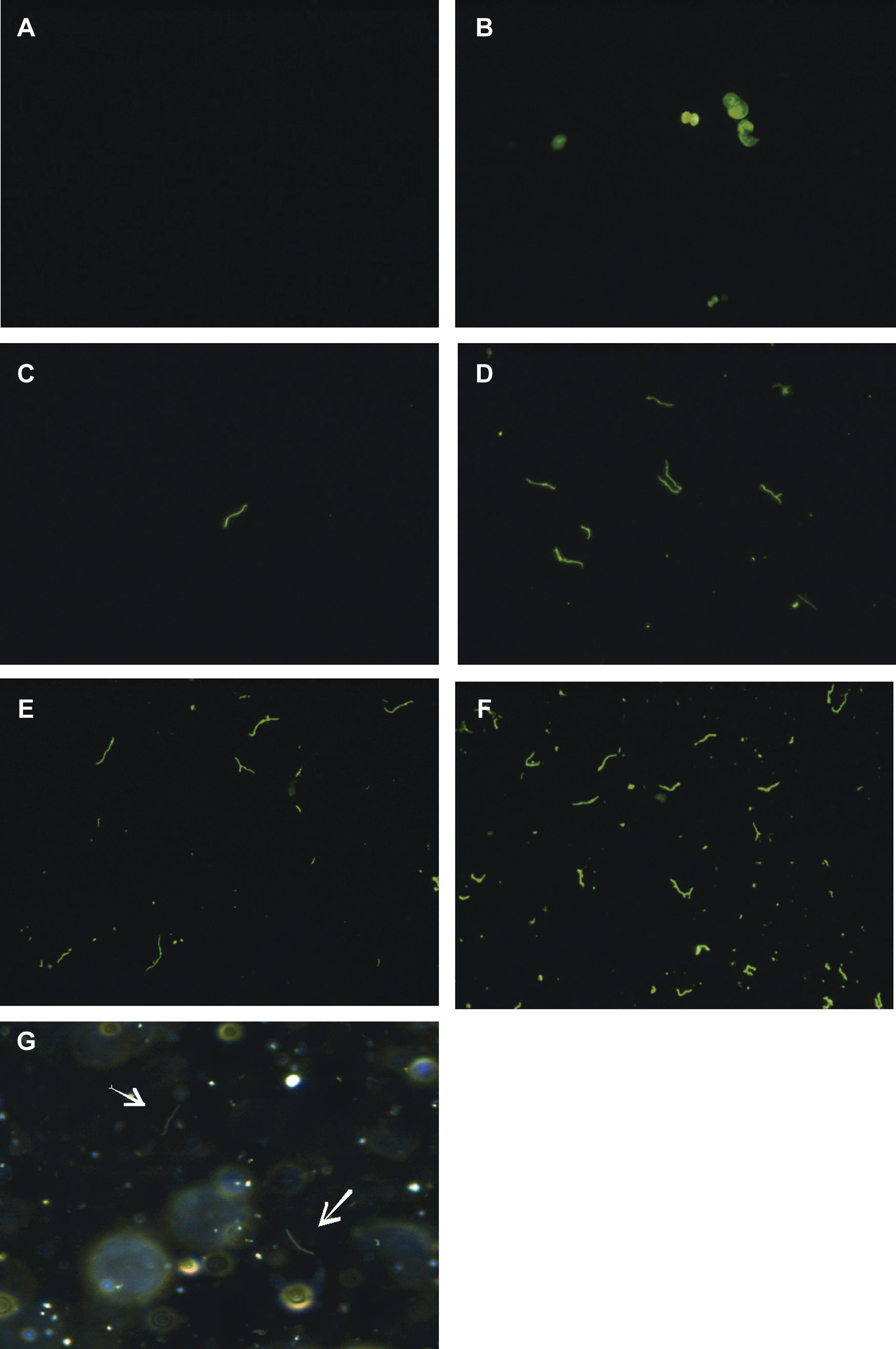
Fluorescence microscopy images of B. burgdorferi in nymphal fecal material detected with FITC-conjugated rabbit anti-B. burgdorferi. Representative images of (
Discussion
There has been limited information published regarding shedding of B. burgdorferi in tick feces. Two previous studies examined tick fecal material for the presence of B. burgdorferi. Benach and associates (1987) were unable to discern any spirochetes present in I. scapularis feces by darkfield microscopy or by culturing; and Gern and colleagues (1996) reported viewing Borrelia by darkfield microscopy in 50% of the I. ricinus fecal samples tested. Our laboratory previously used polymerase chain reaction (PCR) to detect B. burgdorferi DNA in tick feces shed during engorgement (Patton et al. 2011). We did not attempt to visualize the spirochetes because previous studies had executed microscopic analysis in tick feces with varying results. It is also well-established that B. burgdorferi cannot survive outside a host environment; therefore we ventured that desiccated cells would not be observable. However, upon further investigation we observed B. burgdorferi in infected I. scapularis fecal samples using IFA (Fig. 1C–F). We also observed nonmotile Borrelia by darkfield microscopy in PBS-suspended fecal samples (Fig. 1G). Furthermore, we were unable to cultivate Borrelia from the fecal samples, as expected.
One might speculate that the spirochetes observed could have originated from unattached desiccated infected ticks. Our evidence suggests a low probability of desiccated tick contamination, as we recovered 100% of the ticks placed in the capsules from 4 mice. To rule out the possibility that the visualized Borrelia came from the skin lesions where the nymphs fed, we swabbed the plastic capsules that were adhered to the mice and discerned numerous spirochetes in these excrement samples. Collectively, these data support our conclusion that spirochetes were shed with the tick fecal matter.
In this report we expand our previous analysis to show that not only is borrelial DNA detectable, but spirochetes can be visualized in tick excrement. Although there are reports of other bacteria and protozoa causing vector-borne illness through fecal material, B. burgdorferi does not produce an infection via the fecal route. Furthermore, B. burgdorferi in the tick feces was not culturable in either of our studies, and was not motile when visualized with darkfield microscopy. Therefore the spirochetes were presumably non-viable. Spirochete shedding in tick feces is not considered a part of the borrelial transmission process; consequently the biological significance of B. burgdorferi in tick feces is unclear. Nevertheless, it is important to note that spirochetes and their DNA are present in tick fecal material, a fact of which investigators should be aware when performing host/pathogen/tick interface analyses, especially when feeding several ticks in a localized area.
Footnotes
Acknowledgments
The authors thank Gabrielle Dietrich, Marc Dolan, and Joe Piesman for their tick expertise, Barbara J. Johnson and Jeannine Petersen for helpful insights and comments, and members of the DVBD Animal Resources, specifically Andrea Peterson, Lisa Massoudi, Verna O'Brien, and John Liddell, for their assistance.
Author Disclosure Statement
No competing financial interests exist.