
Abstract
Babesia microti, the primary cause of human babesiosis in the United States, is transmitted by Ixodes scapularis ticks; transmission may also occur through blood transfusion and transplacentally. Most infected people experience a viral-like illness that resolves without complication, but those who are immunocompromised may develop a serious and prolonged illness that is sometimes fatal. The geographic expansion and increasing incidence of human babesiosis in the northeastern and midwestern United States highlight the need for high-throughput sensitive and specific assays to detect parasites in both ticks and humans with the goals of improving epidemiological surveillance, diagnosis of acute infections, and screening of the blood supply. Accordingly, we developed a B. microti-specific quantitative PCR (qPCR) assay (named BabMq18) designed to detect B. microti DNA in tick and human blood samples using a primer and probe combination that targets the 18S rRNA gene of B. microti. This qPCR assay was compared with two nonquantitative B. microti PCR assays by testing tick samples and was found to exhibit higher sensitivity for detection of B. microti DNA. The BabMq18 assay has a detection threshold of 10 copies per reaction and does not amplify DNA in I. scapularis ticks infected with Babesia odocoilei, Borrelia burgdorferi, Borrelia miyamotoi, or Anaplasma phagocytophilum. This highly sensitive and specific qPCR assay can be used for detection of B. microti DNA in both tick and human samples. Finally, we report the prevalence of B. microti infection in field-collected I. scapularis nymphs from three locations in southern New England that present disparate incidences of human babesiosis.
Introduction
Improved laboratory assays are needed for better diagnosis of acute human infection and for blood donor screening to prevent transfusion-transmitted babesiosis (Young and Krause 2009, Leiby 2011, Vannier and Krause 2012). Improved assays are also needed to detect B. microti in ticks for vector-based surveillance to identify areas where humans are at risk of exposure to the pathogen. Tick-based surveillance is critical for identification of babesiosis-emerging areas given the lack of clear clinical signs and symptoms that result in underreporting of human cases. Because the number of B. microti sporoblasts within I. scapularis nymphs decreases over time (Piesman et al. 1987a), an assay capable of detecting small numbers of B. microti sporozoites is needed to identify I. scapularis nymphs that are potentially infectious but carry a parasitic load undetectable by currently available assays.
DNA amplification using PCR is recognized as the most sensitive and specific diagnostic method for rapid confirmation of B. microti and other protozoan infections in biological samples. Here we report a novel quantitative PCR assay capable of detecting B. microti DNA in both I. scapularis ticks and human blood samples.
Materials and Methods
Development of quantitative PCR primers, probe, and assay conditions
The BabMq18 assay targets a sequence of the B. microti 18S rRNA gene (GenBank accession number AY144696.1) common to babesia species of the previously described Clade 1 (Goethert and Telford 2003, Nakajima et al. 2009). Primers and probe were designed using both Primer3 (Rozen and Skaletsky 2000) and Primer Express (Applied Biosystems, Foster City CA) and chosen on the basis of GC content and lack of hairpin structures.
To ascertain that the BabMq18 primer/probe combination was specific for B. microti, the DNA target sequence on the 18S rRNA gene was compared with orthologous sequences from other babesia species known to infect humans (B. divergens [BDU16370], B. duncani WA1 [AF158700.1], B. duncani BAB2 [HQ285838.1], B. venatorum [JQ993428.2, JX679174.1], and Babesia sp. KO1 [DQ346955.1]), those found in I. scapularis ticks (B. odocoilei [BOU16369]), or those found in ticks or hosts involved in other enzootic cycles (B. microti Clade 2 [AY144701.1], B. microti Clade 3 [AY144690.1]), using ClustalW (Larkin et al. 2007) and MEGA (Tamura et al. 2011) (Fig. 1).
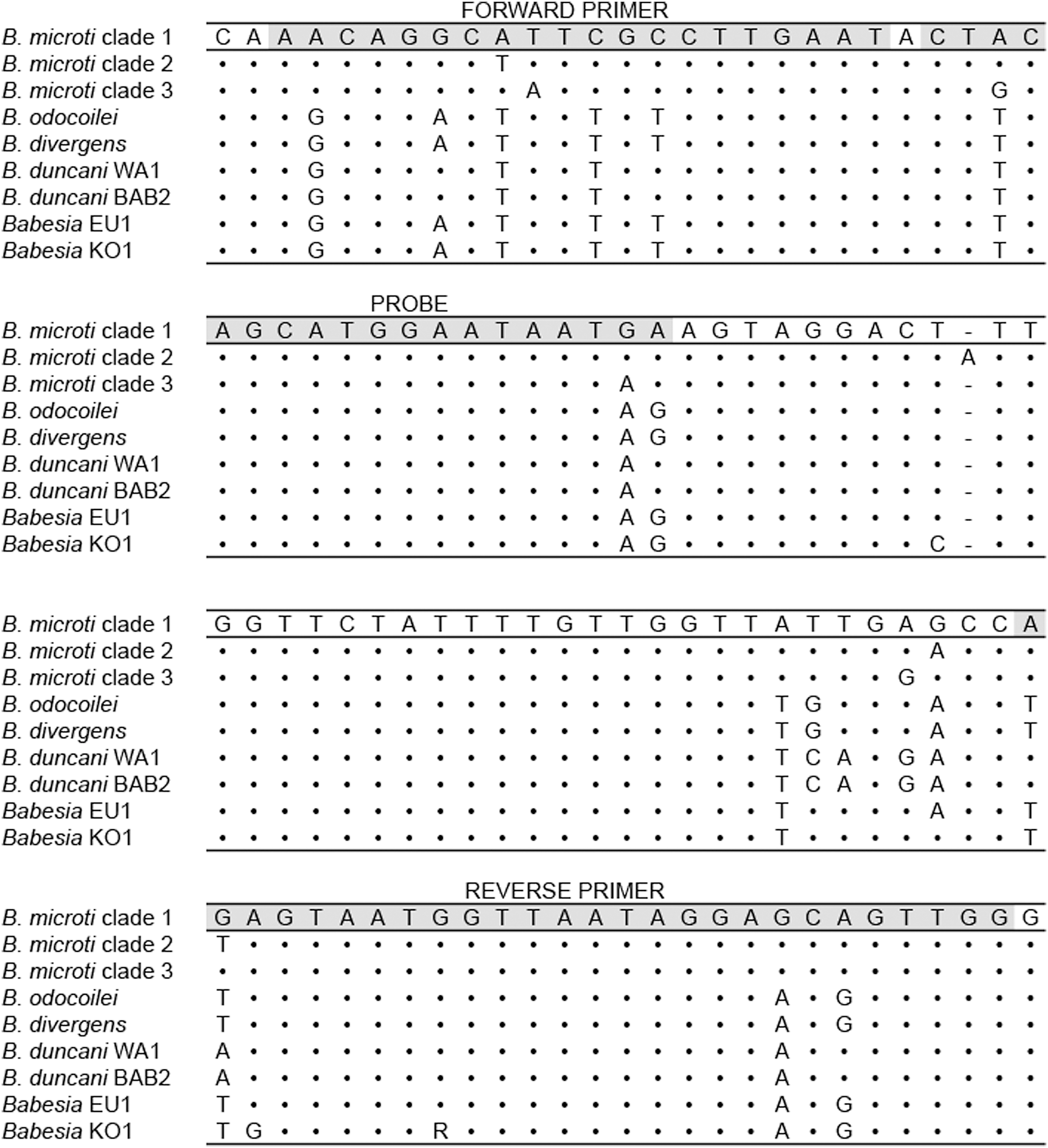
Sequence alignment of other Babesia species compared to a region of the B. microti 18S rRNA gene (GenBank accession number AY144696.1) used in the BabMq18 qPCR assay. Shaded nucleotides represent primer and probe regions; dots represent nucleotides identical to the sequence of interest. Sequence, GenBank accession number, and source: B. microti Clade 1 (AY144696.1, Ixodes scapularis); B. microti Clade 2 (AY144701.1, Procyon lotor); B. microti Clade 3 (AY144690.1, Clethrionomys sp.); B. odocoilei (BOU16369, Odocoileus virginianus); B. divergens (BDU16370, Bos taurus); B. duncani WA1 (AF158700.1, Homo sapiens); B. duncani BAB2 (HQ285838.1, Homo sapiens); Babesia EU1 (B. venatorum) (JQ993428.2, Ixodes persulcatus; JX679174.1, Ixodes ricinus); Babesia KO1 (DQ346955.1, Homo sapiens).
Forward and reverse primers and probe sequences are: Bm18Sf-AACAGGCATTCGCCTTGAAT, Bm18Sr-CCAACTGCTCCTATTAACCATTACTCT, and Bm18Sp-6FAM-CTACAGCATGGAATAATGA-MGBNFQ, respectively. The forward primer starts at position 273 and has a melting temperature (Tm) of 58.4°C, the reverse primer starts at position 350 and has a Tm of 63.1°C, and the probe starts at position 294 and has a Tm of 70.0°C. PCR amplification using these primers produces a 104-bp amplicon. This primer/probe combination is predicted to amplify the DNA of B. microti strains in Clade 1 but not the DNA of other babesia strains or species. The assay was performed using Applied Biosystems 7500 Real-Time PCR machine (Applied Biosystems, Foster City CA). The PCR reaction consisted of 2× Taqman Universal PCR Master Mix (with AmpErase, Applied Biosystems, Foster City CA), 0.9 μM forward and reverse primers, 0.2 μM probe, and 5 μL DNA template in a total reaction volume of 25 μL. Cycling conditions were as follows: 50°C for 2 min, 95°C for 10 min, followed by 40 cycles of denaturation at 95°C for 15 s, and annealing at 59°C for 60 s.
DNA extraction
DNA was extracted from ticks and human blood samples using the Qiagen DNeasy Blood and Tissue Kit (Qiagen, Valencia CA) and a modified protocol (Beati and Keirans 2001). DNA was eluted using 10 mM Tris · Cl (pH 8.5) (Buffer EB, Qiagen, Valencia CA) to a final volume of 120 μL.
Determination of BabMq18 quantitative PCR assay sensitivity
A B. microti–positive control for the BabMq18 assay was constructed by cloning the 104-bp PCR amplicon into a pUC57-Kan plasmid (GENEWIZ, Inc., South Plainfield, NJ). Standard dilutions were developed from laboratory-reared uninfected ticks and uninfected human blood spiked with known amounts of this plasmid DNA (106–100 copy number dilutions), followed by DNA extraction. Individual ticks were spiked with plasmid DNA dilutions immediately after being chilled by liquid nitrogen and pulverized in a microcentrifuge tube at the beginning of DNA extraction. The resulting tick and human standard curves were assessed by analyzing them in triplicate in three independent BabMq18 quantitative (q) PCR tests to determine reproducibility, efficiency of amplification, and the lower limit of B. microti DNA detection.
Determination of BabMq18 qPCR assay specificity
To determine whether the BabMq18 assay would amplify the DNA of Anaplasma phagocytophilum, Borrelia burgdorferi, or Borrelia miyamotoi, we infected laboratory-reared I. scapularis nymphs with these pathogens and then tested five nymphs infected with each pathogen using the BabMq18 assay. We also sought to determine whether the BabMq18 assay would amplify DNA of B. odocoilei, the only other babesia species commonly found in I. scapularis ticks. We collected 91 I. scapularis nymphs from Mansfield, CT, and screened them with a PCR/restriction fragment length polymorphism (RFLP) assay that can distinguish B. microti from B. odocoilei DNA (Armstrong et al. 1998). Samples identified as containing B. odocoilei DNA but no B. microti DNA were submitted to the Yale Center for Genome Analysis for 18S sequencing, and then screened using the BabMq18 assay.
Comparison of the BabMq18 qPCR assay with nonquantitative B. microti PCR assays for detection of B. microti DNA in laboratory-infected I. scapularis nymphs
Ten laboratory-reared I. scapularis nymphal ticks that had been fed on a B. microti–infected P. leucopus mouse were tested using each of the following assays: BabMq18 qPCR assay, a nonquantitative nested B. microti PCR assay (Persing et al. 1992), and a single-round amplification modification of this assay (Gray et al. 2002).
Performance characteristics of the BabMq18 qPCR assay in field-collected I. scapularis and human blood from babesiosis patients
The performance and reproducibility of the BabMq18 assay were further assessed by testing samples of field-collected B. microti–infected and uninfected ticks. The BabMq18 assay was also used to test blood samples from B. microti–infected and uninfected human subjects.
Laboratory-reared I. scapularis nymphs infected with a strain of B. microti isolated from P. leucopus in Connecticut (Anderson et al. 1991) were used to validate the use of the BabMq18 assay for field-collected ticks. I. scapularis nymphs were collected from sites in Nantucket, MA, Mansfield, CT, and Salisbury, CT, by the dragging methodology (Falco and Fish 1992) during June and July of 2010. These areas were chosen to represent a spectrum of B. microti endemicity. Nantucket is an endemic site for human babesiosis where the disease has been reported annually for more than four decades (Western et al. 1970). Mansfield is an emerging site where human babesiosis was first reported in 2002, whereas Salisbury is a nonendemic site where no cases of human babesiosis have been reported (Randall Nelson, Connecticut Department of Public Health, personal communication, 2011).
Blood samples were obtained from 14 B. microti–infected patients from Nantucket, MA, following their presentation with a suspected acute tick-borne illness. A history, physical examination, and specific B. microti, B. burgdorferi, and A. phagocytophilum laboratory tests were performed at the time of acute illness. A definitive diagnosis of babesiosis required the presence of typical babesiosis symptoms and laboratory confirmation of recent babesial infection that included identification of intraerythrocytic babesia parasites by means of a Giemsa-stained thin blood smear. Three of the patients were diagnosed with concomitant babesiosis and Lyme disease infection. Blood was also obtained from five healthy control subjects as part of a biannual serosurvey on Block Island, RI (Krause et al. 2003). Written informed consent was obtained from all study participants in accordance with human experimentation guidelines approved by the Human Investigation Committee at the Yale School of Public Health.
Results
Limit of detection and reproducibility of the BabMq18 PCR assay
The BabMq18 assay detection limit was determined using serial dilutions of uninfected human blood and uninfected I. scapularis ticks spiked with copies of a plasmid in which the PCR amplicon had been cloned. Analysis of each standard curve indicated a detection limit of 101 B. microti gene copies per reaction for the BabMq18 qPCR assay, based on exponential amplification in standard dilutions greater than or equal to 101 copy numbers. The interassay mean cycle threshold (CT) values and standard deviation of the three experiments indicate consistent reproducibility (Table 1). Spiked human blood and tick standard curves are shown in Figure 2, where interassay mean CT values are plotted as a function of the copy number expressed on a log scale. Both standard curves had similar slopes and correlation coefficients: y=−3.609x+39.09, R2 =0.998 and y=−3.597x+38.72, R2 =0.999 for spiked human blood and spiked I. scapularis ticks, respectively (Fig. 2). Unknown samples were considered positive if the CT value was less than or equal to the CT value produced by the 101 standard dilution on each plate.
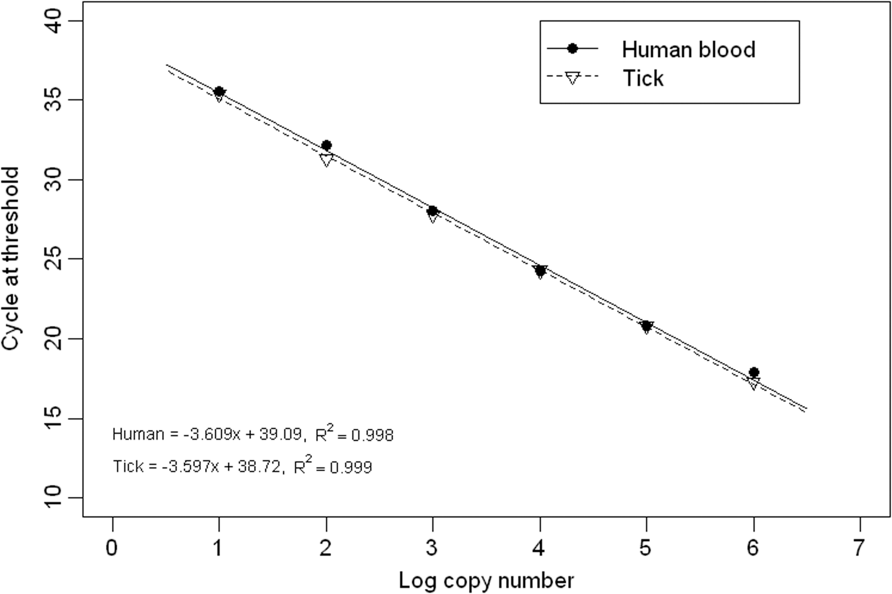
Standard curves produced from uninfected human blood and ticks spiked with B. microti DNA. The plotted values represent the interassay mean (n=9) for each standard dilution.
CT, threshold cycle value; SD, standard deviation; ND, not detected (no CT value generated after 40 cycles).
Determination of BabMq18 qPCR assay specificity
I. scapularis nymphs were infected with either A. phagocytophilum, B. burgdorferi, or B. miyamotoi. For each infection, five tick samples were tested using pathogen-specific PCR assays and were confirmed to be infected with the expected pathogen. The samples were then tested with the BabMq18 assay, and no amplicon was detected in any of these infected ticks. In a separate experiment, B. odocoilei DNA was identified in eight of 91 field-collected I. scapularis using the Armstrong PCR/RFLP assay (Armstrong et al. 1998). Three of these eight samples were successfully sequenced at the Yale Center for Genome Analysis and were confirmed positive for B. odocoilei DNA, but not for B. microti DNA. The BabMq18 qPCR assay was used to test all eight samples containing B. odocoilei and no DNA was amplified.
Comparison of the BabMq18 qPCR assay to nonquantitative B. microti PCR assays for detection of B. microti DNA in laboratory-infected I. scapularis nymphs
Ten laboratory-reared I. scapularis nymphal ticks that had been fed on a single B. microti–infected P. leucopus mouse were tested in triplicate by three separate assays. B. microti DNA was detected in eight of 10 ticks using the BabMq18 qPCR assay, two of 10 ticks using the nonquantitative PCR assay, and none of 10 ticks using the modified nonquantitative PCR assay (data not shown). Using the BabMq18 qPCR assay, B. microti DNA was detected at similar cycle thresholds among triplicate samples of each tick. Two additional nymphal ticks that had been fed on uninfected P. leucopus were used as negative controls in all replicates of each assay; DNA was not amplified in any of these samples. Following amplification with the BabMq18 qPCR assay, the resulting PCR product from five of the infected samples were successfully sequenced and confirmed as B. microti DNA. The other five samples were not sequenced because all of these ticks were fed on the same mouse infected with a single laboratory strain. The BabMq18 assay was found to be more sensitive compared to the non-quantitative B. microti PCR assays.
Performance characteristics of the BabMq18 qPCR assay in field-collected I. scapularis nymphs and human blood from babesiosis patients
We tested whether our BabMq18 assay detects B. microti infection in field-collected nymphal ticks and determined infection prevalence at three study sites. The highest frequency of B. microti–infected ticks was detected at the long-term endemic site on Nantucket Island, MA (7.5%). An intermediate infection rate was found in ticks at the recently endemic site in Mansfield, CT (3.7%). No infection was found in ticks at the nonendemic site in Salisbury, CT (Table 2). Assuming an infection prevalence of 2%, the sample of 150 ticks from Salisbury is large enough to state with greater than 95% confidence that there are no infected ticks at this site. These infection prevalence differences were statistically significant (χ2=13.38, p=0.001). We conclude that the infection prevalence rates detected in ticks by the BabMq18 assay are consistent with the reported rate of prevalence of human babesiosis at these sites.
Fourteen B. microti–infected subjects who experienced typical babesiosis symptoms during the summertime were confirmed to have babesiosis by identification of babesia parasites on Wright- or Giemsa-stained thin blood smears. B. microti DNA was detected in the blood of all ill patients, whereas none was detected in the five healthy control samples using the BabMq18 assay.
Discussion
We have developed a highly sensitive and specific B. microti qPCR assay that successfully amplifies B. microti DNA in both I. scapularis ticks and human blood samples with concentrations as few as 10 gene copies per reaction. The BabMq18 assay does not amplify DNA from B. burgdorferi, B. miyamotoi, and A. phagocytophilum, three pathogens that infect I. scapularis ticks and human hosts, and does not amplify B. odocoilei DNA, a species commonly carried by I. scapularis nymphs.
The probability of detecting B. microti DNA in ticks by laboratory assays is compromised by low numbers of babesia sporoblasts within the salivary glands of unfed ticks (Mather et al. 1990) and parasite diminution during transtadial passage of B. microti in tick vectors (Piesman et al. 1987a). It has been suggested that B. microti parasites are maintained in reservoir mice at much lower concentrations than B. burgdorferi. The natural infection of P. leucopus by B. microti rarely exceeds 0.1% parasitemia (Mather et al. 1990). An advantage of the BabMq18 qPCR assay over nonquantitative assays is the potential to determine the minimal dose of B. microti in ticks that is necessary for transmission to a mammalian host.
The B. microti BabMq18 qPCR assay detects lower levels of B. microti DNA than conventional PCR assays and therefore provides a more sensitive determination of B. microti infection in I. scapularis ticks and human blood. It can detect as few as 10 copies per reaction, which corresponds to two gene copies per microliter of whole human blood and 240 gene copies per whole nymphal tick. The B. microti genome contains two copies of the 18S rRNA gene (Cornillot et al. 2012), and therefore (assuming haploid form) this assay is able to detect one B. microti parasite per microliter of human blood and 120 B. microti parasites per whole nymphal I. scapularis tick. Human blood likely contains only the haploid form; thus, the detection of one parasite per microliter of whole human blood corresponds to an actual parasitemia of 0.00002%.
On the basis of sequence alignment, it is unlikely that BabMq18 qPCR will amplify DNA from other babesia species that infect humans in geographic regions beyond the northeast and northern midwest, including B. duncani, B. divergens, B. venatorum, and Babesia sp. K01. A recently developed qPCR (Teal et al. 2012) developed for detection of B. microti infection in humans reported sequence alignments with similar frequencies of mismatches to the other human babesia species as our assay. The vast majority of human cases of babesiosis in the United States are caused by Clade 1 B. microti. Although a few cases of B. duncani have been described in the northern Pacific coast and three cases of B. divergens–like organisms have been described in the United States (Kjemtrup and Conrad 2000, Vannier and Krause 2012), neither pathogen has been described in babesiosis-endemic areas of the Northeast or northern Midwest where B. microti is prevalent, nor have they been found in I. scapularis ticks.
Previous studies on the prevalence of B. microti in ticks have either analyzed adult ticks, which are not important vectors (Piesman et al. 1987b), or have used salivary gland staining, which cannot easily distinguish between B. microti and B. odocoilei (Armstrong et al. 1998). Our data suggest that previous investigations of B. microti infection prevalence in field-collected I. scapularis ticks using conventional B. microti PCR (Armstrong et al. 1998, Varde et al. 1998, Adelson et al. 2004, Hamer et al. 2007) have underestimated actual B. microti infection prevalence in these ticks. An accurate assessment of the prevalence of B. microti–infected I. scapularis ticks may help identify areas where human babesiosis is emerging or where cases may be underreported.
In summary, we have developed a highly sensitive and specific quantitative B. microti qPCR assay that is superior to nonquantitative B. microti PCR assays and can be used for detection of B. microti in both I. scapularis ticks and in human samples.
Footnotes
Acknowledgments
This project was funded in part by the generous support of the Gordon and Llura Gund Foundation (P.J.K.); the G. Harold and Leila Y. Mathers Charitable Foundation (D.F.); the US Department of Agriculture–Agricultural Research Service Cooperative agreement no. 58-0790-5-068 (D.F.); and by the National Institute of General Medical Science (NIH) grant no. R01GM105246-01 (M.D.). We would like to thank Dr. Sam Telford III, Professor in the Department of Infectious Disease and Global Health at the Cummings School of Veterinary Medicine at Tufts University, for his generous donation of ticks from Nantucket.
Author Disclosure Statement
No competing financial interests exist