
Abstract
A total of 167 ticks collected from humans in Istanbul (Turkey) in 2006 were screened for Rickettsia species, and nested PCRs targeting gltA and ompA rickettsial fragment genes were carried out. Rickettsia monacensis (51), R. aeschlimannii (8), R. conorii subsp. conorii (3), R. helvetica (2), R. raoultii (1), R. africae (1), R. felis (1), and other Rickettsia spp. (2), were detected. To our knowledge, these Rickettsia species (except R. conorii) had never been reported in ticks removed from humans in Turkey. The presence of R. africae also had not been previously described, either in Hyalomma ticks or in any European tick species. In addition, R. aeschlimannii and R. felis had not been found associated with Rhipicephalus bursa specimens. The presence of human pathogenic Rickettsia in ticks removed from humans provides information about the risk of tick-borne rickettsioses in Turkey.
Introduction
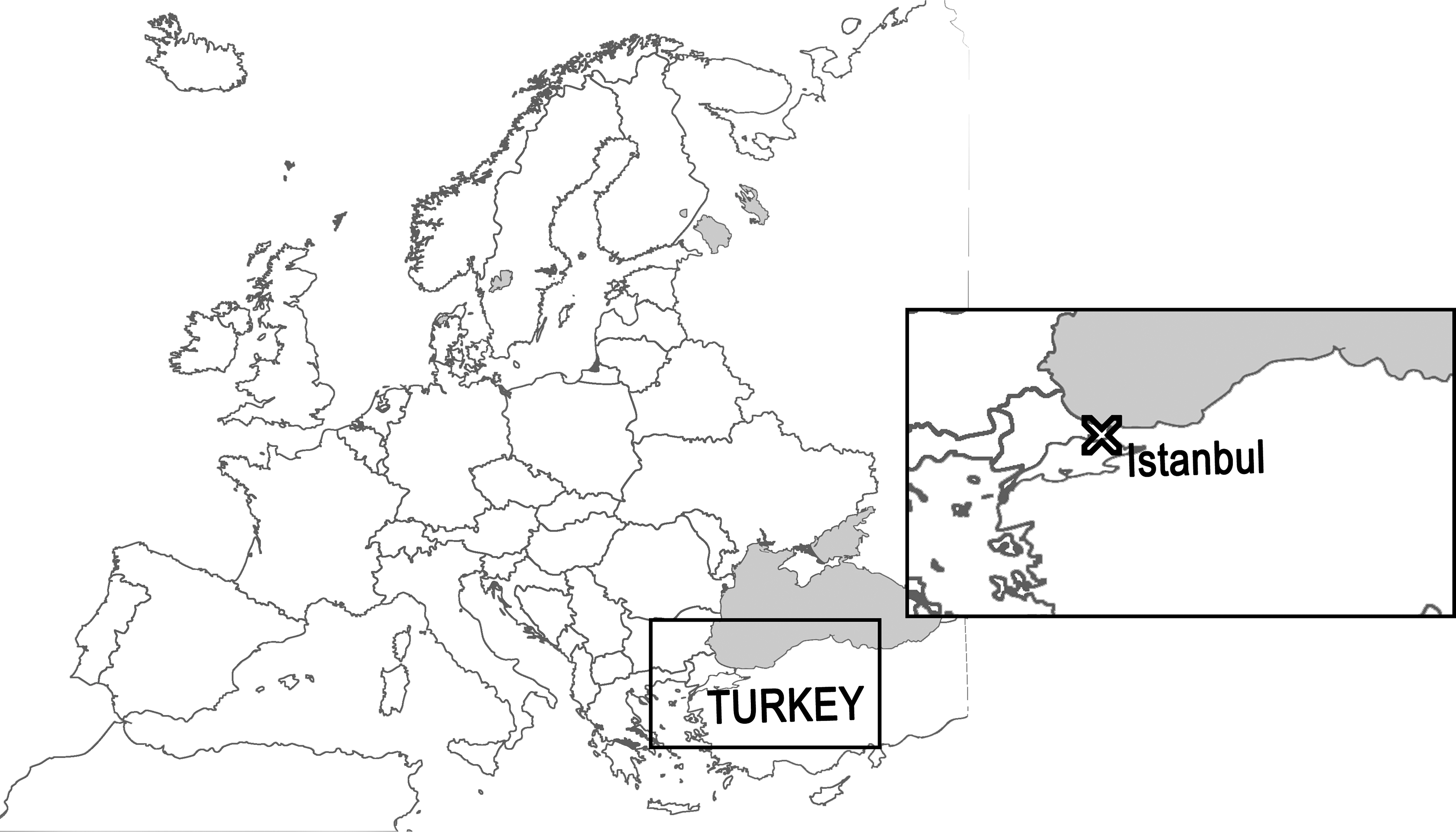
Map of Europe showing the location of Istanbul.
Materials and Methods
In 2006, a total of 1054 ticks were removed from humans in more than 25 hospitals in Istanbul (Turkey), as part of a project supported by the Ministry of Health, Istanbul Branch (Vatansever et al. 2008). Among them, 167 were randomly chosen for this study. The ticks were classified by taxonomic keys (Walker et al. 2003; Apanaskevich 2003; Estrada-Peña et al. 2004). In addition, specimens whose phenotypic classification was not conclusive were also classified by genotypic methods (PCR of the 16S rDNA gene; Black and Piesman 1994). DNA was individually extracted with the Qiagen Dneasy blood/tissue kit (Qiagen, Hilden, Germany), according to the manufacturer's tissue protocol instructions. DNA extracts were used as templates for PCR assays targeting the 16S rDNA gene for tick classification as cited above, as well as the rickettsial citrate synthase (gltA), and 190-kDa protein antigen (ompA) genes. PCR primer pairs, size of the amplicons (bp), and annealing temperatures of the assays, are shown in Table 1. Two negative controls, one of them with template DNA but without primers, and the other with primers and containing water instead of template DNA, as well as positive controls (a tick extract and Rickettsia slovaca, respectively) were included in all PCR assays. Rickettsia spp. were screened by gltA PCR. Positive samples were further tested by ompA PCR, and the obtained amplicons were sequenced for the differentiation of the SFG rickettsia species. However, gltA amplicons were only sequenced from samples that yielded negative PCR results for ompA. Nucleotide sequences were compared with those available in GenBank using the National Center for Biotechnology Information (NCBI; Bethesda, MD) Basic Local Alignment Sequence Tool (BLAST) search engine (
Results
Studied ticks belonged to the following species: Ixodes ricinus (n=130), Rhipicephalus sanguineus group (n=19), Hyalomma aegyptium (n=11), Hyalomma spp. (n=4), Hyalomma marginatum (n=2), and Dermacentor marginatus (n=1). Sixty-nine out of 167 samples (41.3%) tested positive for the presence of Rickettsia spp. Table 2 shows details about the Rickettsia species detected and Rickettsia-infected tick species, as well as the highest percentages of identity with a validly-published Rickettsia species. According to ompA nucleotide sequences, R. monacensis, which was detected in 49 I. ricinus (70%) ticks, was the most prevalent genospecies. On the one hand, R. aeschlimannii was found in 8 tick specimens (12%): 5 H. aegyptium, 2 H. marginatum, and 1 Rhipicephalus bursa. On the other hand, R. conorii subsp. conorii was detected in 3 R. bursa (4%) specimens, whereas R. africae, R. felis, and R. raoultii appeared in 1 H. aegyptium, 1 R. bursa, and 1 D. marginatus, specimens, respectively. According to gltA nucleotide sequences (from samples with negative ompA PCR results), R. helvetica (n=2; 3%), and R. monacensis (n=2; 3%), were detected in 4 I. ricinus ticks. In addition, 2 specimens of the Rhipicephalus genus harbored unidentified Rickettsia spp. that showed gltA sequences with the same percentage of identity with more than one validly-published rickettsia. These sequences shared 99.7% identity with R. raoultii (DQ365804), R. aeschlimannii (U59722), Rickettsia japonica (U59724), or Rickettsia heilongjiangensis (AF178034), among other rickettsia species. Unfortunately, analysis of alternative genes to identify rickettsial species could not be performed, since all DNA was used to investigate other tick-borne bacteria (data not shown).
Same identity with more than one validly published Rickettsia species.
The GenBank accession numbers as well as the highest percentages of identity with Rickettsia species are also included.
N, nymph; F, female; M, male.
Discussion
To date, R. conorii was the unique confirmed tick-borne rickettsia species known in Turkey (Kuloglu et al. 2004). Thus cases of MSF have been reported from this country, and R. conorii has been amplified in ticks by PCR (Kuloglu et al. 2004). Our study confirms this fact, as well as the circulation of other rickettsias different from R. conorii in ticks removed from humans in Istanbul. We report here the presence of these pathogenic tick-borne bacteria: R. conorii subsp. conorii, R. monacensis, R. aeschlimannii, R. helvetica, R. raoultii, R. africae, and R. felis.
In this study, we have found R. conorii subsp. conorii in R. bursa specimens attached to humans. This tick species belongs to the same group as R. sanguineus, which is the current vector of MSF. These tick species can be easily confused, since their morphological differentiation at some stages is still unclear. The detection of R. conorii subsp. conorii in R. bursa corroborates data from a previous study with ticks collected on livestock in Turkey (Christova et al. 2003). We think that R. bursa could also be a vector of R. conorii, since we have observed R. bursa attached to patients who subsequently developed MSF (Oteo and Portillo 2012). Furthermore, our data have shown the presence of R. helvetica and R. monacensis in I. ricinus ticks. These rickettsia species, mainly transmitted by I. ricinus, have been associated with MSF-like diseases previously reported in Europe (Nilsson et al. 1999; Fournier et al. 2000; Jado et al. 2007). In addition, I. ricinus is the most prevalent tick species attached to humans in Istanbul analyzed in our study (Vatansever et al. 2008). According to our data, R. raoultii has been amplified in D. marginatus. This rickettsia species had previously been detected in Dermacentor reticulatus and D. marginatus that bit humans who developed R. slovaca-like infection or TIBOLA/DEBONEL symptoms (Mediannikov et al. 2008). Moreover, we have found R. aeschlimannii in H. aegyptium and H. marginatum, as previously reported, as well as in one R. bursa specimen. To our knowledge, this is the first detection of R. aeschlimannii in R. bursa, although this rickettsia had been amplified in Rhipicephalus spp. (Bitam et al. 2009; Mediannikov et al. 2010). Human infection by R. aeschlimannii was first reported in Africa (Raoult et al. 2002), and it has been associated with European ticks, although there are no recognized endemic cases of the disease (Oteo et al. 2005; Oteo and Portillo 2012).
Our data have revealed the presence of R. felis in R. bursa. Molecular evidence of R. felis in ticks has been previously reported (Reif et al. 2009), but not in R. bursa or in ticks collected in Europe. Nevertheless, fleas are the only known vector of this potential tick-borne pathogen.
R. africae is responsible for ATBF, an infection that has been reported in Africa, and as an imported disease in Europe (Jensenius et al. 2003; Oteo et al. 2004). Until now, R. africae had been associated with Amblyomma and Rhipicephalus species (Kelly et al. 1996; Portillo et al. 2007; Mediannikov et al. 2010). In our study, we found R. africae in H. aegyptium. This tick species is not a known vector of R. africae, but our results demonstrate the circulation of this rickettsial pathogen in the studied environment.
All rickettsia species found in this study are associated with human disease. The presence of several tick-borne pathogens, their vectors, and reservoirs, as well as the evidence of human tick bites, suggest that some of these tick-borne infections are circulating in Turkey and may be misdiagnosed. This information should be taken into account for the correct diagnosis and treatment of tick-borne infections in Turkey.
Footnotes
Acknowledgments
Tick collection and identification studies were supported by TUBITAK-SBAG (108S171). Detection of Rickettsia species was supported by a grant from the “Instituto de Salud Carlos III” (EMER 07/033), and Ministerio de Ciencia e Innovación (Spain). Fundación Rioja Salud awarded a grant to A.M. Palomar for her doctoral thesis (FRS/PIF-01/10).
Author Disclosure Statement
No competing financial interests exist.