
Abstract
The tick-borne bacterium ‘Candidatus Neoehrlichia mikurensis' has recently been recognized as a human pathogen in Europe and appears to be the second most common pathogenic bacterium in Ixodes ricinus ticks in central Europe, second to Borrelia afzelii. Here, we investigate the prevalence of ‘Candidatus N. mikurensis' in host-seeking ticks in southern Sweden and the rate of co-infection with B. afzelii. We developed a real-time qPCR assay targeting the groEL gene of ‘Candidatus N. mikurensis' and applied this assay to 949 I. ricinus ticks collected at several locations over 2 years. We found an overall prevalence of 6.0%, which means that Candidatus N. mikurensis is one of the most common tick-transmitted zoonotic agents in this area. Co-infections with both ‘Candidatus N. mikurensis' and B. afzelii occurred in 2.1% of the ticks, which is significantly more than expected under random co-occurrence. The infection intensity (number of bacterial cells) of ‘Candidatus N. mikurensis' was not affected by co-infection with B. afzelii, and vice versa. We conclude that there is a risk for simultaneous transmission of these 2 tick-borne pathogens. The potential medical consequences of this require further investigation.
Introduction
Individual ticks can be infected with multiple pathogens (Swanson et al. 2006). Such co-infections can result in interactions (competition or facilitation) between co-occurring pathogens, which in turn can affect the transmission of pathogens (Ginsberg 2008). For example, Dermacentor andersoni ticks co-infected with Rickettsia rickettsia and R. peacockii only transmit the latter nonpathogenic species; hence, the presence of R. peacockii reduces the occurrence of Rocky Mountain spotted fever caused by R. rickettsia (Macaluso and Azad 2005). Co-infections in ticks can also affect the outcome of infections because multiple infections can exacerbate symptoms in animal or human hosts (Thomas et al. 2001, Belongia 2002). For example, patients with both B. burgdorferi and A. phagocytophilum infections have more symptoms and longer duration of disease than patients infected with only B. burgdorferi (Krause et al. 2012).
‘Candidatus N. mikurensis' and B. afzelii share the same natural hosts (voles and mice) (Andersson and Råberg 2011, Hellgren et al. 2011), and co-infection with these pathogens has recently been shown in ticks (Richter and Matuschka 2012). However, it is not known whether the rate of co-infection deviates from what should be expected under random co-occurrence. Moreover, no study has yet investigated if B. afzelii and ‘Candidatus N. mikurensis' affect each other's infection intensities in co-infected ticks, something that could potentially affect the infectivity from tick to vertebrate host.
Most studies to date have used nested PCR assays (Kawahara et al. 2004, Tabara et al. 2007, Fehr et al. 2010, von Loewenich et al. 2010) or general bacterial primers (Welinder-Olsson et al. 2010) for detection of ‘Candidatus N. mikurensis'. Here we developed a quantitative PCR assay targeting the groEL gene of ‘Candidatus N. mikurensis', and determined infection prevalence and intensity in 949 I. ricinus ticks collected at 4 locations in southern Sweden. We also determined the prevalence and infection intensity of B. afzelii to investigate the rate of co-infections, and whether co-infection affects infection intensities of either pathogen.
Material and Methods
Tick collection
In May–June, 2010, we collected nymphal and adult I. ricinus from four sites in southern Sweden (Fig. 1): A mixed deciduous forest in Istaby, Blekinge (56°1’N, 14°39’E); a mixed deciduous forest in Hagestad, Skåne (55°23’N; 14°8’E); a pine forest at Sandhammaren, Skåne (55°23’N; 14°11’E); and a mixed deciduous forest in Kalvs mosse, Skåne (55°42’N; 13°29’E). Ticks were collected by dragging 0.7-meter2 white cloths over the ground vegetation. In May, 2011, we collected additional ticks from one of the sites, Kalvs mosse. Ticks were stored in 70% ethanol. DNA extraction was done within a few days after collection and carried out manually according to a version of the HotSHOT protocol (Truett et al. 2000). Briefly, each tick was placed in a tube with 200 μL of alkaline lysis reagent (25 mM NaOH, 0.2 mM EDTA) and was incubated at 95°C for 15 min. A metal bead was put in each tube, and tubes were then shaken with a Retsch TissueLyser (Haan, Germany) at 20 Hz for 2 min. Thereafter samples were incubated for another 15 min at 95°C. Finally, 200 μL of 40 mM Tris HCl was added to each tube.
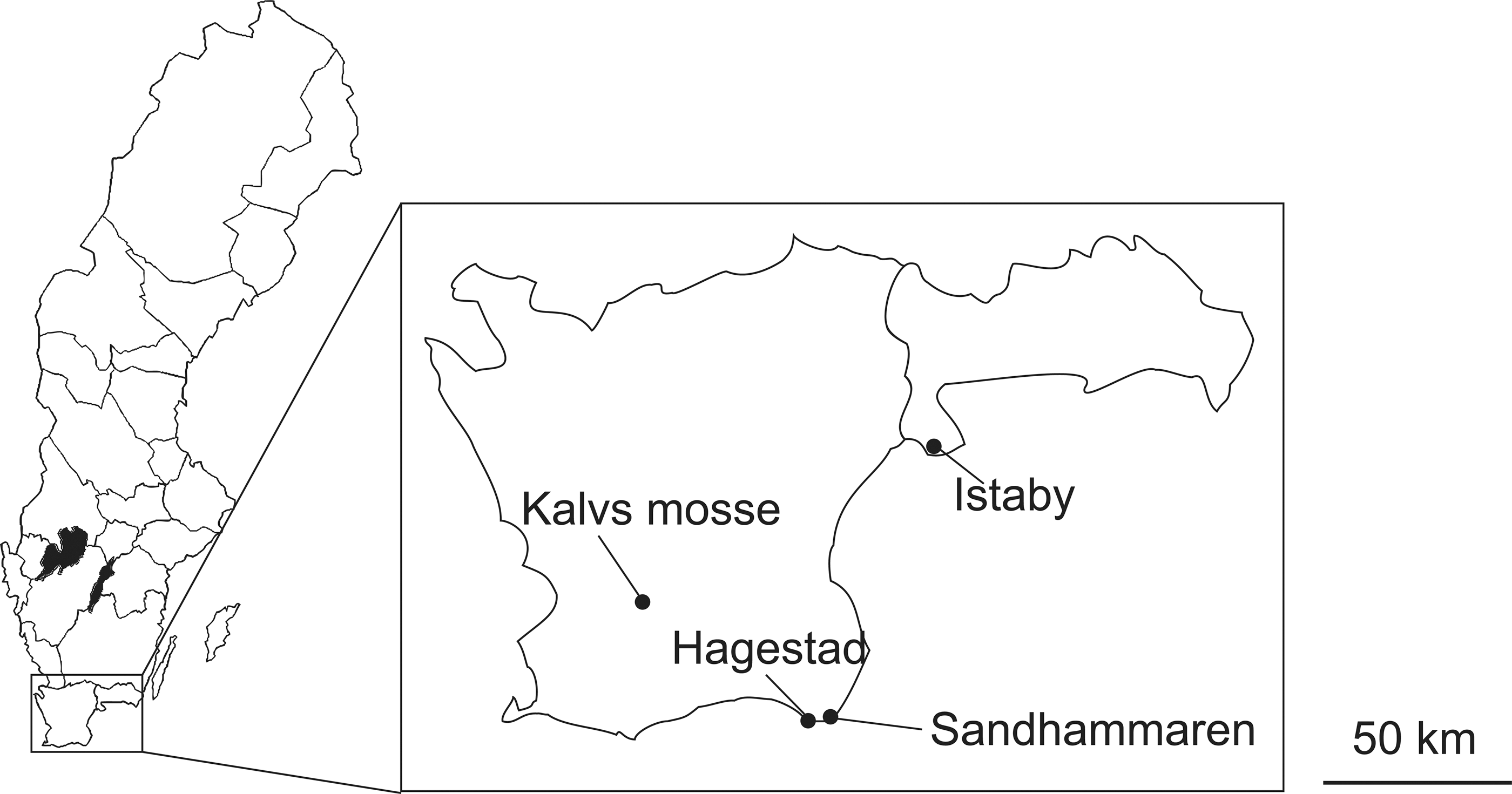
The different sampling areas in southern Sweden.
Primer design and PCR
We aligned the ‘Candidatus N. mikurensis' groEL sequences available at GenBank (AB074461.1, AB084583.1, EU810406.1, FJ966359.1, and HM045824.1) together with the groEL gene from ‘Candidatus N. lotori’ (EF633745.1), A. phagocytophilum strain HZ (NC_007797.1), Ehrlichia chaffeensis strain Arkansas (NC_007799.1), and Bartonella grahamii strain as4aup (NC_012846.1) and identified regions conserved within Neoehrlichia that differed from other aligned species and designed primers in these regions. The forward primer was NeogroELQ_f ACA GCC AAT ACT ACC TAT CCT TGA, and the reverse primer was NeogroELQ_3r ACA TGY AAT CCA CCA CGY AAC T. The amplified fragment was 83 base pairs long, and the nucleotide difference from A. phagocytophilum, E. chaffeensis, and B. grahamii ranged from 16.9% to 34.9%. To ensure specificity of the primers, 16 of the obtained amplicons were sequenced to confirm identity. BLAST searches at GenBank showed, in all 16 cases, highest identity with ‘Candidatus N. mikurensis' sequences. To further validate the specificity of the primers, they were tested against 3 A. phagocytophilum (1 from our study areas in Sweden and 2 from Slovakia) and 5 Bartonella spp. (all from our study areas in Sweden) positive controls, which were not amplified in any case.
Real-time PCR was performed with a Stratagene Mx3000p (Stratagene) using Platinum® SYBR® Green qPCR SuperMix-UDG (Invitrogen, Carlsbad, CA). Each 25-μL reaction contained 12.5 μL of SYBR® Green mix, 1 μL of each primer (10 pmol/μL), 0.1 μL of ROX, and 4 μL of tick lysate (≈40 ng total DNA; range 28–68 ng). The PCR reaction was initially incubated for 1 min at 50°C followed by 2 min at 95°C. Thereafter, 43 cycles consisting of 15 s with 95°C, an annealing step with 60°C for 30 s followed by 72°C for 30 s. A final melting temperature analysis was performed between 55 and 95°C. Samples with a cycle threshold (Ct) value lower than 40 and a melting temperature (TM) between 77.25 and 77.80°C were considered as positive for ‘Candidatus N. mikurensis'.
Infection intensity (defined as number of bacterial cells per tick) was determined by using a standard curve. The template for the standard curve was obtained by amplifying a 524-bp fragment of the groEL gene with a nested PCR assay (von Loewenich et al. 2010). The PCR product was purified with MinElute®PCR Purification kit (Qiagen), diluted, and included as a serially diluted standard (in steps of 1:5) in each run. The concentration of the standard was determined with a Thermo Scientific NanoDrop 2000/2000c Spectrophotometer (Thermo Fisher Scientific, Wilmington, DE). The most concentrated standard contained approximately 4.22×10⁵ groEL copies and had a Ct value of 22. The efficiency of the PCR was between 92.2% and 96% (as calculated with the formula E=10−1/S − 1), where S is the slope of the standard curve). In each run we also included 7 negative controls to check for contamination.
The prevalence and infection intensity of B. afzelii was measured with a real-time qPCR assay using SYBR Green chemistry and targeting the flaB gene, as described elsewhere (Råberg 2012). Briefly, the amplicon was 89 bp long, and the TM was 78.15–78.75°C. A standard curve was obtained by amplifying a longer fragment of the flaB gene. The standard was purified and quantified as described above. The most concentrated standard contained 1.66×10⁴ flaB copies and had a Ct value of 24. The detection limit was 1–5 copies.
Statistical analyses
Analyses of prevalence (proportion of infected ticks) were performed with the Fisher exact test. To test if B. afzelii and ‘Candidatus N. mikurensis' affected the infection intensity of each other, we performed general linear models with infection intensity of B. afzelii against infection status (presence/absence) with ‘Candidatus N. mikurensis' and vice versa. In these models, we also included ”year” as a fixed factor. Infection intensities were log-transformed. All statistical analyses were performed with PASW statistics (version 18, IBM 2009). The repeatability (intraclass correlation coefficient; Sokal and Rohlf 1995) of the number of copies was estimated with R statistics using an analysis of variance (ANOVA)-based method according to Nakagawa and Schielzeth (2010) with 1000 permutations.
Results
Evaluation of the ‘Candidatus N. mikurensis' assay
To investigate the reproducibility of the detection ability, we analyzed 81 tick samples twice. The concordance for infection status (positive/negative) was 100%. The interassay variation of Ct values was 0.12. The repeatability (intraclass correlation coefficient; Sokal and Rohlf 1995) of infection intensities between 2 runs (including only samples positive for Candidatus N. mikurensis, n=57) was 0.87 (p=0.001). The detection limit was determined from the serial dilution of the standard to be approximately 16 copies of the groEL gene.
Infection prevalence
In total, 949 I. ricinus were analyzed, consisting of 399 ticks collected in 2010 (345 nymphs and 54 adults) and 550 ticks collected in 2011 (517 nymphs and 33 adults). In all, 6.0% of the ticks (57/949) were PCR positive for ‘Candidatus N. mikurensis', with 5.9% (51/862) of nymphs and 6.9% (6/87) of adult ticks infected, respectively. Overall 15% (139/949) of the ticks were PCR positive for B. afzelii, with 12% (10/87) of adult ticks and 15% (129/862) of nymphs infected, respectively. There was no significant difference in prevalence of pathogens between adults and nymphs (Fisher exact test: B. afzelii, p=0.43; ‘Candidatus N. mikurensis', p=0.64). For details on prevalence in nymphs and adults at different sites, see Table 1.
n value in parentheses.
To have a more homogeneous data set for analyses of association between ‘Candidatus N. mikurensis' and B. afzelii, we only considered samples from Kalvs mosse (where 70% of the ticks were collected) in the following analyses. The co-infection rate in this dataset was 2.3% (15/660). On the basis of the prevalence of each pathogen, there were significantly more ticks with double infections than would be expected from random occurrence (5.3 expected, 15 observed; Fisher exact test, p<0.001, n=660; an analysis with all samples combined gave similar results [data not shown]).
Infection intensity
In infected ticks, the median number of ‘Candidatus N. mikurensis' groEL copies detected was 3.1×10⁴, ranging from 3.8×10² to 4.6×10⁶. Ct values had a mean of 29 ranging from 23 to 38. The median number of B. afzelii flaB copies detected was 1.7×10⁴, ranging from 12.5 to 39×105. Ct values had a mean of 31, ranging from 22 to 40.
There was no significant difference in infection intensities of B. afzelii between ticks infected with only B. afzelii and ticks infected with both pathogens (F=1.112, degrees of freedom [df]=99, p=0.48). Similarly, there was no difference in infection intensity of ‘Candidatus N. mikurensis' for ticks infected with only ‘Candidatus N. mikurensis' and both pathogens (F=2.073, df=32, p=0.16). There was no significant correlation between infection intensities of ‘Candidatus N. mikurensis' and B. afzelii in co-infected ticks (r=0.223, p=0.42, n=15).
Discussion
The prevalence in this study of 6.0% illustrates that ‘Candidatus N. mikurensis' is a common pathogen in I. ricinus in southern Sweden. Together with findings in central Europe, our results support the notion that ‘Candidatus N. mikurensis' is one of the most common tick-borne pathogens in Europe (Richter and Matuschka 2012). The prevalence in this study is in line with the findings of several other studies; in a recent study, the average prevalence in questing adult I. ricinus collected from vegetation in several European countries was 6.2% (Richter and Matuschka 2012). In the first detection of this bacteria in The Netherlands, the prevalence was 6.6% in 121 I. ricinus collected from roe deer Capreolus capreolus (Schouls et al. 1999), and in Italy, the closely related ‘Candidatus Ehrlichia walkeri’ had a prevalence of 6.5% in 292 I. ricinus ticks (Koutaro et al. 2005).
The B. afzelii prevalence of 15% in this study is slightly higher than in previous studies of ticks from Sweden (4.8% in Fraenkel et al. 2002 and 12% in Wilhelmsson et al. 2010) and elsewhere in Europe (10% in Richter and Matuschka 2012). This could be the result of a difference in sensitivity of detection methods or might reflect that ticks in our study were collected in habitats with abundant rodent populations that are important reservoir hosts with high prevalence of B. afzelii infections (Hellgren et al. 2011).
In total 2.1% of the I. ricinus ticks in this study were infected with both ‘Candidatus N. mikurensis' and B. afzelii. This is very close to the co-infection rates in a cross-sectional study throughout Europe (Richter and Matuschka 2012), where 1.8% of 763 questing adult I. ricinus were co-infected with these pathogens. We did not find any co-infected adult ticks, contrasting with recent findings by Richter and Matuschka (2012), most likely because of the low number of adult ticks in our study. In a meta-analysis of co-infections with A. phagocytophilum and B. burgdorferi, Nieto and Foley (2009) found that the observed rate of co-infection was generally higher than the rate predicted based on the prevalence of each pathogen, showing an aggregated distribution of these two tick-borne pathogens. Similarly, we here report an aggregated distribution where co-infections with both ‘Candidatus N. mikurensis' and B. afzelii are more common than expected from random co-occurrence. The aggregation in the ticks presumably reflects an aggregation in the vertebrate host, because all 20 double infections in this study were found in nymphs, which only have had 1 blood meal before (as larvae). Therefore, they are likely to have been infected with both pathogens from the same host, presuming that no transovarial transmission occured (and ignoring the occurrence of interrupted feeding). Jahfari et al. (2012) recently reported the lack of ‘Candidatus N. mikurensis' in tick larvae, strongly suggesting the absence of transovarial transmission. Therefore, we find it likely that common hosts for I. ricinus larvae, such as rodents, are simultaneously infected with both of these pathogens more often than expected from random co-occurrence and that this aggregation in the vertebrate host is the cause of the aggregation in ticks.
Levin and Fish (2000) showed that most I. scapularis ticks experimentally infected with both B. burgdorferi and A. phagocytophilum were capable of transmitting both pathogens to vertebrate hosts. There was no sign of interaction between these pathogens and co-infected ticks transmitted both pathogens as efficiently as ticks infected with a single pathogen, suggesting that these pathogens are transmitted independently of each other. Similarly, we found no effects of co-infection on infection intensities of ‘Candidatus N. mikurensis' or B. afzelii, suggesting that ‘Candidatus N. mikurensis' and B. afzelii could also be simultaneously transmitted to vertebrate hosts by co-infected ticks, although this remains to be demonstrated.
To date, only a few cases of human infection with ‘Candidatus N. mikurensis' have been described, and the majority of these have concerned immunocompromised individuals (Fehr et al. 2010, von Loewenich et al. 2010, Welinder-Olsson et al. 2010, Pekova et al. 2011). However, it cannot be excluded that human infections are more common than previously known due to the lack of knowledge about the infection and detection methods. The occurrence of co-infection with both ‘Candidatus N. mikurensis' and B. afzelii in vertebrate host and humans therefore requires further investigation.
Footnotes
Acknowledgments
The study was funded by the Swedish research council (to L.R.), Lunds Djurskyddsfond (to M.A.), and The Royal Physiographic Society in Lund (to M.A.). We thank S. Bensch, F. Marlor, and L. Chitimia for comments on the manuscript; M. Lemoine for assistance with R; and B. Víchová for providing A. phagocytophilum-positive controls from Slovakia.
Author Disclosure Statement
No competing financial interests exist.