
Abstract
Echinococcosis or hydatid disease is a parasitic zoonosis acquired by humans through ingestion of viable helminthic eggs of Echinococcus sp. with their food. A hydatid cyst of the spleen is a rare condition, commonly reported in 0.5–8% of patients with echinococcosis. We aim to describe herein an interesting and rare case of splenic hydatid disease diagnosed in a 34-year-old female patient residing in a rural area from Romania, a country endemic for this disorder. The therapy consisted of total splenectomy, and the postsurgical evolution was favorable.
Introduction
Hydatid disease is a major parasitic disorder in Romania. Recent studies performed in its southwestern parts indicating increased incidence in the human population (varying between 2.4 and 4.4 cases per 100,000 inhabitants) during recent years (2004–2010) (Calma et al. 2011, Calma et al. 2012, Moldovan et al. 2012). We describe in this article a rare and interesting case of splenic hydatid disease diagnosed in Romania, a country endemic for this disorder.
Material and Methods
Data were extracted from the medical documents of a patient originating in Caras-Severin County (Romania), who was admitted in 2010 to a university hospital in Timisoara (Romania).
Results
A 31-year-old female patient from a rural region in Romania was admitted in a surgical section with pain in the left upper abdomen (persisting for a couple of weeks) and fatigability. She came with the initial diagnosis of splenic tumor. The physical examination indicated a slender abdomen, mobile at breathing, painful either spontaneously or at palpation in the left upper abdomen, and a palpable spleen. The routine laboratory tests revealed increased leukocyte counts (11,970 cells/mm3) and erythrocyte sedimentation rate (ESR) (20 mm/1 h). The urine tests showed urinary infection with Escherichia coli.
The computed tomography with contrast indicated a cystic uniloculated hypodense tumor, well delimited and measuring 11.5 cm×12.5 cm×10 cm presenting an inferior calcification of the wall (Fig. 1). The tumor exerted a mass effect on the left hemidiaphragm, stomach, pancreatic tail, and left kidney. The surgical intervention removed the spleen. The histopathologic examination of the spleen showed the following aspects: It measured 13 cm×10 cm×7 cm and contained a cystic tumor with a maximum diameter of 14 cm and presenting a 0.3-cm-thick whitish-yellow firm and fibrous wall. The internal germinal layer of the wall presented papillae with parasitic vesicles. Noteworthy was the presence of a chronic granulomatous inflammatory process with numerous gigantic multinucleate cells. The final diagnosis was splenic hydatid cyst. The patient recovered without further complications.
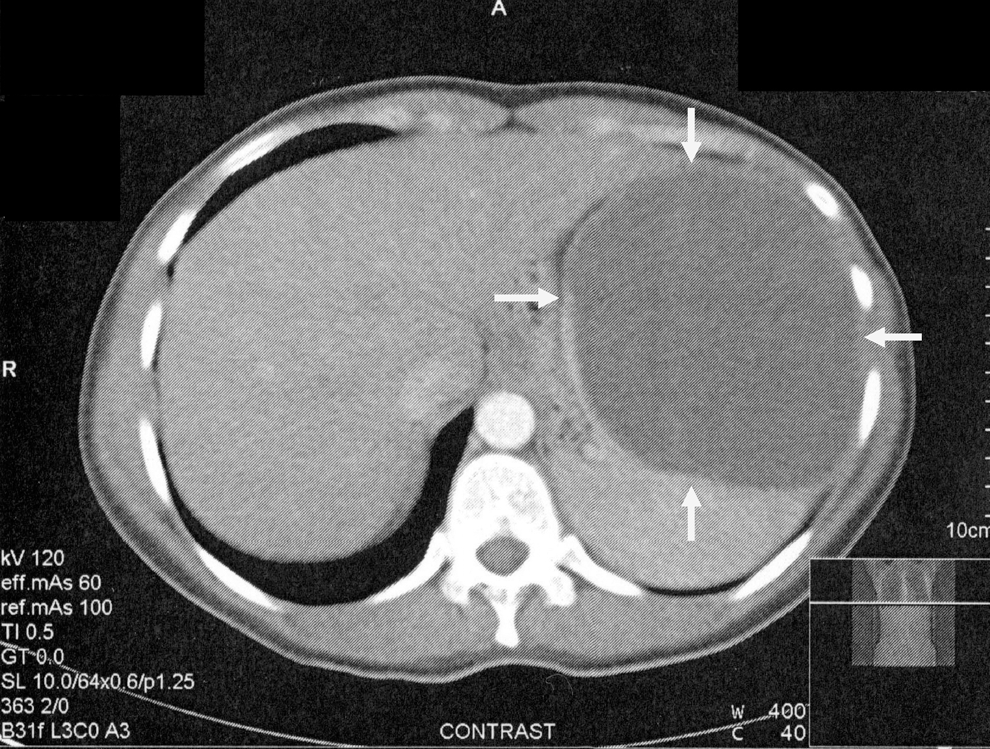
Abdominal computed tomography (CT) scan showing the hydatid cyst of the spleen (marked by arrows).
Discussion
Hydatid cyst of the spleen is a rare condition, being commonly reported in 0.5–8% of patients with echinococcosis (Ramia-Angel et al. 2011). The hexacanth embryos are typically trapped in liver or lung and only infrequently may escape these main filters, reach the spleen, and develop here (Culafic et al. 2010). Additionally isolated splenic involvement is extremely uncommon (Cabadak et al. 2009). Generally, splenic hydatid cysts are asymptomatic and are diagnosed with the occasion of performing imaging techniques for other purposes. When developing, symptoms are moderate and may occur because of the pressure exerted on neighboring organs or from other complications. As it happened in our case, mild discomfort and pain in the left upper abdomen are the most common signs (Akhan and Koroglu 2007). Similarly, according to 2 case studies of splenic involvement in hydatidosis (Culafic et al. 2010, Malik et al. 2011), the female gender seems to be affected with a higher prevalence. Moreover, the age of our patient (31 years old) fits within the adult age range reported in several studies: 30–59 years range reported by Ramia-Angel et al. (2011), 23–45 years reported by Malik et al. (2011), 15–71 years (average 38 years) reported by Ousadden et al. (2010), and 19–75 years reported by Culafic et al. (2010) and is similar to a case reported by Patanvadia et al. (2011) (35 years) and a case reported by Azordegan et al. (2007) (38-year-old female patient).
The patient came from a rural region where the hygienic rules regarding food consumption and handwashing are not strictly followed. In their study on cases of hydatidosis of the spleen, Culafic et al. (2010) found that 60% of the patients originated in rural regions. The rural patients also prevailed in the study undertaken by Ousadden et al. (2010) in Morocco. Our patient had only moderate leukocytosis and a slightly increased ESR, whereas the eosinophil counts were normal. Culafic et al. (2010) revealed in their study that only 25% of their patients presented slightly elevated ESR, leukocyte count, and/or fibrinogen. The therapy in our case consisted in total splenectomy, a procedure used in 60% of the Serbian patients (Culafic et al. 2010), 40.9% of Moroccan patients (Ousadden et al. 2010), 83% of Spanish patients (Ramia-Angel et al. 2011), in 100% of Indian patients (Malik et al. 2011), and in other isolated cases described in the literature (Jahani et al. 2004, Azordegan et al. 2007, Cabadak et al. 2009) all diagnosed with the same condition. Although splenectomy has been considered the therapy of choice for splenic hyatid disease, in recent years more conservative surgical intervention combined with medical therapy to reduce opportunistic postoperative infections have been increasingly preferred. Nevertheless, the medical treatment seems to be ineffective (Patanvadia et al. 2011).
Conclusion
The atypical presentation of cystic echinococcosis (primary splenic giant hydatid cyst) in a rural patient with minimal symptomatology suspected of a splenic tumor makes this case particularly important. To prevent the disease, which represents a major parasitosis with numerous negative repercussions (both on the health of human population and on animal production), significant informative campaigns should be implemented in the near future. On the other hand, practitioners should take this disorder into account more frequently when performing the differential diagnosis of an abdominal or thoracic tumor-like appearance.
Footnotes
Author Disclosure Statement
No competing financial interests exist.