
Abstract
Taiwan is in the subtropical zone and has typhoons every year. Leptospirosis is an endemic disease in Taiwan, and feline leptospirosis in Taiwan remains unknown so far. From January, 2010, to September, 2011, 233 cats in south Taiwan (159 stray cats and 74 household cats) were sampled in this research. Leptospira antibody titer was detected by the serology gold standard, the microscopic agglutination test (MAT). Both serum and urine were examined for Leptospira DNA by polymerase chain reaction (PCR) with two sets of primers. In this study, the serological survey showed 21 (9.3%) examined sera contained antibodies specific for pathogenic Leptospira serogroups. The results of PCR revealed that 25 (19.1%) serum and 80 (67.8%) urine samples were found positive for leptospiral DNA sequences. All products amplified from PCR reactions were sequenced by an automated method for further confirmation. This is the first study concerning the epidemiology of pathogenic Leptospira in stray and household cats' urine, and the results demonstrate that some of the cats are susceptible to pathogenic Leptospira and have the potential to shed pathogenic Leptospira into the environment. This could be an issue of public health.
Introduction
L
Both domestic and wild animals are natural reservoirs and carriers of Leptospira, acting as maintenance or accidental hosts (Felt et al. 2011). In Taiwan, the seroprevalence of leptospirosis was 11.49% (64/557) in small mammals and rodents, 33.6% (395/1175) in cattle, 58.8% (83/141) in swine, and 44.2% (102/231) in stray dogs (Lin 2001, Lai 2004, Lin 2004, Pan et al. 2006). An animal can be the maintenance host of one serovar and serve as an accidental host for another serovar (Levett 2001, Felt et al. 2011). Rodents are generally the maintenance host of serovars belonging to serogroups Icterohaemorrhagiae and Ballum, and dogs may serve as the reservoir host of serovar Canicola (Levett 2001; Millán et al. 2009). In Taiwan, the most prevalent serogroup by the highest titers in the microscopic agglutination test (MAT) were Pomona and Bataviae in small mammals and rodents, Pomona and Shermani in cattle, Shermani in swine, Shermani and Canicola in stray dogs, and Shermani in humans (Lin 2001, Lai 2004, Lin 2004, Pan et al. 2006, Centers for Disease Control of Taiwan 2010).
Limited reports are published on Leptospira in feline. Leptospira seroprevalence of felines was 4.9–33.3% in different countries (Shophet 1979, Agunloye and Nash 1996, Mylonakis et al. 2005, Jamshidi et al. 2009, Millán et al. 2009, Mosallanejad et al. 2011). The serologic evidence above suggests that cats are susceptible to Leptospira. However, attention was seldom paid to significant zoonotic risk of feline leptospirosis, although Larsson et al. proved that cats infected with Leptospira experimentally were able to scatter potential zoonotic Leptospira through their urine for up to 3 months (Larsson et al. 1985).
Cats are one of the most common pets in Taiwan. It had been pointed out that domestic cats may act as a potential source of leptospiral infection of human or other domestic animals (Larsson et al. 1985). Yet there is no information about the incidence of cat leptospirosis in Taiwan. In this study, we detected the DNA sequence of pathogenic Leptospira in feline urine and serum by polymerase chain reaction (PCR), and analyzed the antibodies against L. interrogans sensu lato in felines using MAT. The aim of this research was to determine the prevalence of pathogenic Leptospira in household and stray cats in southern Taiwan. Meanwhile, we detected pathogenic Leptospira DNA sequences in cats' urine, which suggested that cats might disseminate pathogenic Leptospira via urine.
Materials and Methods
Study area
Situated in the subtropical zone, the average rainfall in southern Taiwan is about 200 mm per month. During the typhoon season, June to October, the amount of the rainfall can reach 400 mm per month, according to the records of the Central Weather Bureau of Taiwan. Typhoons that bring in large amounts of rainfall could contribute to the epidemic of leptospirosis in Taiwan (Su et al. 2011). Southern Taiwan is a plains area, and the main labor force is employed in agriculture and animal husbandry. Rice is the principal crop growing along southern Taiwan. Rice needs to be grown in flooded plains called paddies, which is a decent environment for Leptospira. We suspect that the muddy environment will increase the incidence of farmers becoming infected with pathogenic Leptospira.
Sample collection
All cat sample collection and laboratory research for this study were conducted between January, 2010, and September, 2011. We collected samples from three local animal hospitals and five animal shelters around southern Taiwan. Two of the animal shelters were in the countryside and the other shelters and animal hospitals were in urban areas. Cats were anesthetized with Zoletil (Virbac, France) or Dexdometor (Pfizer, USA), with the owners' and the shelters' permission, to ensure that all cats were under low stress during the sample collection process. The sample collection procedure was followed the rules of Animal Protection Act of Taiwan, and this project was approved by Bureau of Animal and Plant Health Inspection and Quarantine, Council of Agriculture, Executive Yuan.
A total number of 233 cats were sampled, including 159 stray cats and 74 household cats. The methodology of this study is illustrated as Figure 1. Due to the various volumes of collected serum, each sample might not have been sufficient for both MAT and PCR. To obtain a sterile urine sample, a 22-gauge needle and 10-mL syringe were used for ventral cystocentesis. Urine was obtained only when the urinary bladder had stored more than 3 mL of urine, which was confirmed by ultrasonography. There were 225 serum samples (155 stray cats and 70 household cats) examined for pathogenic Leptospira antibody titer by MAT. For PCR, 131 serum samples (101 stray cats and 30 household cats) and 118 urine samples (92 stray cats and 26 household cats) were examined for pathogenic Leptospira DNA sequences. Serum samples were obtained from venipunctures of cephalic veins and collected in serum separator tubes. After clotting at room temperature for 15 min, serum was separated and frozen at −20°C.
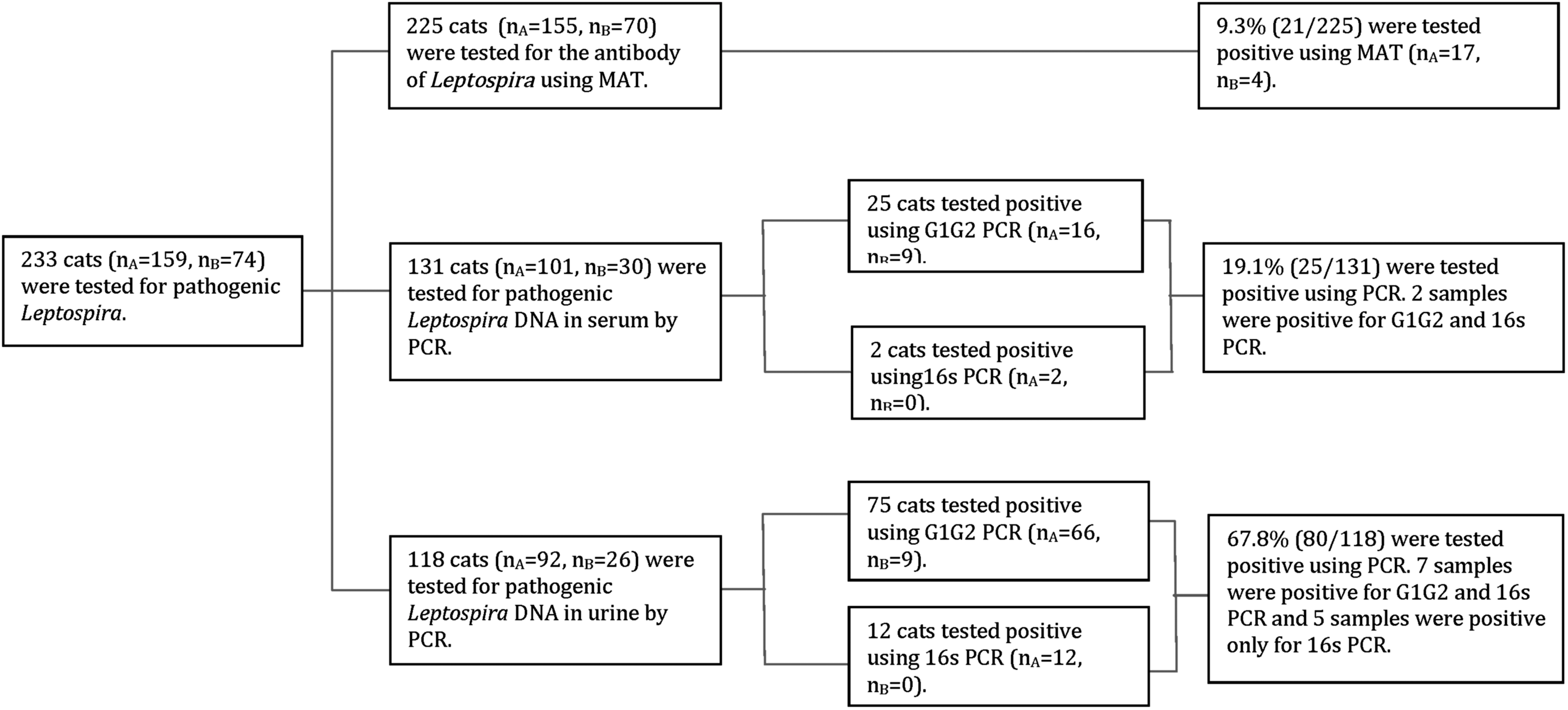
Flow chart of laboratory tests for cat pathogenic Leptospira. MAT, microscopic agglutination test; PCR, polymerase chain reaction.
Laboratory procedures
DNA extraction
Total genomic DNA in serum was extracted using QIAamp DNA Mini Kits (QIAgen, Germany). Urine samples were kept in sterile tubes and centrifuged at 15,000×g for 30 min at 4°C (Branger et al. 2005). Urine sediment was suspended in 200 μL of phosphate-buffered saline (PBS) solution (Harkin et al. 2003). Genomic DNA in urine was extracted by QIAamp RNA Mini Kits (QIAgen, Germany) because this commercial kit is able to inactivate the PCR inhibitors in urine. All the extraction procedures were performed according to the manufacturer's protocols.
Microscopic agglutination test
Serum was examined for antibodies against 11 antigens, shown as Table 1. These antigens were 6- to 10-day-old live cultures of L. interrogans (serovars Canicola, Icterohaemorrhagiae, Pyrogenes, Australis, Pomona, Bataviae, Autumnalis, and Panama), L. borgpetersenii (serovars Javanica and Tarassovi), and L. santarosai (serovar Shermani), as shown in Table 1. The test was performed as described by Cole et al. (1973). Briefly, the serum was diluted separately (1:50–1:6400) and then mixed with the individual Leptospira cultures mentioned above. We performed the MAT test starting from the serum dilution of 1:50. Antibody titers≥1:100 were determined as a cutoff value for positive (Jamshidi et al. 2009, Mosallaneijad et al. 2011).
Polymerase chain reaction
Serum and urine samples were tested using two pairs of primers. One primer set G1/G2 amplified a 285-bp sequence by PCR from strains of all pathogenic Leptospira spp. except L. kirschneri. The reactions were carried out as described previously (Gravekamp et al. 1993). The other pair of primers amplified a 331-bp sequence of the Leptospira rrs (16S) gene. The PCR assay was performed according to Mérien et al. (1992). After the amplifications, 5 μL of PCR products were analyzed using 1% agarose gel that was subsequently stained with HealthView Nucleic Acid Stain (Genomics BioSci & Tech Corp, Taiwan) for detection of the expected product under ultraviolet (UV) light. All of the 285-bp and 331-bp products amplified from PCR reactions were sequenced by an automated method. Because primers G1 and G2 amplified only pathogenic Leptospira, as mentioned previously, samples were considered positive when the diagnostic 285-bp band was observed, whether the amplified products were sequenced successfully or not. The primer set–amplified Leptospira rrs (16S) was not able to distinguish between nonpathogenic L. biflexa and pathogenic Leptospira spp. Therefore, the products of 331 bp must sequence as pathogenic Leptospira or the products would be considered as negative.
Results
Of 225 cats tested for pathogenic Leptospira antibody titer by MAT, 21 cats reacted positively for one or two leptospiral antigens, including 17 stray cats and 4 household cats (Table 2). The titers were ranging from 1:100 to 1:400 are shown in Table 3. For stray cats, 10 were serologically positive against Shermani, five against Javanica, two against Icterohaemorrhagiae, two against Australis, and one against Pyrogenes. Antibodies against more than one leptospiral antigen were found in three cats. One cat had antibodies reacting with Icterohaemorrhagiae (1:100) and Shermani (1:100), one with Australis (1:200) and Javanica (1:100), and the other one with Shermani (1:400) and Australis (1:200). These multiple reactions were not considered to be cross-reactions due to the high dilution titers. No positive titers were obtained with Pomona, Panama, Autumnalis, Canicola, Bataviae, and Tarassovi.
2010–2011 (total 233 cats). 225 cats were tested using MAT (microscopic agglutination test) and 9.3% (21/225) were found to be positive for the leptospiral antibody. 131 serum samples were tested for Leptospira using PCR with two sets of primers. Twenty-five cats were tested positive for pathogenic Leptospira in serum and 118 cats were examined for leptosiral DNA in their urine, and 80 cats were tested positive.
21 cats tested positive for the leptopsiral antibody. 18 cats were positive for only 1 serogroup. 3 cats were positive for 2 different serogroups. (One was positive for Australis 200× and Javanica 100×; one was positive for Shermani 100× and Icterohaemorrhagiae 100×; the other one was positive for Shermani 400× and Australis 200×).
positive rate within 238 cats.
MAT, microscopic agglutination test.
Among 131 serum samples tested for pathogenic Leptospira DNA by PCR with two sets of primers, 19.1% (25/131) were positive for pathogenic Leptospira DNA in serum (16 stray cats and 9 household cats). As for urine samples, 67.8% (80/118) were positive for pathogenic Leptospira DNA (71 stray cats and 9 household cats) (Table 2). All of the 285-bp (n=100) and 331-bp (n=88) bands were confirmed by DNA sequencing, as shown in Table 4. Of n the 100 products amplified with G1/G2 primers, 82 products (12 serum samples and 70 urine samples) were sequenced as L. interrogans. The other 18 samples failed to be sequenced due to inadequate PCR product concentration, although these 18 samples showed the expected 285-bp products generated by the G1/G2 primer set. We still considered these 18 samples as positive for pathogenic Leptospira DNA, because the primer G1/G2 is specific for pathogenic Leptospira. Of the 88 331-bp sequences of the Leptospira rrs (16S) gene, 53 samples were sequenced with the following results: L. interrogans (n=12), L. kirschneri (n=2), Brevundimonas intermedia (n=3), Pasturella pneumotrpica (n=1), and uncultured bacterium (n=35) (Table 4). The remaining 35 samples of 16S PCR failed to be sequenced because of the low copy numbers, and these samples were considered as negative because of the inability to differentiate between pathogenic and nonpathogenic Leptospira by amplification of the Leptospira rrs gene.
135 samples were sequenced successfully and 53 were sequenced unsuccessfully. The sequenced results of 285-bp products were 82 L. interrogans. Only 14 of 331-bp products were found as L. interrogans (n=12) or L. kirchneri (n=2).
Discussion
Pathogenic Leptospira thrives in warm and humid weather, therefore leptospirosis is an endemic disease in Taiwan, especially after the typhoon and flood seasons (Su et al. 2009). This is the first study of feline leptospirosis in southern Taiwan using serological and PCR detection. Southern Taiwan was chosen for the study area because this area has suffered more floods than other areas of Taiwan. Rice farms in southern Taiwan may also increase the incidence of leptospirosis.
The results of serological survey showed that 21 cats had antibodies against L. interrogans (serovars Icterrohaemorrhagiae, Pyrogenes, and Australis), L. santarosai (serovar Shermani), and L. borgpetersenii (serovar Javanica) in dilutions≥1:100. The lowest serum dilution used in Shophet's study was 1:24. The author indicated that for some serovars animals may shed Leptospira through urine with a low or undetectable antibody titer. Therefore, low serum dilutions in the MAT test should be included to estimate the seroprevalence of cats (Shophet 1979). The antibody titer of 1:100 was considered as a cutoff value for positive reaction in this study to rule out the cross-reaction at low titers. The positive MAT results of cats in this study may indicate their previous exposure to Leptospira, because cats in Taiwan were not routinely vaccinated against Leptospira. The seroprevalence of leptospirosis in cats is different from place to place. Leptospiral prevalence was reported to be 4.9% (5/102) in stray cats in Ahvaz (Mosallanejad et al. 2011), 27% (30/111) in stray and household cats in Tehran (Jamshidi et al. 2009), 20.0% (5/25) in feral cats in Spain (Millán et al. 2009), 33.3% (33/99) in household cats in a feline hospital population in Greece (Mylonakis et al. 2005), and 9.3% (21/225) in cats in southern Taiwan in this study. However, these differences may be a consequence of different cutoff values and serovars panels. The sources of the cats' infection could be wildlife, contaminated water, cohabitating dogs, and rodents (Green et al. 2011). Mosallanejad et al. suggested that different environmental factors related to the duration of survival of leptospires may affect the prevalence of leptospirosis (Mosallanejad et al. 2011).
Ten stray cats in the present study had antibodies against Shermani, and this serogroup was also found in rodents, cattle, swine, stray dogs, and humans in Taiwan (Lin 2001, Lai 2004, Lin 2004, Pan et al. 2006, Centers for Disease Control of Taiwan 2010); hence Shermani could be one of the most widespread serogroups in Taiwan. The serogroup Javanica was diagnosed in five stray cats in this study, and this serogroup was also diagnosed previously in rodents and stray dogs in Taiwan (Lai 2004, Pan et al. 2006). Three sera of household cats in this survey had titers to Icterohaemorrhagiae. Out of 231 stray dogs in Taiwan, which had never been vaccinated against Leptospira, 17 also had antibodies reacting with Icterohaemorrhagiae (Lai 2004). In view of these finding, cats might infect Leptospira directly by ingesting contaminated prey (Shophet and Marshall 1980) or indirectly by the water contaminated by domestic animals, rodents, and stray dogs (Green et al. 2011). However, more studies need to be done to clear out the pathogen transmission between domestic animals, rodents, dogs, and cats in Taiwan.
As shown in Table 2, there were 25 serum samples found positive for Leptospira DNA. The duration of leptospiremia in cats is very short because of the rapid rise of leptospiral antibodies (for review, see Larsson et al. 1985). The short occurrence of leptospiremia makes detection of Leptospira DNA in serum a last-resort method to diagnose leptospirosis. Leptospiral culture of feline urine was used to monitor the shedding of Leptospira in experimentally and naturally infected cats. None of Leptospira was isolated in Agunloye's and Nash's investigation in cats in Scotland (Agunloye and Nash 1996). In Larsson's experiment, two cats inoculated with L. interrogans canicola had the presence of L. interrogans detected by urine bacterial cultures between 2nd and 10th week after inoculation (Larsson et al. 1985). In our study, Leptospira DNA sequences were detected using PCR in 80 urine specimens. The detection rate of Leptospira DNA in cats' urine was considerable, whereas urine culture for leptospires was not done in this study and the viability of leptospires in cats' urine was unknown. More stray cats (n=71) were detected Leptospira DNA in urine than household cats (n=8). This could be due to the fact that stray cats are at a higher risk of exposure to the environmental infections. The detection rate of urine using PCR (67.8%) was obviously higher than the results of serum using PCR and MAT, 19.1% and 9.3%, respectively. In view of these data, the prevalence of Leptospira DNA in cat urine may be underestimated if only MAT was used for detecting leptospirosis.
Our sequence results presented here confirm the previous description of Gravekamp et al. that the G1/G2 primers are specific for pathogenic Leptospira, but DNA from L. kirschneri is unable to be amplified using this set of primers (Gravekamp et al. 1993). Palaniapan et al. claimed that L. biflexa can be amplified using G1/G2 primers (Palaniappan et al. 2005), but L. biflex was not amplified in our study. To detect L. kirschner, the other primer set was used to amplify sequences from the Leptospira rrs (16S) gene. Fourteen 331-bp products were sequenced as L. interrogans (n=12) and L. kirchneri (n=2), but 39 331-bp bands were sequenced as Pasturella pneumotropica (n=1), Brevundimonas intermedia (n=3), and uncultured bacterium (n=35). Therefore, we conclude that the primer-amplified sequence from the Leptospira rrs (16S) gene was not specific for Leptospira and sequencing was necessary for further confirmation.
As shown in Table 5, 85 cats were examined with all three methods used in this study, including MAT and PCR of serum and urine. Some animals infected with Leptospira may shed the spirochete with an undetectable or low serological titer, as reported by Shophet in 1979; this was again confirmed in this investigation. In the experiment of feline leptospirosis, no positive result was obtained for the blood cultures of Leptospira due to the early rise of antibody after infection, and the urine culture was positive 14 days after inoculation. In our data, 13 cats had Leptospira DNA detected both in serum and urine at the same time. Reinfection might be one of the reasons that Leptospira presented in serum and urine simultaneously. One household cat not only had antibodies against Icterohaemorrhagiae but also was positive for Leptospira DNA in both serum and urine. It is possible that the low titer of antibody (1:100) was not enough to eliminate the antigens in serum. In Larsson's experiment, no urine culture was positive in the cats inoculated with Icterohaemorrhagiae (Larsson et al. 1985). In our study, this household cat with a titer of 1:100 antibodies against Icterohaemorrhagiae had DNA of L. interrogans detected in its urine.
In the 85 cats, 43 cats tested positive for leptospiruria. 12 cats tested positive for leptospiruria and leptospiremia, but tested negative or leptospiral antibodies.
Seldom were clinical signs noticed in cats infected with Leptospira despite the presence of leptospiremia and leptospiruria (Green et al. 2011). However, three cats naturally infected with Leptospira were reported by Arbour et al. (2012) to have polyuria, polydipsia, uveitis, and lameness. These investigators said that clinical signs might develop eventually after a long period of infection (Arbour et al. 2012). No clinical signs were observed in all the cats involved in this study. It was a pity that we did not persistently monitor the cats with evidence of leptopsiral infection.
This study demonstrates that cats in southern Taiwan were exposed to pathogenic Leptospira and the DNA sequences of Leptospira were found both in serum and urine. Clinical signs might not be noticed in cats with leptospirosis. This evidence indicated that cats can be naturally infected by Leptospira, and cats could be the potential source of human and animal Leptospira infection. However, the viability of Leptospira in cat urine was unknown because urine culture was not done in this study.
Footnotes
Acknowledgments
This study was approved and sponsored by the Bureau of Animal and Plant Health Inspection and Quarantine, Council of Agriculture, Executive Yuan. We followed the rules of the Animal Protection Act of Taiwan for sample collection procedures. We thank Dr. Min-Liang Wong (Department of Veterinary Medicine, College of Veterinary Medicine, National Chung-Hsing University, Taichung, and Taiwan) and microbiologist John P. Thibideau for his critical appraisal.
Author Disclosure Statement
All authors declare that no competing financial interests exist.