
Abstract
The seroprevalence of Rift Valley fever (RVF), brucellosis, and Q fever among domestic ruminants on the southeastern shore of Lake Chad was studied. The study area consisted of two parts, including mainland and islands. On the mainland, the study was conducted in nine randomly selected villages and camps. On the islands, samples were collected from all four available sites. A total of 985 serum samples were collected and 924 were analyzed using enzyme-linked immunosorbent assay (ELISA) for RVF. A total of 561 samples collected from islands were analyzed using ELISA for Q fever and both ELISA and Rose Bengal tests (RBT) for brucellosis. The apparent RVF seroprevalence by species was 37.8% (95% confidence interval [CI] 34.2–41.3) in cattle, 18.8% (95% CI 12.3–25.2) in goats, and 10.8% (95% CI 3.0–18.5) in sheep. For brucellosis and Q fever, only cattle samples from islands were analyzed. For Q fever, the apparent seroprevalence was 7.8% (95% CI 5.6–10.1). For brucellosis, the RBT showed a prevalence of 5.7% (95% CI 3.8–7.6), and ELISA showed 11.9% (95% CI 9.3–14.6) with a kappa value of 0.53 showing a moderate agreement between the two tests. This study confirms the presence of the three diseases in the study area. More research is required to assess the importance for public health and conservation of the Kouri cattle breed.
Introduction
I
Lake Chad is a breeding area of choice for ruminants (cattle, goats, and sheep) and especially for the endemic Kouri breed, which is in danger of extinction not only because of its existence only in this region but also because of health interventions and disease control. Hence, the Lake Chad region is also an ecological niche for vectors, such as mosquitoes that could be the source of transmission of zoonotic diseases such as RVF, which has been reported in Chad.
Rift Valley fever
Most RVF outbreaks have occurred in the East African region. However in North and West African regions, numerous outbreaks of RVF were reported in different countries during the last two decades. An outbreak was reported in the Senegal River basin in southern Mauritania and northern Senegal in 1987, causing a high rate of abortion ruminants and more than 200 human deaths in Mauritania alone (Jouan et al. 1988). The disease was reported in Egypt in 1993, in Cameroon among goats in 2003 (LeBreton et al. 2006), and in Sudan in 2007 (Hassan et al. 2011). An unexpected outbreak was reported in the northern Sahelian region of Mauritania in 2010 after a heavy rainfall (El Mamy et al. 2011), whereas the most recent outbreak in Africa occurred in Mauritania in October, 2012, resulting in the death of 13 people among 30 infected human cases (WHO 2012).
Despite the health risk and economic impact on humans and animals and its presence in neighboring countries, the epidemiological situation of the disease remains obscure in Chad, although epizootic reports exist in humans where RVF virus (RVFV) was isolated (Durand et al. 2003). An analysis of samples collected from ruminants at slaughterhouses in Abeche and N'Djamena, Chad, showed a prevalence of 10.7% in sheep, 8% in goats, and 4% in cattle (Ringot et al. 2004). However, there have been no further studies on the disease or its possible endemic status in some parts of the country.
Q fever
In Chad, Q fever is among the diseases neglected by both public and animal health surveillance systems. In the early 1950s, a study showed a relatively high seroprevalence of Q fever (35–75%) among specific groups of humans (meat sellers, breeders, and butchers) who had close contact with livestock in southern Chad (Giroud et al. 1951). Another study showed a 3.5% seroprevalence of Q fever in human serum collected from N'Djamena, Chad. The same study mentioned some positive microagglutunation reactions in cattle, goats, sheep, and camels (Maurice et al. 1968). A study conducted by Schelling et al. in 2003 to assess the seroprevalence of Q fever and brucellosis in nomadic pastoralists and their livestock in the region of Chari Baguirmi in Chad showed a prevalence rate of 1% in humans, 80% in camels, 4% in cattle, 13% in goats, and 11% in sheep and reported that being a camel breeder was a risk factor for Q fever (Schelling et al. 2003).
Brucellosis
In sub-Saharan Africa, bovine brucellosis remains one of the most widespread livestock diseases. It is responsible for considerable economic losses through its negative impacts on livestock production, including late-term abortion, birth of weak calves, and infertility (Akakpo 1987, Corbel 1997, Bronsvoort et al. 2009, Sanogo et al. 2013). An epidemiological study of cattle, goats, sheep, camels, and zebu brucellosis was conducted in seven countries in tropical Africa from 1977 to 1983. It showed an average seroprevalence of 22.5%, distinguishing one group of countries where the prevalence was high (Niger, Rwanda, and Togo at 30–41%) and another where it was relatively low (Benin, Burkina Faso, and Cameroon with 10–12%) (Akakpo 1987). However, contemporary assessments indicate a lower prevalence than that presented by Akakpo. Serological evidence of its existence has been demonstrated in many studies in sub-Saharan countries like Côte d'Ivoire (Sanogo et al. 2013), Uganda (Kashiwazaki et al. 2012), Ethiopia (Asmare et al. 2013), and Togo (Dean et al. 2013), and in particular some neighboring countries to Chad, notably from the southwest where the study area borders Nigeria (Alausa et al. 1976, Bertu et al. 2012, Mai et al. 2012).
In Chad, brucellosis remains a neglected disease. However, evidence of its existence has been mentioned in some studies. The study of LeFevre in 1970 showed a seroprevalence of more than 15% among goats and 10 human cases confirmed by blood culture (LeFévre et al. 1970). Another seroepidemiological survey of brucellosis in abattoir personnel in N'Djamena, Chad, was conducted in the early 1990s, reporting around 14% seropositivity among 107 abattoir workers compared to zero seropositivity among blood donors (Massenet et al. 1993). Later studies conducted by Delafosse et al. on cattle in the region of Abeche, Chad, showed a true prevalence of 2.6%, with herd prevalence estimated at 20%. The main risk factors identified for brucellosis were the ethnic Arabic group herding by the children of the herdsmen. (Delafosse et al. 2002). A confirmed case of Brucella melitensis with Plasmodium falciparum co-infection was reported in a young veterinary researcher returning to France from Chad after working in close contact with farmers and their animals for more than 3 months in 2004 (Badiaga et al. 2005). Earlier work by Schelling et al. in 1999 and 2000 on Chadian pastoralists and their animals demonstrated the presence of brucellosis in humans (with a prevalence of 4%) and animals (0.4% in camels, 7% in cattle, and 0% in small ruminants) (Schelling et al. 2003).
The objectives of our study were to: (1) Estimate the apparent seroprevalence of RVF, brucellosis, and Q fever among domestic ruminants on the southeastern shore of Lake Chad, (2) characterize the seroprevalence and identify its main risk factors, and (3) identify additional training and quality assurance to assure IRED is equipped and trained to do monitoring and surveillance of these three diseases in collaboration with the Swiss Tropical and Public Health Institute in Basel (Switzerland).
Materials and Methods
Study area
The study area consisted of two parts, including the mainland and islands in the zones of responsibility of the veterinary posts of Bol and Baga-Sola in the administrative region of Lake Chad, Chad (Fig. 1). In the mainland, nine different villages and camps were randomly selected from existing camps and villages using the Excel function for random selection. Four camps/villages belong to the veterinary post of Bol (Yira Wari, Kalamina, Sawa, and Yoko) and five belong to the veterinary post of Baga-Sola (Kafia, Ballom Bassari, Bibi, Liya Koura1, Liya Koura2, and Boul-wanda). Meanwhile, samples were collected from all four available islands (Centre de Sauvegarde de la race Kouri 1 (CSK1), CSK2, Blarigui, and Tchika).
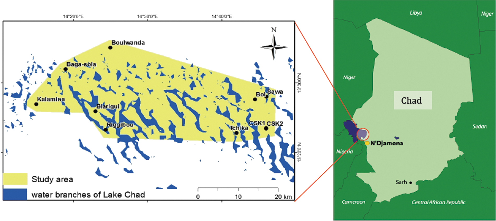
Study area.
Target population, sampling, and data analysis
The sample selection method consisted of three sampling stages. (1) The veterinary posts were designated as the primary sampling level, (2) the camps or villages constituted the second level, and (3) the tertiary level was the eligible animals (goats, sheep, and cattle from both sexes aged of 1 year or more). Animals were randomly sampled in every herd. Eligible animals were randomly sampled according to a previous local study on rinderpest serology (Rapport final de l'enquête sérologique sur la peste bovine au Tchad, LRVZ, 2009). A total of 985 blood samples were collected with dry vacutainers tubes for sera. All samples collected were tested for RFV using competitive enzyme-linked immunosorbent assay (ELISA) for detection of specific antibodies against RVFV (ID Screen Rift Valley Fever Competition [Multi Species]). Because of some limitations in reagent availability, only samples collected from cattle from islands were further tested for Q fever, using indirect ELISA (ID Screen Q-Fever Indirect Multi-Species), and for brucellosis using the Rose Bengal test (RBT) and indirect ELISA (ID Screen Brucellosis Serum Indirect Multi-Species). We need to mention that the small ruminants (goats and sheep) are usually kept far from islands. For ELISA, samples were tested in duplicate and the mean optical density (O.D.) value of each sample correlated to positive and negative controls (percentage of positivity [PP]) was used to interpret the results according to the manufacturer's rules of test validity and results interpretation. However, the RBT was performed by observing the agglutination by the naked eye and results were confirmed by ELISA afterward. We did not calculate the true prevalence because tests were not previously validated for Chad. Further work is needed to isolate and characterize local positive samples of those pathogens to validate the tests (ELISA and RBT) for the country and the region.
The statistical analysis of the results was performed using STATA software (v. 12, StataCorp, College Station, TX). A logistic model was used to analyze risk factors such as sex, age, category, and location.
Results
RVF apparent seroprevalence
The highest RVF apparent seroprevalence was among cattle (37.8% with 95% confidence interval [CI] 34.2–41.3%) and the lowest was among sheep (10.8% with 95% CI 3.0–18.5%). The apparent seroprevalence among goats was 18.8% (95% CI 12.3–25.2%) (Table 1, Fig. 2). In cattle, the females were almost two times more likely than males to be infected. The older the cow, the more likelihood of infection, with those aged 10 years or more almost five times more likely to be infected than the baseline age group, which was between 1–3 years. However, being on islands seemed to be protective. Animals on the islands were more than three times less likely to be infected than animals on the mainland (Table 2).
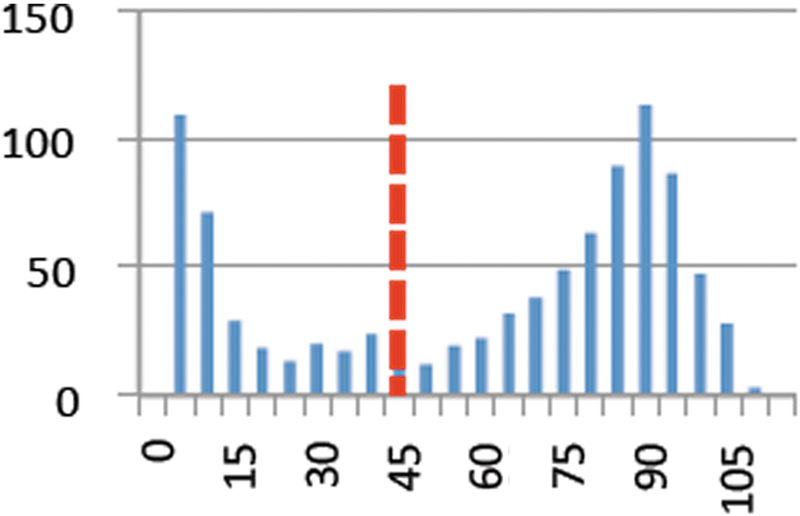
Optical density distribution. Rift Valley fever (RVF) in cattle. The dotted line shows the cutoff value recommended by the manufacturer. Color images available online at
RVF, Rift Valley fever; SE, standard error; CI, confidence interval.
RVF, Rift Valley fever; ELISA, enzyme-linked immunosorbent assay; Pos, positive; OR, odds ratio; CI, confidence interval; LRT, likelihood ratio test.
Q fever apparent seroprevalence
In total, 561 samples from cattle on islands were analyzed using indirect ELISA for Q fever. The overall apparent seroprevalence was 7.8% (95% CI 5.6–10.1%) (Table 3, Fig. 3). However, gender was one of the most important risk factors. The females were more than three times more likely to be infected than males. The highest odds ratios were observed among the animals aged from 7 to 9 years old (age group 3), where the animals seemed to be more than seven times as likely to be infected than those aged 1–3 years (Table 4).
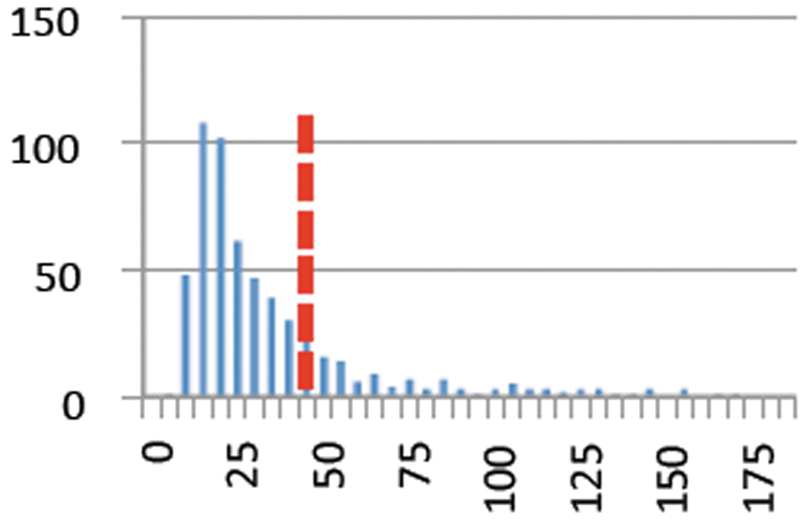
Optical density distribution. Q fever in cattle. The dotted line shows the cut-off value recommended by the manufacturer. Color images available online at
SE, standard error; CI, confidence interval.
ELISA, enyme-linked immunosorbent assay; Pos, positive; OR, odds ratio; CI, confidence interval; LRT, likelihood ratio test; NA, not applicable.
Brucellosis seroprevalence
All samples tested for Q fever were further analyzed for brucellosis using the RBT and indirect ELISA. The RBT showed an overall prevalence of 5.7% (95% CI 3.8–7.6%), whereas the ELISA showed an overall prevalence of 11.9% (95% CI 9.3–14.6%) (Table 5, Fig. 4). The kappa statistics for interrater agreement showed an agreement of 92.3% between the two tests (Table 6). The results of the logistic regression model to study the potential risk factors were similar to those for Q fever. Age had a high influence on the odds of infection. Among animals aged 10 years or more (age category 4), the odds were almost 34 times more likely to be infected than those aged 1–3 years. The increased odds ratio of infection was more than four times higher among females than males (Table 7).

Optical density distribution. Brucellosis in cattle. The dotted line shows the cut-off value recommended by the manufacturer. Color images available online at
ELISA, enzyme-linked immunosorbent assay; SE, standard error; CI, confidence interval.
RBT, Rose Bengal test; ELISA, enzyme-linked immunosorbent assay; SE, standard error; Prob, probability.
RBT, Rose Bengal test; ELISA, enzyme-linked immunosorbent assay; Pos, positive; OR, odds ratio; CI, confidence interval; LRT, likelihood ratio test; NA, not applicable.
Discussion
The present study was conducted to establish the seroprevalence status of three neglected zoonotic diseases. Those are RVF, Q fever, and brucellosis. The National Surveillance Network for Animal Diseases (REPIMAT) is not currently monitoring these diseases, and very few studies were carried out in the last two decades in the country (Massenet et al. 1993, Delafosse et al. 2002, Durand et al 2003, Ringot et al 2003, Schelling et al. 2003). Our study showed serological evidence of these pathogens in the study area, especially RVF where the situation seems to be endemic, contrary to the expected status of an epidemic disease that occurs after heavy rainfall. The apparent seroprevalence of RVF was found to be higher in cattle than among goats and sheep. This could be explained by the fact that cattle live longer and are thus more exposed to the infection than the sheep and goats. In addition, the small ruminants are mostly kept relatively far from the lake compared to cattle. However, it has been shown in the literature that both cattle and small ruminants have a similar sensitivity to RVF infection.
Q fever seems to be also an issue in the area where the mosquitoes persist throughout the whole year. The apparent seroprevalence of 7.8% found in our study indicates that the infection is present in the area, especially if we take into account the cases of abortions reported by farmers usually after the end of the rainy season (between November and March). Further investigations should be done to know more about the current status of the infection, both among cattle and small ruminants.
Brucellosis is known to be endemic in most areas in Chad. The southeastern shore of Lake Chad is not an exception. However, in the present study, we found an apparent seroprevalence of 11.9%, and the females were more likely to be infected than males. That could be due to the effect of age, which is correlated to sex in all three diseases. The majority of the old cattle are females. In addition to what the farmers report about abortion, after discussion with them in the field, they also recognize the presence of carpal hygromas in their herds, which is a typical symptom of brucellosis.
Finally, we believe that our study will contribute to raise awareness about possible infection among humans in the study area. In Chad, more than 70% of the population lives in rural areas. Human/public health is linked to animal health in the rural areas where farmers/villagers are in direct contact with the animals. The concept of “one health” introduces a new approach that demonstrates a significant positive health and economic impact when veterinarians and physicians work in close collaboration (Zinsstag et al. 2007). In 1987, cases of hemorrhagic fever in the south of Mauritania were diagnosed initially as an outbreak of yellow fever, before being corrected a few days later after it was confirmed by veterinary services that there was a RVF outbreak in livestock (Digoutte 1999). Joint interventions in public and animal health sectors have also demonstrated their effectiveness, such as the joint vaccination campaign organized in 2002 in Chad (Bechir et al. 2004, Schelling et al. 2007, Zinsstag et al. 2011).
Conclusion
In summary, our study highlights clearly the presence of these three zoonoses in the study area. Our results were based on serological analysis to detect immunoglobulin G (IgG) antibodies of these diseases. Further investigations are needed to detect possible IgM antibodies, which would indicate recent infection, and possibly to isolate and characterize the pathogens, in particular for RVF and brucellosis. The human aspect and the vectors implicated in the transmission of infection should also be investigated to better contribute to the efforts for improvement of the local population health conditions and welfare.
Footnotes
Acknowledgments
The authors acknowledge: The Institut de Rechercehe en Elevage pour le Dévéloppement (IRED) ex Laboratoire de Recherches Vétérinaires et Zootechniques (LRVZ) and the AfriqueOne-IRED consortium Project funded by Wellcome Trust in N'Djamena, Chad, for funding the study; the Swiss Tropical and Public Health Institute (Swiss TPH) in Basel, Switzerland, for academic support; and those who contributed to data collection and laboratory analysis—Théophile Ngarlembaye, Tchari Doungous, Fatima Abdelrazak, and Sokoum Issa.
Author Disclosure Statement
No competing financial interests exist.
Author contributions were: M.F. Abakar, principal investigator; J. Zinsstag and N.B. Naré, study design, methodology and supervision; J. Hattendorf and E. Schelling, support for statistics and data analysis; and I.O. Alfaroukh, support for project management.