
Abstract
Coxiellosis caused by Coxiella burnetii is a cosmopolitan zoonosis, which causes significant losses through abortions and stillbirths in small ruminants. A cross-sectional seroprevalence study was conducted in two major sheep and goat farming districts of Punjab (Layyah and Muzaffargarh), Pakistan. In total, 542 small ruminants (271 sheep and goats each) of both sexes (60 males and 482 females) of different age groups from 104 flocks (52 flocks of either species) were randomly selected for the collection of sera and related epidemiological information. The sampling plan was devised at the expected prevalence of 50%, confidence interval (CI) of 95%, and error margin of 5%. A commercial indirect enzyme-linked immunosorbent assay (iELISA; ID Vet) was used to test the samples for the presence of both phase I and II antibodies. A high herd level prevalence (73.1%, 95% CI 63.5–81.3) was recorded in the studied districts. Individual level seroprevalence was recorded as 30.8% (95% CI 26.9–34.9). Higher value was recorded in females (32%) when compared with males (21.7%). Higher prevalence (34.8%, 95% CI 21.4–50.2) was observed in animals of 1 year (nulliparous) than to primiparous (24.8%, 95% CI 17.4–33.5) and multiparous (32.3%, 95% CI 27.6–37.3) animals. Univariable analysis indicated that caprine species (odds ratio [OR] 1.96, p = 0.22), females (OR = 1.70, p = 0.104), infestation with ticks (OR = 234.39, p < 0.001), abortion history (OR 1.96, p = 0.14), retention of fetal membranes (OR 1.50, p = 0.35), keeping a single breed in a herd (OR 1.50, p = 0.56), and mixed feeding management (OR 1.37, p = 0.33) were the variables found associated with high prevalence of antibodies to C. burnetii. The study indicates that seroprevalence of coxiellosis was high in the studied small ruminant population and further studies are required to discern its epidemiology more precisely.
Introduction
Q
Historically, no extensive study has been conducted in Pakistan to determine seroprevalence of coxiellosis in ruminants especially sheep and goats. A solitary report on occurrence of Q fever in southern parts of Pakistan could be traced back in the literature (Ahmed 1987). However, to date, no information on coxilleosis was available from northeast Paksitan. The aim of this study was to determine the apparent seroprevalence and possible risk factors associated with coxiellosis in sheep and goats in two district of Pakistan viz., Layyah and Muzaffargarh employing indirect enzyme-linked immunosorbent assay (iELISA).
Materials and Methods
Study population and sudy settings
According to Livestock Census of Pakistan (Anonymous 2006), the districts of Muzaffargarh and Layyah, Punjab, Pakistan harbored 1.06 and 0.505 million heads of goats and sheep, respectively. Muzaffargarh lies at latitude 30° 20′ N and longitude 71° 05′ E, whereas Layyah is situated at latitude 30.57° 36.72′ N and longitude 70.56° 32.28′ E. The climate of Muzaffargarh is characterized by comparatively higher temperature (°C) and frequent winds during summer compared to Layyah (°C) However, in winter relatively low temperature prevail in both districts (°C).
Sample collection
For this study, 542 small ruminants (271 sheep and goats each) belonging to 104 flocks (52 herds of each species) were randomly selected from seven different locations of the districts Layyah and Muzaffargarh (Table 1 and Fig. 1). The sample size was calculated for an expected disease prevalence of 50% (unknown prevalence) at 95% confidence interval (CI) and 5% error margin (Thrusfield 2007). Survey Toolbox software was used for the random selection of herds and animals (Cameron 1999). Study animals belonged to both sexes (60 males, 482 females) and different age groups based upon their reproductive status (less than and equal to 1 year = 46; above 1 and below 2 years of age = 181; between 2 and 3 years of age = 143 and above 3 years of age = 172). Additionally, a questionnaire was also designed, pretested, and information was collected after directly interviewing the owners about possible risk factors including breed, herd size, age and purpose of flock, feeding and watering technique, pregnancy status, parturient history, history of reproductive problems including abortion, stillbirth, delivery of week lambs and kids, and retained placenta. Animals were examined carefully for the infestation of ticks and information was recorded accordingly. Samples were collected through jugular vein puncture in evacuated blood collection tubes (Improvacuter, Shanghai International Hamburg Holding, gmbH, Germany). Immediately after collection, tubes were marked for flock identity, placed vertically in a cool box packed with gel freezing pads, and shipped to the laboratory for further processing. Sera were centrifuged (4500 rpm for 10 min) and stored in cryovials (Cryo.S™; Greiner Bio-one, GmbH Frickenhausen, Germany) until testing.
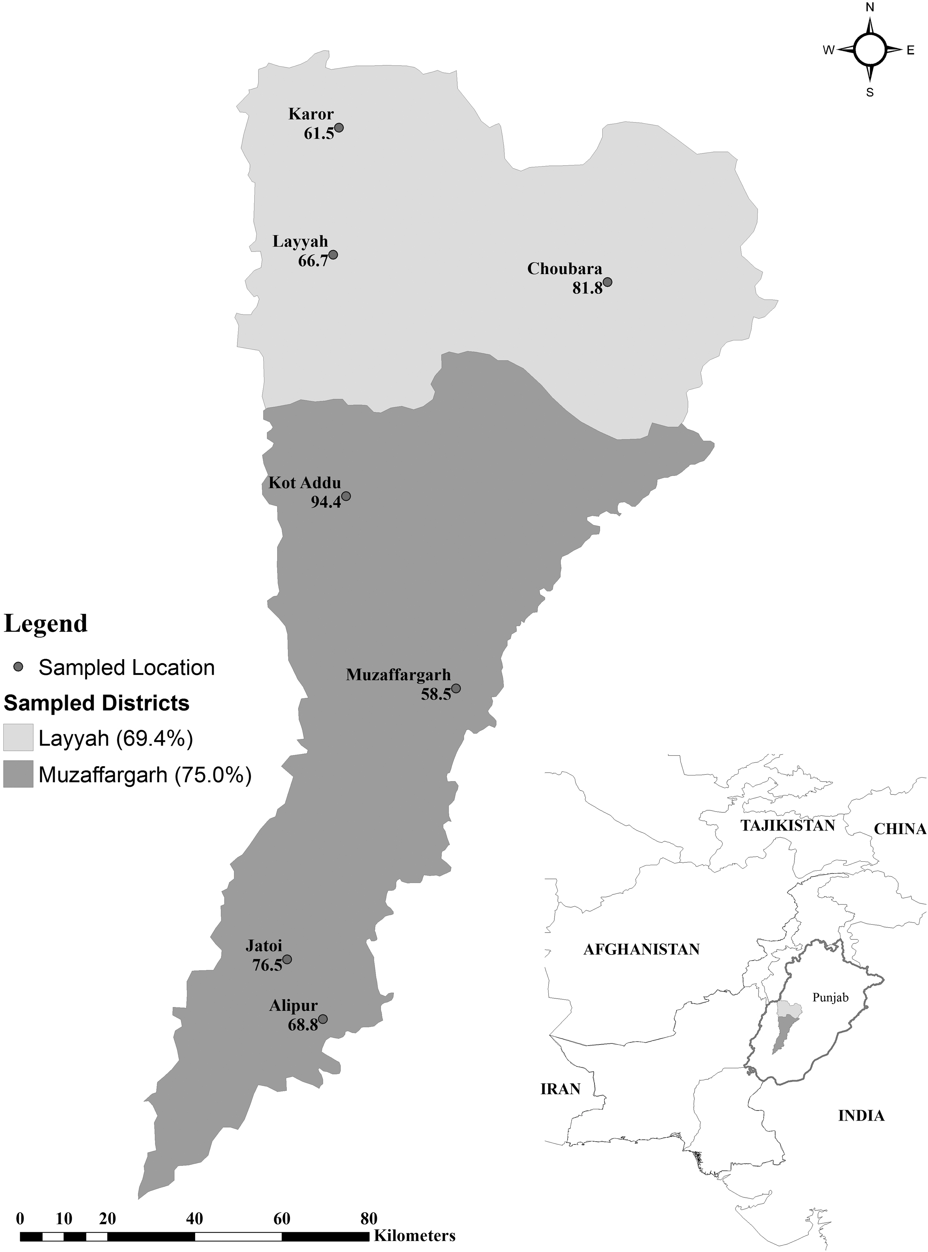
Choropleth map showing herd level prevalence of Q fever/Coxiellosis in small ruminants of two districts of Punjab, Pakistan.
Individual prevalence was not significantly different in various areas, χ2 = 7.742, 6 df, p = 0.258. Herd prevalence was not significantly different in various areas, χ2 = 6.627, 6df, p = 0.357.
CI, confidence interval; OR, odds ratio.
Serological testing
All sera were tested for the presence of specific phase I and II antibodies against C. burnetii using an iELISA kit for coxiellosis (ID Vet, France) following the instructions of the manufacturer. Optical density (OD) values were calculated at a wavelength of 450 nm. Sample/positive percentages (S/P%) for each serum sample were determined after adjusting to the negative control using the following formula (ODsample − ODnegative)/ODpositive – ODnegative) × 100. The S/P% values thus obtained were divided in different classes, as described by the manufacturer: negative (S/P ≤ 40%), doubtful (40% >S/P ≤ 50%), and positive (50% < S/P). All positive and equivocal samples were retested.
Statistical analysis
Prevalence (%) and respective 95% CI were calculated. Chi-square test was performed to calculate the significance of association (p < 0.05) between seroprevalence and various variables. Further univariable analysis by applying chi-square analysis was conducted and odds ratio (OR) was computed by using IBM SPSS Statistics 17.0 for Windows® (IBM Corporation).
Results
Seroprevalence of C. burnetii with reference to different locations, individual and herd level variables is presented in Tables 1 and 2. A herd level seroprevalence of 73.1% (95% CI 63.5–81.3) was recorded and varied from 58.8% to 94.4% among seven locations (p = 0.258). Higher prevalence was observed in Muzaffargarh (75.0%) when compared to Layyah (69.4%), p = 0.54. Within herds individual prevalence was found to be 30.8% (95% CI 26.9–34.9) and varied from 23.6% to 38.2% in seven locations (p = 0.357). Prevalence was not significantly (p = 0.104) different between the two sexes but a higher value was observed in females (32%) compared to males (21.7%). Generally, higher prevalence (34.8%) was observed in animals of the age group of 1 year (nulliparous) compared to primiparous (24.8%) and multiparous (32.3%) animals.
A high percentage (97.9%) of animals infested with ticks (p < 0.001), having history of abortion (77.5%) and retention of fetal membranes (76.8%) were seropositive for C. burnetii antibodies. A marginally lower seroprevalence was recorded in animals with history of stillbirth (72.9%). Seroprevalence of anti C. burnetii was higher in herds where a single breed of animals was kept (76.2%) and a mixed feeding system (pasture and stall feeding) was practiced (74.5%).
Univariable analysis
Univariable analysis was performed to understand the relationship of twelve individual and herd level variables with the seroprevelance of C. burnetii (Table 2). Out of these only tick infestation (OR 234.39) was found statistically (p < 0.001) associated with higher seroprevalence of C. burnetii. Other than this, herds located in Muzaffargarh (OR 1.32, p = 0.54), caprine species (OR 1.25, p = 0.227), female sex (OR 1.70, p = 0.104), abortion history (OR 1.96, p = 0.139), retention of fetal membranes (OR 1.50, p = 0.357), keeping a single breed in a herd (OR 1.50, p = 0.556), and mixed feeding management (OR 1.37, 0.327) were the other variables found associated with higher prevalence of C. burnetii in this study. However, their association was not statistically significant (p > 0.05).
Multivariable analysis
Individual and herd level variables yielding Wald p ≤ 0.2 at univariable analysis were entered into multivariable regression models. However, no variable was found significantly associated with seroprevalence of C. burnetii (p > 0.05) except tick infestation. Therefore, no multivariable model was left to present.
Discussion
European outbreaks seemingly associated with sheep and goats have enhanced awareness of Q fever as an important emerging zoonosis (Georgiev et al. 2013). The organism can survive for extended periods of time in the environment because of its ability to confront heat, drying, and various disinfectants (Çekani et al. 2008). Serological assays are efficient tools for screening of herds for different diseases and among the commercially available serological tests for the detection of C. burnetii antibodies, ELISA is preferred due to its higher sensitivity and specificity (Rousset et al. 2007). To the best of authors knowledge, this is the first study of coxiellosis in small ruminants of Pakistan where sheep and goat flocks were investigated separately. However, lack of sample collection from mixed flocks of sheep and goats was the shortcoming of our study and limited our analysis to highlight the variations possibly associated with other variables rather than ruminant species.
In this study, a very high herd level prevalence (73.1%) ranging from 58.8% to 94.4% in seven locations of Muzaffargarh and Layyah was observed. The higher prevalence found in the district Muzaffargarh might be a result of the prevailing climatic conditions. These findings are parallel with previous studies documenting an obvious association of transmission of C. burnetii with dry weather, low vegetation, dust, and strong winds (Aitken et al. 1987; van der Hoek et al. 2011). This difference within herds and various locations might be attributed to prevailing management and hygiene practices or environmental factors that were not covered by our questionnaire. All studies being conducted in rural/remote areas on coxiellosis have revealed that underprivileged hygienic conditions could be an augmenting factor in the transmission of C. burnetii (Lyytikäinen et al. 1998). Additionally, quantity of feed and restocking, lambing/kidding rate, and the number of visiting or working professionals at a farm are also responsible for spreading of C. burnetii in a population (Schimmer et al. 2011). The higher herd prevalence might be associated with aforementioned management factors.
The results of this study revealed that goats were more often seropositive than sheep corroborating the findings of recent studies (Rodríguez et al. 2010; Van den Brom et al. 2013). Nevertheless, some authors have documented a higher prevalence in sheep (Hatchette et al. 2002; Kshash 2012; Anastácio et al. 2013). Differences in the intrinsic vulnerability to C. burnetii among goats and sheep have not been documented in the literature (Klaasen et al. 2014). The observed difference between goats and sheep seen in this study might be attributed toward different management practices and a higher number of goat samples tested.
In literature, many studies could be found reporting on the association of seroprevalence and age of ruminants by ELISA (Kennerman et al. 2010; McCaughey et al. 2010; Ruiz-Fons et al. 2010). In this study, it is evident that age was not significantly associated with seropositivity. However, seroprevalence increased with age and shows the animals under study might have been exposed to C. burnetii at a very young age, which remains valid for most of their life. These findings may reflect horizontal transmission and maintenance of C. burnetii in adult population (Astobiza et al. 2012). This association may simply be explained by the higher contagion risks due to contact with infected animals or coxiellae during lifetime (Ruiz-Fons et al. 2010). Moreover, in this study an IgG-based antibody test was employed, thus probably demonstrating previous exposure to C. burnetii (McCaughey et al. 2010). Animals can remain seroreactive for many years after the acute infection has been resolved (McQuiston et al. 2002).
Among all samples tested sera from females were more often positive (32%) when compared to those of males (21.7%), but the results were statistically not significant. These findings are concomitant with the results of previous studies (Cetinkaya et al. 2000; Sakhaee and Khalili 2010) documenting higher prevalence in females than males. The higher seroreactivity in females might be caused by an increased susceptibility of pregnant ewes/goats and continuous shedding of bacteria into the surroundings following normal delivery or abortion through placenta, vaginal discharge, amniotic fluid, fetal membranes, and milk (Sakhaee and Khalili 2010).
A not significantly higher prevalence was also observed in animals with a history of abortion (77.5%) and retention of fetal membranes (76.8%). These results are consistent with the findings of other authors (Asadi et al. 2013; Cetinkaya et al. 2000; Vaidya et al. 2010) describing a higher prevalence associated with reproductive disorders. The presence of specific antibodies against C. burnetii in the serum samples from two different districts (Laiyah and Muzaffargarh) points to the fact that C. burnetii could be one of the causative agents of reproductive disorders in sheep and goat flocks of Pakistan. Nonetheless, additional studies, particularly molecular investigations, are necessary to identify the etiological factors involved in these reproductive disorders.
Statistically, a significant association (p < 0.001) was recorded between tick infestation and the presence of anti C. burnetii-specific antibodies. These findings are in agreement with the results of previous researchers (Ioannou et al. 2009; Angelakis and Raoult 2011) who documented the significant role of ticks in the transmission and maintenance of the disease among animals and humans.
Conclusion
The results of this preliminary study show that coxiellosis is prevalent in the sheep and goat population of Pakistan. Additional studies are necessary to typify the epidemiology of the infection more precisely.
Footnotes
Author Disclosure Statement
No competing financial interests exist