
Abstract
Dengue fever is currently the most prevalent disease caused by mosquito-borne flaviviruses. Despite being potentially fatal, there are no specific antiviral therapies for Dengue virus (DENV) infections. Therefore, early, accurate, and rapid diagnosis plays an important role in proper patient management. In this study, we evaluated the performance of a probe-based real-time RT-PCR (rRT-PCR) assay against that of a conventional RT-PCR assay in three sample cohorts from Pakistan (n = 94) and Singapore (first cohort; n = 559, second cohort; n = 123). The Pakistan cohort also included a comparison with virus isolation. The rRT-PCR assay showed relatively lower overall sensitivity (20.2%) in the Pakistan cohort than that in first (90.8%) and second (80.5%) Singapore cohorts. Surprisingly, the overall sensitivity of rRT-PCR assay was lower compared with the virus isolation (26.6%) among Pakistan samples, indicating a high percentage (79.8%) of false negatives due to rRT-PCR assay. The analysis of sequences of failed and successful DENV isolates indicated mismatches in probe binding regions as the likely cause of rRT-PCR assay failure. Our observations testify the importance of utilizing a combination of methods for dengue diagnostics and surveillance. We emphasize that a thorough understanding of the genetic composition of local DENV populations as well as regular monitoring of the performance and reviewing of probe/primer sequences are essential to maintain a consistently high diagnostic accuracy of PCR-based assays.
Introduction
I
Real-time RT-PCR (rRT-PCR) assays provide rapid detection and typing of DENV serotypes concurrently using the fluorescent-based reporter chemistries. This improvement has allowed high-throughput screening with quantification of viral load in clinical samples. Despite these advantages of rRT-PCR assays, false positive and false negative results are likely due to cross-contamination and probe-template incompatibility arising from genetic heterogeneity of DENV populations (Reynes et al. 2003; Chien et al. 2006). Complementarity between primers/probes and template is crucial for the success of rRT-PCR assays as primer-template mismatches could reduce the priming efficiency and impair quantification (Stadhouders et al. 2010).
Materials and Methods
In this study, we evaluated the performance of a probe-based serotype-specific rRT-PCR assay (Lai et al. 2007) compared to virus isolation in a previously described cohort of 94 DENV-positive sera obtained from dengue-suspected patients in Pakistan between 2006 and 2011 (Koo et al. 2013). The presence of DENV RNA in 94 sera was confirmed by a serotype-specific conventional RT-PCR assay as described previously (Lanciotti et al. 1992, Koo et al. 2013). The rRT-PCR assay used in this study is faster than the conventional RT-PCR assay and is described in detail elsewhere (Lai et al. 2007). The virus isolation was carried out as described previously (Koo et al. 2013). To determine rRT-PCR assay performance within a large cohort of dengue-positive sera (first Singapore cohort), we analysed 559 retrospective test results generated by Environmental Health Institute (EHI) Diagnostics during 2011–2014 using the same rRT-PCR and conventional RT-PCR assays. All test sera (n = 559) were confirmed to be positive for DENV by an NS1 antigen detection assay (Standard Diagnostics, Inc., Republic of Korea). Moreover, the performance of conventional RT-PCR and rRT-PCR assays was evaluated in a diverse panel of DENV isolates (second Singapore cohort; n = 123) detected in Singapore during 2013–2014. The panel included isolates belonging to 12 DENV genotypes/clades of all four serotypes.
The rRT-PCR assay uses a pair of pan-dengue (outer) primers and a pair of serotype-specific probes (inner). To determine whether the outer primers suffered from inefficient annealing, we tested failed sera of both Pakistan and Singapore cohorts by using a SYBR Green-based rRT-PCR protocol as described elsewhere (Lai et al. 2007) using only the outer primers. The SYBR Green assay has shown similar detection sensitivity as the rRT-PCR assay (Lai et al. 2007). Reactions were performed using the same annealing temperature (60°C) and thermal platform (LightCycler 2.0) as in the rRT-PCR assay. The presence of any mismatches in probe-binding regions was confirmed by sequencing the 3′ untranslated region (UTR) of all four serotypes detected in failed (n = 49; 32 DENV-2, 17 DENV-4) and successful (n = 120; 36 DENV-1, 60 DENV-2, 19 DENV-3 and 5 DENV-4) sera from Singapore (first cohort; n = 34, second cohort; n = 123) and Pakistan (n = 12) as described elsewhere (Koo et al. 2013).
All sera used in this study were collected from patients after obtaining their informed consent. The usage of sera collected in Pakistan and Singapore was approved by the Ethics Review Committee, Aga Khan University, Pakistan and the Institutional Review Board of National Environmental Agency, Singapore (IRB003.1), respectively.
Results
Of 94 DENV positives by conventional RT-PCR in Pakistan cohort, only 32 sera were positive by virus isolation (n = 13), rRT-PCR assay (n = 7), or by both methods (n = 12). It is likely that high detection sensitivity of the conventional RT-PCR assay was due to its seminested protocol. Having known that the rRT-PCR assay is more sensitive than virus isolation (Lai et al. 2007), the failure of virus detection by rRT-PCR assay in 13 sera [DENV-2 (n = 11), DENV-3 (n = 1), and DENV-4 (n = 1)], confirmed to be positive by virus isolation, suggested that the rRT-PCR assay generated false negative results. The highest failure rate of rRT-PCR assay was among DENV-2 and DENV-4 sera (Table 1). The phylogenetic analysis of envelope (E) gene sequences revealed that all DENV-2 and DENV-4 Pakistan strains that failed in the rRT-PCR assay belonged to the Indian subcontinent lineage of cosmopolitan genotype and genotype I, respectively (Koo et al. 2013).
Conventional RT-PCR and rRT-PCR assays have been described in detail elsewhere (Koo et al. 2013, Lai et al. 2007, Lanciotti et al. 1992).
The median crossing point values for the rRT-PCR assay are shown in brackets.
Sensitivity of virus isolation and rRT-PCR assay was calculated compared to conventional RT-PCR assay by dividing the number of positives by virus isolation and rRT-PCR assay, respectively, by the number of positives by conventional RT-PCR assay and presented as a percentage value.
Percentage of false negatives was calculated against conventional RT-PCR by using the following formula:
DOF, days of fever at sample collection; NA, not available.
Of 559 NS1 positives in the first Singapore cohort, DENV serotypes were confirmed in 523 (93.6%) sera (80 DENV-1, 400 DENV-2, 23 DENV-3, and 20 DENV-4). Surprisingly, the rRT-PCR assay failed to detect viral RNA in 48 (9.2%) of 523 sera, which were successfully typed by conventional RT-PCR. The typing failed in remaining 36 sera by both PCR assays, most likely to be due to low viremia. The rRT-PCR assay showed the lowest sensitivity (30%) among DENV-4 sera in the Singapore cohort (Table 2). Likewise, in the Pakistan cohort, DENV-2 strains that failed in the first Singapore cohort belonged to Indian subcontinent lineage of cosmopolitan genotype, whereas DENV-4 belonged to genotype II. Moreover, the extended evaluation of conventional RT-PCR and rRT-PCR assays using a second cohort of 123 DENV isolates, of all four serotypes detected in Singapore, also confirmed the failure of rRT-PCR assay in detecting DENV-2 of the Indian lineage of cosmopolitan genotype (13/13 failed) and genotype I (2/2 failed) and II (9/10 failed) of DENV-4 (Table 3), achieving an overall sensitivity of 80.5%.
All sera were confirmed to be positive for DENV by an NS1 antigen detection assay.
Sensitivity and percentage of false negatives of rRT-PCR assay were calculated against the conventional RT-PCR assay as stated in Table 1.
Cp, crossing point value; NA, not available.
DENV genotype and clade classification are as described before (Lee et al. 2012).
The positioning of mismatches is according to the first 5′ end base of the 3′ UTR of reference sequences for DENV-1 (NC001477), DENV-2 (NC001474), DENV-3 (NC001475), and DENV-4 (NC002640) from GenBank.
The positions of pan-dengue (outer) primers and probes in 3′ UTR are as follows:
Pan-dengue forward primer–5′TTGAGTAAACYRTGCTGCCTGTAGCTC3′–145-171 (DENV-1)/130-156 (DENV-2)/125-151 (DENV-3)/56-82 (DENV-4)
Pan-dengue reverse primer (complementary)–5′GARAGACCAGAGATCCTGCTGTCTC3′–375-399 (DENV-1)/363-387 (DENV-2)/354-378 (DENV-3)/299-323 (DENV-4)
DENV-1 upstream probe (complementary)–5′ACATAACGCAGCAGCGGGGC3′–266-285
DENV-1 downstream probe (complementary)–5′CAACACCAGGGGAAGCTGTATCCTG3′–287-311
DENV-2 upstream probe–5′CTTACAAATCGCAGCAACAATGGG3′–248-271
DENV-2 downstream probe–5′ GCCCAAGGTGAGATGAAGCTGTAGTC3′–273-298
DENV-3 upstream probe–5′GCCCGAGCACTGAGGGAAGCT3′–263-2 83
DENV-3 downstream probe–5′ACCTCCTTGCAAAGGACTAGAGGTTATAGG3′–286-315
DENV-4 upstream probe–5′ATCACTGACAAAACGCAGCAAAAG3′–180-203
DENV-4 downstream probe–5′ GGCCCGAAGCCAGGAGGAAG3′–208-227
indicates strains that failed detection by the rRT-PCR assay.
The SYBR Green assay detected DENV RNA in all sera of Pakistan and Singapore cohorts that failed in rRT-PCR assay, yielding 100% sensitivity and thereby demonstrating successful annealing of outer primers of the rRT-PCR assay to the template. Sequence analysis revealed mismatches in both primer and probe-binding regions of failed and successfully detected strains (Figs. 1 and 2 and Table 3). For comparison, the mismatch pattern of a selected panel of DENV-2 and DENV-4 isolates that were either successfully detected or failed in the rRT-PCR assay is shown in Figures 1 and 2. In outer primer pair, mismatches were found only in the forward primer binding region mainly located either at the 5′ end or middle of the primer sequence (Fig. 1 and Table 3). On the other hand, in DENV-2 failed samples, multiple mismatches were found at the 5′ end of upstream (FL) probe and the 3′ end of downstream (LC) probe, whereas they were detected within the 5′ half of only the FL probe in failed DENV-4 samples (Figs. 1 and 2 and Table 3).
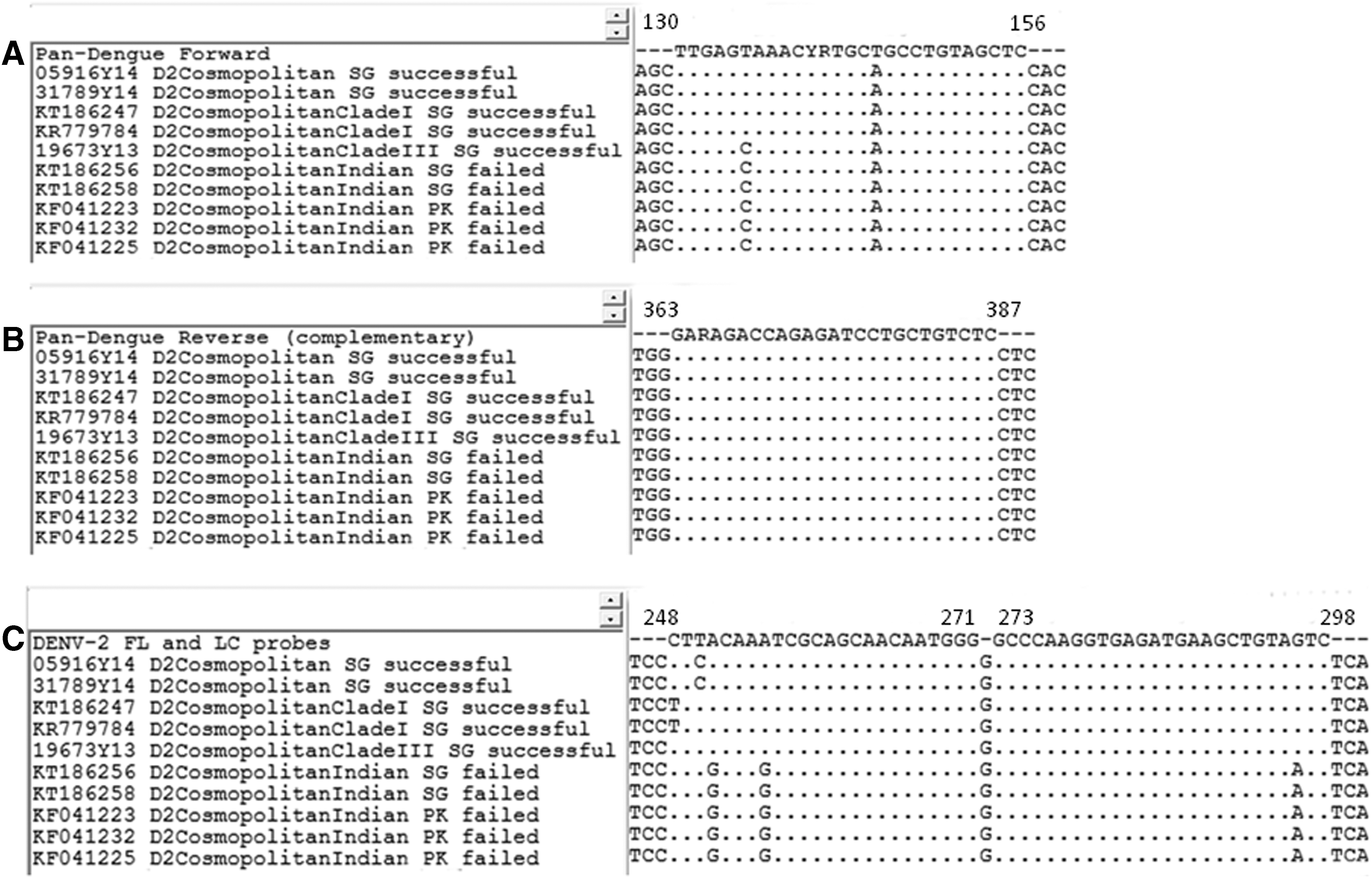
Alignment of pan-dengue (outer) primer and probe binding regions of 3’ UTR of DENV-2 isolates that were either successfully detected or failed in rRT-PCR assay.
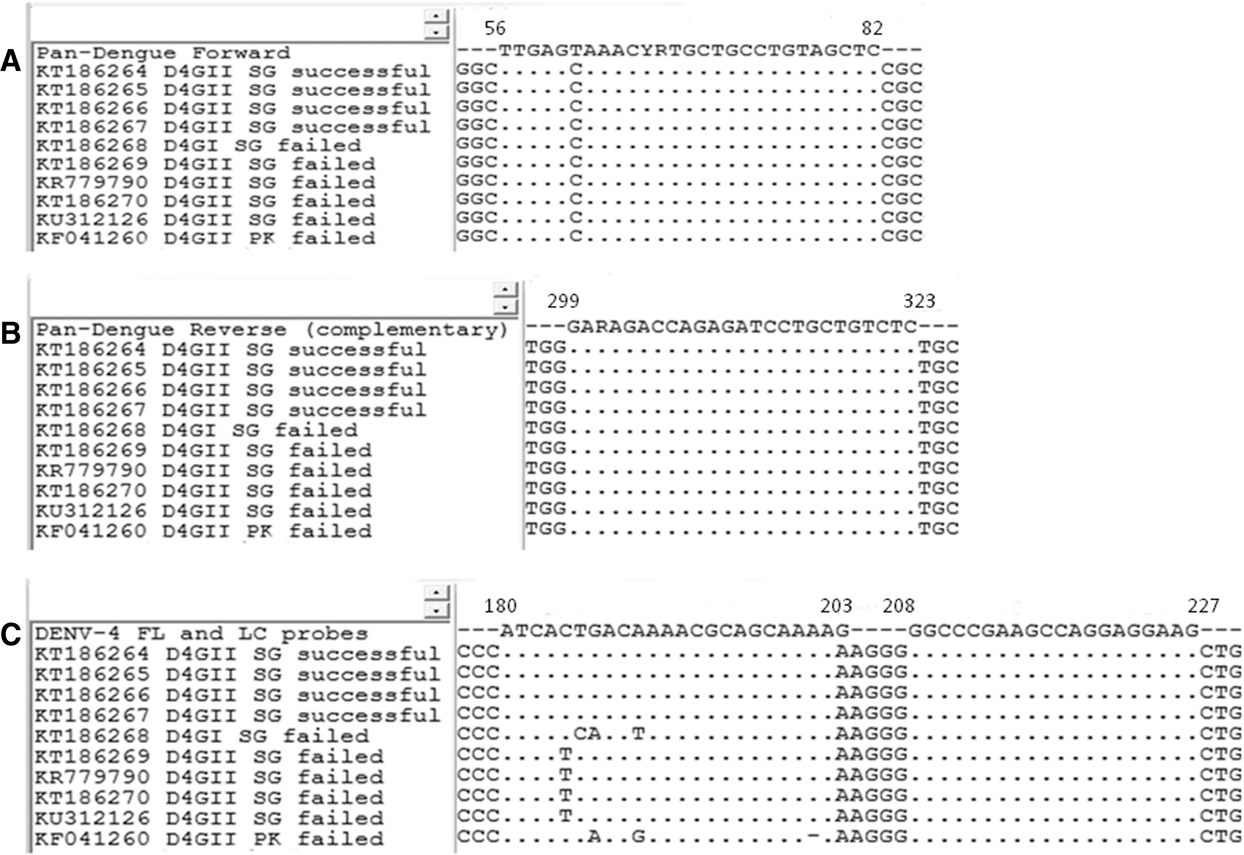
Alignment of pan-dengue (outer) primer and probe binding regions of 3′ UTR of DENV-4 isolates that were either successfully detected or failed in rRT-PCR assay.
Discussion
The rRT-PCR assay used for the detection and typing of DENV in our study sera utilizes hybridization probes in a single-tube format. The assay differentiates DENV serotypes by using a pair of pan-dengue (outer) primers and four pairs of serotype-specific Fluorescence Resonance Energy Transfer (FRET) probes (inner), flanking a 258 bp region of the 3′ UTR of DENV (Lai et al. 2007). In each pair of serotype-specific probes, the upstream probe holds a donor dye (Fluorescein) and the downstream probe carries an acceptor dye (RedLC). Hybridization of serotype-specific probes to template during the annealing step permits the energy transfer from the donor dye to the acceptor dye based on the physically close association of two probes, emitting a fluorescent signal detectable at different wavelengths. Accordingly, the successful annealing of both probes is essential to generate a positive signal. Therefore, given the amount of template that exceeds the detection threshold, one of the possibilities for rRT-PCR assay failure is inefficient template annealing of either outer pan-dengue primers or inner probes.
The successful amplification of viral RNA from failed and successful sera by using pan-dengue primers in a SYBR Green-based assay indicated that observed mismatches in outer primers did not have a substantially negative impact on template annealing. This observation indicated that probe mismatches were the likely cause of false negative results generated by the rRT-PCR assay. In general, virus strains with single mismatches in the probe sequences were successfully detected, suggesting that probe annealing could tolerate up to one mismatch in their binding regions. In the presence of more than one mismatch, failures of detection appeared to depend on the type of mismatch. For example, in all DENV-2 samples that failed detection, there were two mismatches at the 5′ end of the upstream probe (Fig. 1). Both mismatches were adenine to guanine (A-G) substitutions. On the other hand, two mismatches involving cytosine to thymine (C-T) and cytosine to guanine (C-G) in one of the DENV-2 samples did not result in detection failure. Similarly, 13 DENV-1 genotype III samples that were successfully detected, despite having two substitutions, possessed C-T mismatches (Table 3). Previous evidence has shown that A-G mismatches tend to have a more detrimental effect on RT-PCR amplification success than C-T mismatches (Klungthong et al. 2010, Stadhouders et al. 2010).
This report demonstrates that even subtle differences between probe and template sequences can lead to false negative results in PCR-based dengue diagnostics assays. A similar observation has previously been made among DENV-1 strains in Cambodia (Reynes et al. 2003) that emphasized the need to continuously improve and validate PCR-based assays to overcome potential detection failures due to genetic variability of virus strains (Reynes et al. 2003, Chien et al. 2006). In practice, this is especially applicable to fast evolving RNA virus populations such as DENV, in which high mutation rates tend to increase the possibility of introducing mismatches into probe/primer binding regions. Our observations testify the importance of utilizing a combination of methods, instead of relying on molecular methods alone for dengue diagnostics and surveillance, to minimize the underestimation of true disease burden and improper clinical management. For instance, the laboratory-based dengue surveillance program in Singapore utilizes the detection of NS1 antigen as the first-line diagnostic assay. All NS1-positive sera are subjected to typing of DENV serotypes to assess the potential risk of outbreaks (Lee et al. 2010). Both rRT-PCR and conventional RT-PCR assays described in this study are being used at EHI diagnostics for serotyping purposes; rRT-PCR assay for the rapid detection of DENV serotypes and the conventional RT-PCR assay to retest NS1 positive, but rRT-PCR-negative sera.
Another important observation is the requirement of a thorough performance evaluation of molecular assays in local settings before such assays are used for disease diagnosis and surveillance. For example, our findings showed that the performance of rRT-PCR assay differed among virus lineages and genotypes. The assay failed to detect the Indian subcontinent lineage of cosmopolitan genotype, which is dominant among Pakistan DENV-2 strains (Koo et al. 2013), but is relatively uncommon in Singapore (Lee et al. 2012). On the other hand, the dominant DENV-2 strains in Singapore, clades I, II, and III of cosmopolitan genotype (Lee et al. 2012) were successfully detected by the assay. The sequence variations in probe/primer binding regions of dominant DENV-2 populations in Pakistan and Singapore explained the difference in assay performance between two cohorts. This study, therefore, provides a real-life example of molecular assay failure, which was captured as a result of utilizing a “combination assay” approach in routine DENV surveillance. Our findings emphasize that a thorough understanding of the genetic composition of local DENV populations as well as regular monitoring of the performance and reviewing of probe/primer sequences are essential to maintain a consistently high diagnostic accuracy of PCR-based assays.
Availability of supporting data
DENV-2 and DENV-4 3′ UTR sequences generated during this study were deposited in GenBank database [GenBank: GU370051, KF041218, KF041221-223, KF041225, KF041231-235, KF041237, KF041260, KJ806774, KJ806799-800, KP685236, KR779782, KR779784-785, KR779790, KT186247-271].
Footnotes
Acknowledgments
The study was funded by Aga Khan University Research Council Grant and National Environment Agency, Singapore. The funding sources of this study had no role in the study design, data collection, data analysis, data interpretation, writing of the report, or in the decision to submit the article for publication.
Author Disclosure Statement
No competing financial interests exist.