
Abstract
Fascioliasis causes significant economic losses to the cattle industry and is considered a reemerging zoonosis. In Caldas-Colombia, an increase of bovine fascioliasis was detected at the Manizales Municipal Slaughterhouse, which is a potential risk to public health. The ecoepidemiology of human fascioliasis was analyzed in a region of bovine fascioliasis in Caldas-Colombia. The risk factors were studied. Samples were taken from 111 people who were directly related to the bovine milk production process. The immunoglobulin G frequency of Fasciola hepatica was determined in serum. A seriate stool test and a molecular analysis were conducted on those with positive results to look for parasite eggs and DNA, respectively. 6.3% of the samples were positive for the presence of antibodies; none was positive for the presence of eggs, while two samples showed a weak amplification band of the 124-bp DNA fragment of F. hepatica. Fifty-seven percent of the positive samples came from places located at 2026 meters above sea level (masl); 71% of people testing positive had been recently dewormed. Also, 86% had been in contact with cattle and handled grass and excrement. They eat salads and drink untreated water from the springs or ravines of the area. An outbreak of human fascioliasis was detected in Caldas, associated with risk factors for the disease. Clinical trials to confirm the presence of the parasite and implement public health control measures are required.
Introduction
F
The increased number of cases have led scientists to consider fascioliasis a health problem worldwide. It is hard to diagnose in the acute or invasive stage. The stool test, the enzyme-linked immunosorbent assay (ELISA) to detect antibodies, the angiography, and the retrograde cholangiopancreatography are methods to diagnose this disease. The sensitivity and specificity of these methods are appropriate to identify the parasite or, at least, the immune response to it. Hepatic surgical procedures may incidentally find the parasite or confirm clinical suspicions (Senates et al. 2014). In 2007, the World Health Organization (WHO) confirmed that there are molecular tests used for research, but they have not been validated yet to use to diagnose the disease.
A bovine fascioliasis outbreak was detected in Caldas through the confiscation of bovine livers from the Manizales Municipal Slaughterhouse and by a study of bovine fascioliasis in the region, with prevalences of 12.3% by coprologic analysis, 19.1% by ELISA test, and 67.2% by PCR (unpublished data). This study presents the results of the assessment of human fascioliasis prevalence in this outbreak by searching for eggs, specific immunoglobulin G (IgG), and parasite DNA through conventional PCR.
Materials and Methods
Areas with bovine milk production systems were selected; they were located at 2026 and 3721 masl, in the midsouth region of the Caldas Department that had previous records of fascioliasis in bovine cattle (Giraldo et al. 2011). Twenty-six livestock facilities were previously chosen to be georeferenced and geographically divided into four areas with similar environmental characteristics. In addition, 111 blood samples without anticoagulant were obtained from people who worked in the bovine milk production process. They agreed to participate in the study by signing an informed consent that was approved by the medical ethics committee of the Faculty of Health Sciences of Universidad de Caldas. The samples were processed at the Microbiology Laboratory of Universidad de Caldas-Colombia.
Surveys
Only essential information was gathered. Every participant of the study took the survey, which considered several demographic variables, such as age, gender, actual residence, previous places of residence, actual and past diseases, and water supply sources. Information regarding the handling of animals, use of deworming products, and eating habits were also obtained. The design of this survey was based on the risk factors associated with fascioliasis (Esteban et al. 2002).
Immunological analysis
An indirect ELISA technique (BIOX Diagnostics) to detect bovine F. hepatica was used to determine the IgG, and this technique was standardized for humans with positive sera donated by Dr. Pedro Luis Ortiz Oblitas, from Universidad Nacional de Cajamarca in Peru. An anti-human IgG conjugated to peroxidase (Sigma-Aldrich) was used. The results were interpreted according to the instructions from the manufacturers of the test. Prevalence was calculated based on the number of people with IgG, considering the total individuals who were assessed.
Stool analysis
People who tested positive by immunological test underwent a seriate stool procedure using the modified sedimentation technique of Dennis (López et al. 2008). To identify eggs from F. hepatica, the parameters described by Boray et al. (1983) and Valero et al. (1999) were considered. The samples were positive when containing at least one F. hepatica egg.
Molecular analysis
The DNA was extracted from the stool samples using the fecal kit Ultraclean (MO BIO Laboratories), and the concentration and purity were determined by absorbance readings at 230, 260, and 280 nm.
A repetitive genome sequence of 124 pb miracidium was amplified, present in about 15% of the F. hepatica genome. The amplification was carried out by conventional PCR using the primers Fsh1 (5′-GAT-CAA-TTC-ACC-CAT-TTC-CGT-TAG-TCC-TAC-3) and Fsh2 (5′-AAA-CTG-GGC-TTA-AAC-GGC-GTC-CTA-CGG-GCA-3′) (Kaplan et al. 1995, Caron et al. 2010, 2014). These primers were compared with human, animal, plant, and different human parasite genes using the basic local alignment search tool, available at
The Kapa High Fidelity HotStart PCR kit (Kapa Biosystems) was used during the amplification process. The following amplification protocol was followed: Initial denaturation at 95°C for 3 min, 35 cycles of denaturation at 98°C for 20 s, annealing at 53°C for 15 s, extension at 72°C for 15 s, and a final extension at 72°C for 1 min. These reactions were done in a DNA Engine BIO-RAD PTC 200 machine. The amplified product was examined by electrophoresis with 2% agarose gels and ethidium bromide as dye.
Statistical analysis
The SPSS 18, Inc., software was used. The chi-squared test was used to determine the degree of association with risk factors. A value of p < 0.5 was considered significant. The odds ratio and confidence intervals of 95% were applied to determine the association between seropositivity and risk factors.
Results
Twenty (18%) females and 91 (82%) males between 15 and 71 years agreed to participate in this study. In addition, 25% of them had lived on the same plot of land for more than 7 years. Of the 111 serums that were analyzed by the immunologic test, 7 (6.3%) were positive for IgG against F. hepatica (Fig. 1), 5 (2 females and 3 males) with ages ranging between 26 and 35 years. All positive samples were tested again to confirm the results. No statistical significance was found concerning gender. Three stool samples were obtained from each person with Fasciola antibodies; no eggs were found. Two samples with 124-bp fragment (Fig. 2) have been achieved in the PCR.
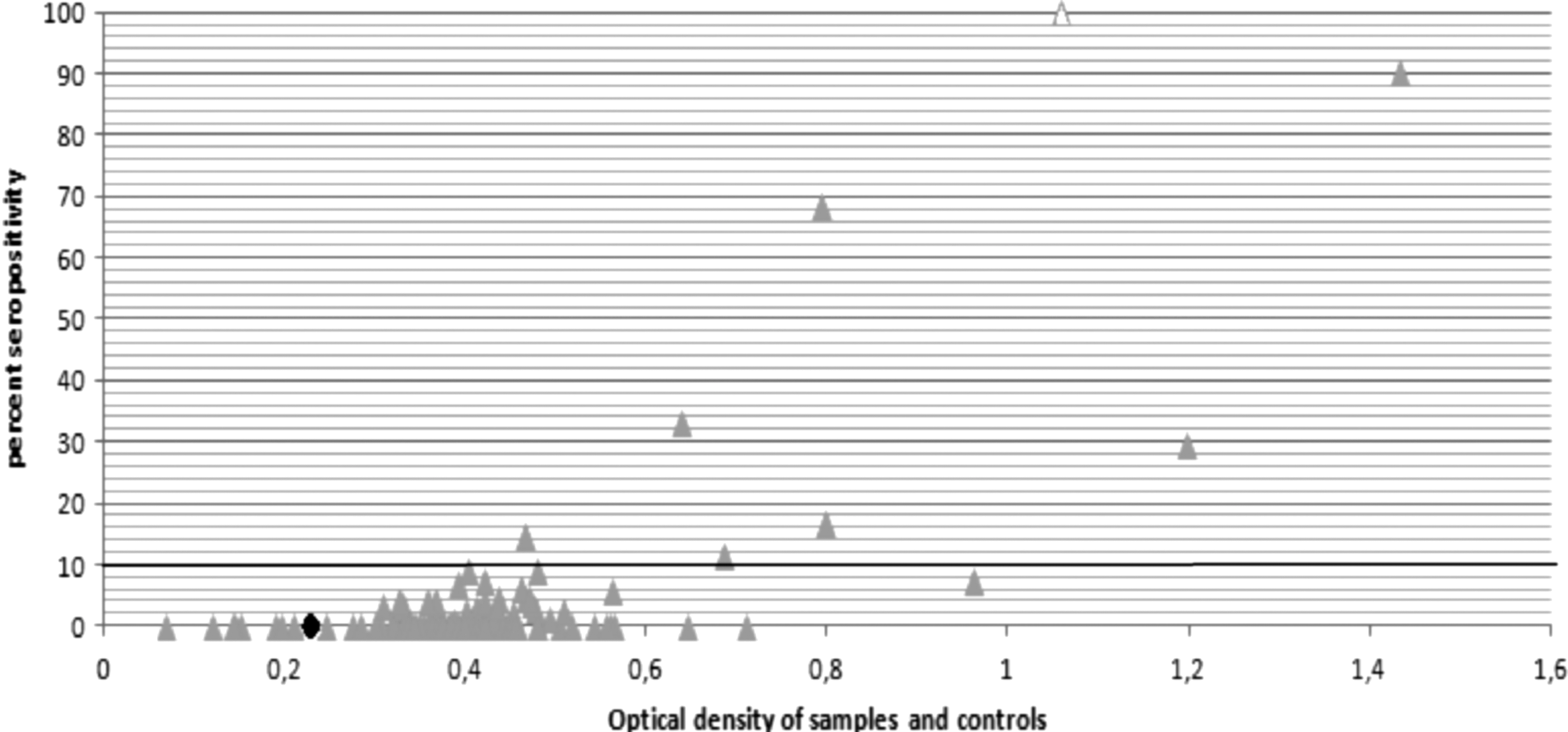
Distribution of samples and controls according to optical density and percent of seropositivity. The black line shows the cutoff from which the samples were positive, obtained according to manufacturer's test. ♦: Negative control. ▵: Positive control.

Electrophoresis of amplification products of 124-bp fragment of Fasciola hepatica. F2–F10, human samples with and without antibodies against Fasciola; note the faint bands of columns F8 and F9. PM, molecular weight ladder (Hipperladder 25 bp) ranging from 25 to 500 bp. CP, positive bovine sample for F. hepatica from the stool test and PCR. CN, negative bovine sample for F. hepatica and PCR.
Eighty-six percent of people lived at a height between 2300 and 3200 masl. Four (57%) had antibodies against F. hepatica; however, no statistically significant differences were found. Most people who tested positive (6/7) came from the municipality of Villamaría (Caldas), where a previous study shows a frequency of bovine fascioliasis of 19.1% by ELISA test (unpublished data).
Six of those with positive results had a habit of eating salads. Only two persons mentioned that they also ate watercress and other aquatic plants. Fifty-seven percent of them defecate in the open field. Other risk factors, such as handling grass and excrement, contact with cattle, and eating pieces of grass, were engaged in by 86% of the people with positive results. No statistical significance was found when analyzing risk factors.
The farms where the people work have flood zones, and therefore use water channels to drain water, enhancing the presence of snails, which thrive in areas with low water flow and high oxygenation. In the study, all people with positive results drink untreated water from springs and ravines that may be contaminated with these sources of water.
One of the most commonly referenced symptoms was malaise and fever; 71% of those with positive samples presented both. Only two of those with positive samples showed several of the following manifestations of fascioliasis: upper right side pain (right hypochondrium), jaundice, white feces, and dark urine. No statistically significant differences were found when relating the symptoms to fascioliasis.
Some of the people who were surveyed frequently used deworming products; albendazole and metronidazole were the most common, and 71% of those that tested positive used such medications recently.
Discussion
The presence of hosts, such as bovines, Lymnaea columella, and Nasturtium officinale in the north of South America cause outbreaks of fascioliasis in animals and people who coexist in this region. The areas of this study are similar to the Bolivian Plateau, where the prevalence of the parasite in humans has been reported in more than 60% of the population (Marcos et al. 2005). The Cajamarca and Mantaro Valleys, which are hypoendemic and hyperendemic, respectively (Ortiz et al. 2000), are the same. The seropositivity of this study was below that of these reports.
The frequency of the disease based on gender shows that the females were more affected (10%) than males (5.5%). This result is similar to that obtained by Amer et al. (2011) in the Nile delta, which may be related to the consumption of untreated water or raw vegetables included in the diet of the families and workers of these parcels.
The clinical manifestations of fascioliasis in humans are unspecific. In nonendemic areas such as ours, this disease is not considered part of the differential diagnosis. Several studies have proven that the most common method to diagnose fascioliasis before the symptoms appear is the ELISA test, in which sensitivity and specificity levels are good (Salimi-Bejestani 2005, Marcos et al. 2007, Espinoza et al. 2010, Valero et al. 2011).
Another factor to consider is that the frequent and recent use of deworming products in individuals with positive results could effectively remove the parasite, but the immunological trace remains, which could stay for years. This finding is a possible explanation for the negative results shown by the stool tests. Other authors also observed this phenomenon (Salimi-Bejestani et al. 2005, Valero et al. 2011).
Other factors associated with the absence of eggs are highly infective forms, which increase the time a parasite needs to reach sexual maturity and delay the appearance of eggs in feces (Espino 1997). Due to the uterine size of the trematode, the oviposition period may take place 250 days after the infection (Valero et al. 2006), and the degree of infection may be small and sporadic with few adult individuals, which means low oviposition. However, the release of antigens induces the production of antibodies before oviposition (Salimi-Bejestani et al. 2005).
Open-air water irrigation channels to supply homes in Peru are considered the primary source of infection in endemic areas, and this is thought to be a risk factor for human fascioliasis, which is worse when there are some parts where the water flow is slower (Marcos et al. 2007). The area studied had a water circulation system that uses pipes or hoses that avoid mollusk development; however, these sources of water may contain vectors of the parasite that release cercariae and therefore contaminate these sources, thus risking the health of the people that drink it directly.
The weak bands in electrophoresis gels of PCR products confirm the presence of F. hepatica eggs and suggest the possibility of contamination from water sources by defecating near the ground and the absence of sewage treatment facilities, which enhance the biological cycle of the trematode (Mas-Coma 2011, Monteiro et al. 2013).
In Colombia, the species of molluscan intermediate hosts associated with F. hepatica are Galba truncatula (Müller 1774), Lymnaea columella (Say 1817), L. diaphana (King 1830), L. viatrix (Orbigny 1835), L. cubensis (Pfeiffer 1839, Cruz-Mendoza et al. 2011), L. bogotensis (Pilsbry 1935), and Pseudosuccinea columella (Say 1817, Velásquez 2006).
Lymnaea columella and Succinea sp. were found in the region studied. They are located in the vegetation present and migrate during the day, particularly in the hours in which sunlight is weaker. These findings coincide with the statements of Prepelitchi (2011), who mentioned that the frequency of Lymnaea in plants depends on the humidity level of the environment. In this case, the daily migration could be caused by the effect of solar radiation that produces desiccation. In addition, at higher heights above sea level, the evapotranspiration processes are accelerated due to the low atmospheric pressure and high solar radiation. Succinea sp. has been reported in Mexico where fascioliasis has a 16% prevalence in humans (Cruz-Mendoza et al. 2011). This frequency is higher than that observed in this study.
Based on the results, we conclude that the municipality of Villamaría-Caldas-Colombia has an outbreak of human fascioliasis, which agrees with a high prevalence of bovine fascioliasis, which affects people who are exposed to risk factors associated with the infection. More studies, particularly clinical studies, are needed to confirm the presence of the parasite in this group of inhabitants.
Footnotes
Acknowledgment
The authors thank the Universidad de Caldas for financing this research.
Author Disclosure Statement
No competing financial interests exist.