
Abstract
Bacteria of the genus Bartonella have been described in multiple mammalian hosts with many species capable of causing disease in humans. Cats and various species of rats have been reported to play a role as vertebrate hosts to a number of Bartonella spp. This study aimed to identify Bartonella spp. in Western Australia, Dirk Hartog Island (DHI), and Christmas Island (CI) and to investigate the presence of potential arthropod vectors. Feral cats were collected from CI (n = 35), DHI (n = 23) and southwest Western Australia (swWA; n = 58), and black rats were collected from CI (n = 48). Individuals were necropsied, ectoparasites were collected by external examination of carcasses, and splenic tissue was collected for polymerase chain reaction analysis to detect Bartonella DNA. Bartonella henselae DNA was detected from two cats and Bartonella koehlerae DNA from one cat in southwest WA, but Bartonella DNA was not identified in cats on DHI or CI. Bartonella phoceensis (28/48 = 58.3%) and a novel Bartonella genotype (8/48 = 16.7%) based on the internal transcribed space region were detected in the spleens of black rats on CI. Detection of Bartonella spp. in each location corresponded to the presence of ectoparasites. Cats from southwest WA harbored four species of fleas, including Ctenocephalides felis, and black rats on CI were infested with multiple species of ectoparasites, including mites, fleas, and lice. Conversely, cats on Dirk Hartog and CI were free of ectoparasites. This study has identified the DNA of Bartonella species from island and mainland swWA with some (B. henselae and B. koehlerae) of known zoonotic importance. This study further extends the geographical range for the pathogenic B. koehlerae. The association of Bartonella with ectoparasites is unsurprising, but little is known about the specific vector competence of the ectoparasites identified in this study.
Introduction
B
Bartonella infection has been described in a large number of mammalian species, including livestock, domestic pets, and wildlife (Saisongkorh et al. 2009). However, the geographical distribution and epidemiology of many Bartonella species, including the vectors involved in their transmission, are not fully understood. The infection and replication of Bartonella within arthropod vectors have only been shown to definitively occur in three species, B. quintana (sand flies), B. henselae (fleas), and B. schoenbuchensis (lice) (Chomel et al. 2009), although epidemiological research supports the role of ticks and lice as competent vectors (Billeter et al. 2008). Reported prevalence rates of Bartonella in cats (Felis catus) vary markedly between populations and geographical locations, presumably related to rate of infestation with vectors such as Ctenocephalides felis (Assarasakorn et al. 2012, Guptill 2012).
In Australia, Bartonella species have been isolated from a wide range of mammalian species, including domestic cats (B. henselae) (Flexman et al. 1995) and a number of rodent and murine species (B. coopersplainsensis, B. rattiaustraliensis, and B. queenslandensis) (Saisongkorh et al. 2009). Newly described species of Bartonella have also been identified in native mammals in southwest Western Australia (swWA) (Kaewmongkol et al. 2011a, 2011b, 2011c, 2011d).
Domestic cats represent an important reservoir in the life cycle of at least three Bartonella species (B. henselae, B. clarridgeiae, and B. koehlerae), and feral and stray cats are more likely to be bacteremic than domesticated individuals (Chomel et al. 2006). Importantly, at least four Bartonella species isolated from cats (B. henselae, B. clarridgeiae, B. koehlerae, and B. quintana) and seven species from rodents (B. grahamii, B. elizabethae, B. vinsonii subsp. arupensis, B. volans, B. tribocorum, B. rattimassiliensis, and B. washoensis) are confirmed or suspected human pathogens (Ellis et al. 1999, Castle et al. 2004, Saisongkorh et al. 2009, Chomel and Kasten 2010, Kosoy 2010). It has been hypothesized that all Bartonella species have the potential to cause human Bartonellosis (Gil et al. 2010, Lin et al. 2010).
This study aimed to identify the presence of Bartonella species in Western Australia and two offshore islands with close wildlife/human interface and/or high conservation priorities, Christmas Island (CI) and Dirk Hartog Island (DHI), and to investigate the potential importance of feral cats and black rats (Rattus rattus) as reservoir hosts for Bartonella species.
Materials and Methods
Study locations
Samples were collected from three geographically and climatically distinct locations; mainland swWA, CI, and DHI (Fig. 1). The swWA is a large ecoregion, located south of a line from Geraldton (28°46′28″S 114°36′32″E) to Esperance (33°51′40″S 121°33′31″E). Samples were collected from 12 different locations within swWA encompassing urban land use, agricultural farmland, Mallee and Jarrah forest, and plain lands. This region has a Mediterranean climate characterized by hot dry summers and cool wet winters.
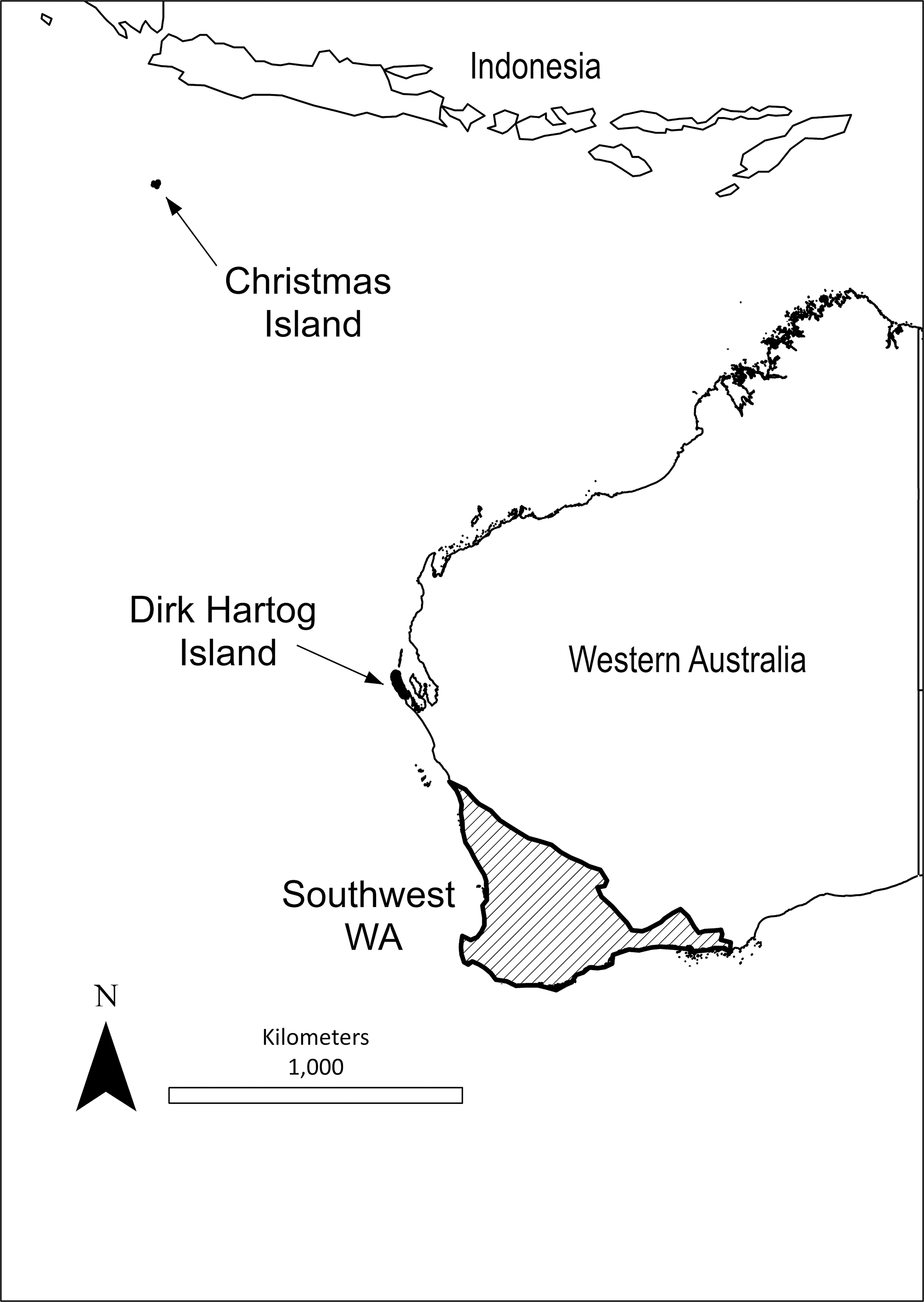
Map showing the geographical distribution of the three study sites sampled in this study; Christmas Island, Dirk Hartog Island, and southwest Western Australia.
CI is an Australian territory, located in the Indian Ocean (10°29′S, 105°38′E) ∼360 km south of the Indonesian capital, Jakarta. The island has an equatorial climate with distinct wet and dry seasons, and year-round high humidity. A large proportion of the island is dense tropical rainforest National Park (∼70%) and mining lease area (∼20%) with the remainder being settled land.
DHI (25°50′S 113°05′E) is an inshore island located to the west of Shark Bay. The vegetation is sparse with low open shrub land and sand dunes. The island experiences a semiarid climate region. This island was previously under pastoral lease, but has now been returned to National Park status with an emphasis on ecotourism.
Sample collection and measurements
Cat cadavers (n = 116) were collected from CI (n = 35), DHI (n = 23), and swWA (n = 58). Cats from CI and DHI were sourced from Department of Parks and Wildlife management programs, and cats from swWA were obtained during community-coordinated culling programs from 12 locations. Rats (n = 48) were collected from CI concurrently with the cats. Weight was recorded for all carcasses and used to determine age category for cats (kitten ≤1.5 kg and adult cat >1.5 kg) according to the method previously described by Algar et al. (2003). It was not possible to obtain accurate age estimates or determine age category for rats.
All cadavers were placed straight away into individually sealed plastic body bags then stored at −20°C. Spleen tissue was collected at necropsy and preserved in 70% ethanol.
Ectoparasite identification
An external examination was conducted on all carcasses for the presence of ectoparasites before necropsy, and the body bag was closely examined for ectoparasites that may have fallen off the carcass. Ectoparasites were identified to genus and species using morphological techniques and keys (Fritz and Pratt 1947, Voss 1966, Roberts 1970).
DNA extraction
DNA was extracted from rat and cat spleens using Qiagen spin columns, tissue procedure, according to the manufacturer's instructions (Qiagen). Negative controls were used in the polymerase chain reactions (PCRs) with the inclusion of PCR water instead of genomic DNA.
PCR conditions
Primers specific to the 16S-23S internal transcribed space (ITS) of Bartonella species were used to screen samples. Primers used were from the 16S-23S ITS region, 438s (5′-GGT TTT CCG GTT TAT CCC GGA GGG C-3′) and 1100as (5′-GAA CCG ACG ACC CCC TGC TTG CAA AGC A-3′) from Beard et al. (2011). PCRs were performed in an optimized 25 μL reaction volume containing 1× PCR buffer (Fisher Biotech), 2.0 mM MgCl2, 0.2 mM dNTPs, 0.02 U/μL Taq polymerase (Fisher Biotech), 1 μL template DNA, 6 μL of cresol red, and 1 μM of each primer. The reactions were run under the following conditions: one denaturing cycle at 95°C for 2 min, followed by 55 cycles at 94°C for 15 s, 66°C for 15 s, and 72°C for 18 s, before a final extension cycle at 72°C for 30 s. Amplified DNA fragments were visualized on a 1.5% agarose gel by electrophoresis.
DNA purification and sequencing
The tip elution method was used for extracting and purifying PCR products (Yang et al. 2013). Positive bands were sliced from the gel and the fragment placed in a 100-μL filter tip (with the tip cut off) within a 1.5-mL Eppendorf tube. This tube was then spun at 20,000 g for 5 min. The filter tip was discarded and the eluent retained for sequencing.
The purified DNA was sequenced using an ABI prism Terminator Cycle Sequencing kit (Applied Biosystems) according to the manufacturer's instructions on an Applied Biosystems 3730 DNA Analyzer. Sequencing results were compared against available sequences in GenBank using BLAST search. Multiple sequence alignments were constructed using additional isolates from GenBank. Distance trees were constructed using MEGA version 6 (Tamura et al. 2013). Genetic distances were calculated in MEGA using the Kimura 2 parameter model.
Statistical analyses
Prevalence was expressed as proportion (%) of animals infested (ectoparasites) or positive for Bartonella (PCR). Ectoparasite overall prevalence was expressed as proportion (%) of animals positive for at least one ectoparasite. The 95% confidence intervals for overall prevalence were calculated using Jeffrey's method (Brown et al. 2001).
Statistical analyses were performed using the software IBM SPSS Statistics, version 21 (IBM). Relationships between categorical host factors (gender, ectoparasite recovery, lice recovery, mite recovery, flea recovery, and tick recovery) and the presence of rat Bartonella species were analyzed using Pearson's chi-square and Fisher's exact tests. Statistical analyses were not performed for prevalence of Bartonella and host factors in cats due to a low prevalence.
Results
Ectoparasites
The details of the ectoparasites recovered from rats and cats are shown in Table 1. Black rats were infested with zero to three ectoparasite species with 85% of rats infested with at least one species. Of the 41 rats infested with ectoparasites, 18 were parasitized by one species, 19 by two species, and 4 by three species. The cats from swWA were parasitized with up to two species of ectoparasites per host with 55% of cats infested with at least one species. Of the 32 cats infested with ectoparasites, 26 were infested with one ectoparasite species and 6 with two. No ectoparasites were found on cats from CI and only lice were recovered from a single cat on DHI (Table 1).
swWA, southwest Western Australia; DHI, Dirk Hartog Island; CI, Christmas Island.
Bartonella species and prevalence
The prevalence of Bartonella in rat and cat spleen samples is shown in Table 2. Bartonella DNA was identified in spleen tissue from 36 rats from CI and 2 kittens (B. henselae and B. koehlerae) and 1 adult (B. henselae) from swWA (Table 2).
Overall, four species of Bartonella were identified by sequencing from two sites (CI and swWA), specifically B. henselae, B. koehlerae, B. phoceensis, and an unknown Bartonella species (Table 2). Blast results showed a 99% similarity with B. phoceensis, 99% similarity with B. koehlerae, and 100% similarity with B. henselae. The unknown Bartonella species A had 91% similarity to Bartonella species SE-Bart-D previously reported by Loftis et al. (2006) from a Rattus norvegicus flea (Xenopsylla cheopis) in Egypt. The sequences identified in the present study have been deposited in GenBank under the following accession numbers: KU170606 and KU240393-KU240430. These sequences were then constructed into a dendogram along with sequences of known rat and cat Bartonella species obtained from GenBank (Fig. 2). Of the 28 rats that B. phoceensis was detected in, only 6 were used in the phylogeny reconstruction.

Phylogenetic relationship of Bartonella species detected in this study inferred by distance analysis of 16s-23s internal transcribed space sequences (indicated in bold). Percentage support (>50%) from 1000 pseudoreplicates from neighbor-joining analyses using bootstrapping is indicated at the left of the supported node.
No association of Bartonella infection with host gender or recovery of ectoparasites was identified in rats.
Discussion
This study has provided new information about Bartonella infections in Australia. In general, members of the genus Bartonella are recognized as important emerging pathogens on a global scale. However, the occurrence and distribution of Bartonella species in rats, in particular, have not been well studied in Australia, and there are no data pertaining to these bacteria in feral species in island communities off the coast of Australia (Branley et al. 1996, Dillon et al. 2002, Fournier et al. 2002, Barrs et al. 2010).
Both species of Bartonella identified in cats from swWA in this study (B. henselae and B. koehlerae) have been reported previously in cats overseas (Branley et al. 1996, Droz et al. 1999, Maruyama et al. 2001, Fournier et al. 2002). B. henselae has been identified in humans and felids in Australia and is considered the leading cause of cat scratch disease and zoonotic Bartonellosis worldwide (Branley et al. 1996, Dillon et al. 2002, Fournier et al. 2002, Saisongkorh et al. 2009, Barrs et al. 2010, Kaewmongkol et al. 2011b). Identification of B. koehlerae was unexpected and is the first report of this species from a Southern Hemisphere country. B. koehlerae has been identified in small numbers of cats (seven in total) from California, France, Israel, and Thailand (Boulouis et al. 2005, Assarasakorn et al. 2012, Fleischman et al. 2015), from rodent fleas in Afghanistan (Marié et al. 2006), feral pigs in North Carolina (Beard et al. 2011), and from a dog in Israel (Ohad et al. 2010). The identification of B. koehlerae in a cat from swWA therefore expands the known geographical distribution of this zoonotic pathogen. The epidemiology of B. koehlerae, including potential reservoirs and mode of transmission, is not well described. Of note is the wide diversity of symptoms that have been attributed to B. koehlerae in humans, including fatigue, insomnia, memory loss, decreased tactile sensation, hallucinations, and endocarditis (Breitschwerdt et al. 2010).
Interestingly, Bartonella species were identified in rats, but not cats, from CI. Rodents are recognized as reservoirs for zoonotic species (Saisongkorh et al. 2009, Tsai et al. 2010) and the close relationship between rodents and humans globally highlights the importance of rats as a source of zoonotic infection (Castle et al. 2004). Bartonella, including B. phoceensis, have been previously reported in rodents in Asia, including Thailand and Indonesia (Billeter et al. 2008, Tsai et al. 2010). Given the close geographical proximity of CI to southeast Asia, the finding of B. phoceensis in rats may be explained by rats arriving at the island over the years on ships from nearby Indonesia. Since B. phoceensis has been identified in multiple rodent species, this highlights a potential risk for its spread into Australian rodent populations if it was to be introduced onto mainland Australia. Furthermore, identification of a potentially novel Bartonella species (unknown Bartonella spp. A) in rats from CI adds to the diversity of Bartonella species currently described. To determine if this isolate represents a novel species, further molecular phylogenetic study would need to be conducted using more than one gene locus (La Scola et al. 2003, Lin et al. 2010).
Bartonellosis is a vector-borne disease (Gundi et al. 2004) and is commonly reported from tropical environments (Chomel et al. 2006) like CI. The distribution of suitable arthropod vectors most likely explains the Bartonella prevalence observed in this study. Our detection of Bartonella species in rats, but not cats, on CI was supported by our observation that cats on the island did not appear to have any ectoparasites. The reason for this is unclear—the cats were placed into plastic bags that were sealed immediately after euthanasia. This procedure is expected to trap any ectoparasites for later identification, as was the case on DHI and in swWA. Lice infestations were identified in rats from CI, including Hoplopleura pacifica, an ectoparasite for which transmission of B. phoceensis has been previously reported in various small mammal species (Reeves et al. 2006, Billeter et al. 2008, Tsai et al. 2010). Dissimilar to the situation on DHI and CI, flea and tick infestations were common in cats from swWA where B. henselae and B. koehlerae were identified at low prevalence. Transmission by the cat flea (C. felis) has previously been reported for B. henselae and it is suspected to be a vector of B. koehlerae (Chomel et al. 2006, 2009).
Despite over 20 years of research, the modes of transmission are still not well understood for many Bartonella species. Fleas appear to play a significant role in the transmission of multiple Bartonella species and ticks have been suggested as competent vectors (Tsai et al. 2011a). While it is possible for cat fleas to carry Bartonella species among cats, the vector competency has only been established for B. henselae (Guptill 2012). One of the challenges in describing the epidemiology of Bartonella with respect to transmission is differentiating the presence of Bartonella spp. DNA due to a previous blood meal rather than implying vector competence in those arthropods. Almost all the ectoparasites identified in this study have previously been associated with Bartonella species, including B. tribocorum, B. elizabethae, B. queenslandensis, B. rochalimae, B. tamiae, B. rattimassiliensis, B. phoceensis, B. henselae, and B. koehlerae (Tsai et al. 2011a). The diverse nature of ectoparasite species recovered from rats and cats in this study suggests that further research into vector competency of these ectoparasites is required.
Conclusion
This study identified Bartonella species in cats from mainland Western Australia and black rats from CI, including those with pathogenic and zoonotic potential. Additionally, we report B. koehlerae in Australia for the first time. The findings suggest that rodents as well as cats can be mammalian reservoirs for these vector-borne infections and highlight the need for preventive measures with regard to public health and conservation management.
Footnotes
Acknowledgments
The authors would like to thank Dr. Gunn Kaewmongkol and Louise Pallant for advice on molecular analysis and Dr. Kaewmongkol for providing positive controls. The authors would also like to thank the Murdoch University Veterinary Trust and Weston Fernie award for providing funds to support this research.
Author Disclosure Statement
No competing financial interests exist.