
Abstract
The article describes the isolation of a cowpox virus (CPXV) isolate originating from a horse. The skin of a foal, aborted in the third trimester, displayed numerous cutaneous papules. The histological examination showed A-type inclusion bodies within the lesion, typical for CPXV infections. This suspicion was confirmed by real-time PCR where various organs were analyzed. From skin samples, virus isolation was successfully performed. Afterwards, the whole genome of this new isolate “CPXV Amadeus” was sequenced by next-generation technology. Phylogenetic analysis clearly showed that “CPXV Amadeus” belongs to the “CPXV-like 1” clade. To our opinion, the study provides important additional information on rare accidental CPXV infections. From the natural hosts, the voles, species such as rats, cats, or different zoo animals are occasionally infected, but until now only two horse cases are described. In addition, there are new insights toward congenital CPXV infections.
T
There are only two reported cases in horses. One case involved a 7-year-old CPXV-infected-Arabian horse in Zwickau (Germany) with typical signs including pox lesions distributed throughout the body surface (Pfeffer et al. 1999). There was a second CPXV case documented in Germany in 2001. The animal was a premature foal with weakness, low body temperature, and gasping respiration. However, the animal did not display any typical pox lesions (Ellenberger et al. 2005). In addition to these CPXV cases, there was also one Horsepox virus (HPXV) case documented in Mongolian horses in 1976 (Tulman et al. 2006).
In this case report, we describe a CPXV infection of a pregnant warm blood mare that resulted in an abortion. The female foal was aborted within the third trimester (weight 19.5 kg) at the end of February 2015. Macroscopic evaluation revealed multifocal and occasionally coalescing brown-reddish papules with a maximum diameter of 1.2 cm distributed throughout the entire body surface including the mucocutaneous junctions and the oral cavity. There were also multifocal central hemorrhages present, and there were several larger lesions covered by crusts (Fig. 1A).
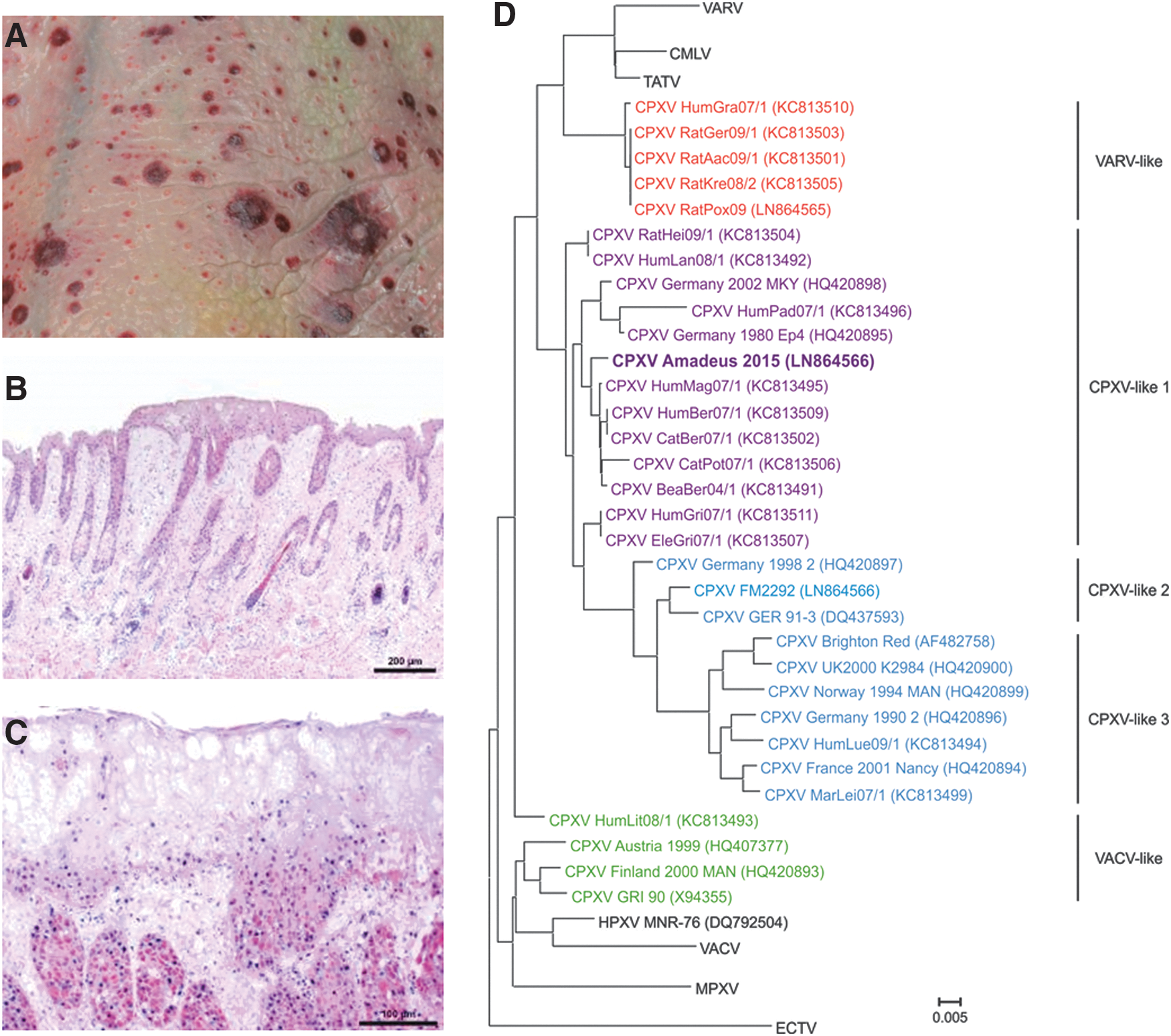
We collected tissues from various organs including the placenta, lung, liver, spleen, thymus, and skin for further pathological, virological, and molecular investigations. The tissue samples for histopathology were fixed in 4% formalin and embedded in paraffin. The tissue sections were cut 5 μm thick, dewaxed, and stained with hematoxylin and eosin following standard protocols. The microscopic examination of the epidermis revealed multifocal thickened regions with swollen or degenerated keratinocytes and multifocal hemorrhages. The basal epidermal layers and follicular epithelia contained numerous cytoplasmic eosinophilic inclusion bodies (Fig. 1B, C). These A-type inclusions or Downie bodies are typical of CPXV infections.
DNA was extracted from all organs and tested by real-time PCR using a protocol that can differentiate between CPXV and other OPV (Maksyutov et al. 2015). The Cq value (Cq = cycle of quantification) is the cycle in which fluorescence increased above background. The Cq values measured were less than 22 in all organs. The low Cq values indicate there is high CPXV genome copy number. The highest viral load was detected in a skin sample, which had a Cq value of 12.
The DNA extracted from skin was analyzed using de novo sequencing of the complete genome by next-generation sequencing with a MiSeq reagent kit v2 and the Illumina MiSeq (Illumina, San Diego, CA). This analysis generated a complete genome consisting of 222,069 bp. A phylogenetic analysis was performed for all full-length genome sequences using IQ-Tree (v. 1.2.2) using a best fit model (TVM+I+G4). The novel isolate was a CPXV clustering within the “CPXV-like clade 1.” The sequence is clearly distinguished from the HPXV isolate MNR-76 (DQ792504), which clustered with Vaccina virus (VACV) (Fig. 1D).
Virus was isolated from skin using the African green monkey liver cell line Vero76 (Collection of Cell Lines in Veterinary Medicine CCLV; Friedrich-Loeffler-Institut, Greifswald-Insel Riems, Germany). The isolate was named “CPXV Amadeus 2015” (LN879483).
Interestingly, the mare did not display any clinical signs during pregnancy. However, there were OPV antibodies detectable in the serum by immunofluorescence assay. The serum was collected 10 weeks after abortion (titer ≥1:500). The epitheliocoreal placenta of horses is impermeable to macromolecules such as immunoglobulins (Furukawa et al. 2014). Therefore, the fetus was not protected by maternal antibodies.
Our findings indicate this is a very rare case of an equine CPXV infection resulting in a spontaneous abortion. There is another report of a foal born 29 days prematurely that showed clinical signs of a CPXV infection. The foal was euthanized on the sixth day of life. The foal did not display any cutaneous pox lesions, and the case was clearly associated with streptococcal septicemia. Therefore, the authors assumed the foal was infected orally after birth (Ellenberger et al. 2005). The first case of a stillbirth caused by CPXV was described in 2001 for an Asian elephant housed in a zoo. The stillborn calf was mature and showed generalized pox lesions. There were no clinical signs observed in the mother or the other elephants of the group (Wisser et al. 2001).
In this case report, we describe for the first time a congenital CPXV infection in a horse that was lethal. Interestingly, lesions of this generalized infection appear in different stages in infected animals (Schmiedeknecht et al. 2010). This result conflicts with the smallpox phenotype observed in humans. Other poxvirus infections such as congenital Ectromelia virus (ECTV) infections are endemic in farms of silver foxes and minks in the Czech Republic. The adult animals did not show clinical signs (Mahnel et al. 1993). In addition, fetal VACV infections have been reported previously, and this rare complication of smallpox vaccination has resulted in fetal or neonatal death (Cono et al. 2003).
Previous studies have demonstrated that voles are the definitive reservoir of CPXV. Thus, it is possible that the pregnant mare grazed or received supplemental feeding that contacted hay or straw contaminated by urine, feces, or exhaled droplets from CPXV-infected voles.
The phylogenetic analysis showed the new isolate grouped with other CPXV strains found in a specific area of Berlin (Dabrowski et al. 2013). Additional information regarding the exact location of infection would allow a direct relationship between these CPXV strains to be determined. It is currently unclear whether CPXV infections in horses are frequently caused by contaminated hay and straw. Furthermore, it is unknown why the infection in this specific case resulted in an abortion with severe clinical signs and typical poxvirus lesions in the fetus. Thus, further studies of CPXV seroprevalence in horses may improve our understanding of CPXV epidemiology in spill-over hosts such as the horse.
Footnotes
Acknowledgments
We thank Doris Junghans and Mareen Lange for their excellent technical assistance. We also thank Silvia Kühnel (Pferdeklinik, Seeburg) for providing the serum of the mare. In addition, we are very thankful to Dr. Angele Breithaupt (Freie Universität Berlin, Berlin) for critical reading and comments. This project was partially funded by the German Research Foundation SPP1596 project BE5187 awarded to M.B.
Author Disclosure Statement
No competing financial interests exist.