
Abstract
Ehrlichia ewingii is the causative agent of human and canine granulocytic ehrlichiosis. Since its discovery in 1970, little work has been done to characterize the pathogen or study the transmission dynamics due to the inability to grow the agent in vitro. The aim of this study was to assess the suitability of multiple cell lines and media formulations for propagation of E. ewingii in cell culture. In this study, we present an overview of attempts to isolate E. ewingii from the buffy coat of goats naturally infected by Amblyomma americanum ticks, as well as a methodology for maintaining the pathogen for up to 16 weeks in culture. The most promising results were seen with HL-60 cells differentiated by the addition of 1.5% DMSO to the media and supplemented with 8 mM
Introduction
E
To date, E. ewingii has never been isolated in cell culture and, consequently, has not been studied as extensively as most other human and canine ehrlichial pathogens. Cell lines composed of the target cells (neutrophils and eosinophils) or derived from the reservoir or vector species are natural candidates for successful in vitro cultivation. HL60, a human promyelocytic cell line, has previously been used to cultivate another granulocyte-infecting pathogen, Anaplasma phagocytophilum (Goodman et al. 1996), but thus far, attempts to culture E. ewingii in this same cell line have been unsuccessful (Anderson et al. 1992). Attempts have also been made using a canine monocytic cell line (DH82) (Anderson et al. 1992) and tick cell lines (ISE6) (Yabsley et al. 2011), both of which have been used to culture other Ehrlichia spp. The majority of these attempts have used small amounts of blood or buffy coat from infected animals, usually dogs. Goats are competent laboratory reservoirs of E. ewingii, readily becoming ill when infested with very few infected ticks, and have the advantage of providing a larger volume of blood than smaller animals (Loftis et al. 2008). Goats are also refractory to infection with E. chaffeensis preventing coinfection concerns when using wild ticks that may contain both pathogens (Loftis et al. 2008). The purpose of this study was to evaluate the suitability of infected goat blood and different cell lines for the propagation of E. ewingii.
Materials and Methods
A list of cell lines and media formulations used in this study is shown in Table 1. Cell lines were chosen based on host reservoir status, the target cell type, or previous use in the culture of other Ehrlichia spp.
See Table 2 for media descriptions.
NS, not suitable.
To obtain infected blood for culture isolation, ∼100 adult A. americanum were fed on each of two 1-year old goats (one each in 2011 and 2013) as previously described for rabbits (Troughton and Levin 2007). Ticks were collected from the vegetation at the Rum Creek Wildlife Management Area (WMA) in Forsyth, GA. This field site had been sampled multiple times from 2009 to 2011 with a mean E. ewingii infection prevalence of 3.9% in adult ticks (Killmaster et al. 2014). The tick-infested goats were monitored for clinical signs of infection, and blood and sera were collected every other day for polymerase chain reaction (PCR) and indirect fluorescent antibody test (IFA). Once goats became febrile and PCR positive for E. ewingii, ∼80 mL of heparinized blood was collected from each infected goat for the following 2 weeks. Blood was centrifuged at 1000 g for 20 min, and the buffy coat was removed and either inoculated immediately into cell culture or frozen in liquid nitrogen. Two methods of cryopreservation were used: fetal bovine serum with 10% DMSO and Sucrose Phosphate Glutamate solution (Eremeeva and Dasch 2015), supplemented with 0.005 M MgCl2○6H2O and Renografin (13.6 mL/L). Cells in six-well plates were inoculated with 0.7 mL of buffy coat per well. Cells in 25 cm2 flasks were inoculated with 1–2 mL of buffy coat. Cultures were placed in an incubator overnight, and inoculums were removed and replaced with fresh media the following morning.
Cell lines were obtained from the American Type Culture Collection, with the exception of the AAE2 cells, which were generously provided by Dr. Uli Munderloh. Most cultures were maintained at 37°C with 5% CO2, and media were changed twice weekly. AAE2 cells were kept at 34°C and in a microaerophilic environment. Cell cultures were monitored by PCR and microscopy, using both an immunofluorescence assay and Diff-Quik staining for observation of morulae. PCR was performed using a qPCR assay with primers targeting the 16s rRNA gene of Anaplasmataceae, as previously described (Karpathy et al. 2016). All tests were performed with 0.2–0.5 mL of cells either resuspended in media or scraped from the monolayer. Due to low levels of infection, the use of supernatant to monitor cultures was not reliable.
Blood of goats fed upon by wild ticks was tested by PCR for a variety of A. americanum-borne pathogens (Killmaster et al. 2014) and found to be positive only for E. ewingii. Sera from two dogs and two goats, which seroconverted to Ehrlichia in previous E. ewingii transmission trials, were used as the primary antibody for the IFA. FITC-labeled goat or dog conjugates (KPL) were used for secondary antibodies. Blood samples from all four animals were also tested using a multiplex PCR assay for Rickettsia spp., E. chaffeensis, and E. ewingii, as well as by nested PCR for the Panola Mountain Ehrlichia, and found positive only for E. ewingii.
FBS, fetal bovine serum.
Results and Discussion
Under the conditions described above, most of the cell lines used (Table 1) were not permissive to E. ewingii infection. E. ewingii DNA was detectable in the KG-1 cell line for up to 4 weeks postinoculation and in the Thp-1 cell line for up to 6 weeks, but morulae were not observed in either cell line upon microscopic examination. The HL60 cell line was able to maintain the bacteria for 16 weeks, as evidenced by DNA detection and observance of morulae by both staining methods. During this time, the amount of E. ewingii in the cultures hovered around the threshold of detection for the PCR assay, cycling between positive and negative with higher Ct values (37–40). Morulae were observed in less than 0.1% of cells, and multiple morulae in a single cell were not seen. In subsequent experiments, 1.5% DMSO was added to medium at 9 weeks postinoculation to induce differentiation of promyelocytic HL60s into partially or fully differentiated neutrophils. After addition of DMSO, E. ewingii began to proliferate at more sustainable levels for a short period of time (up to 2–4 weeks) before the cells and/or bacteria died. During that time period, Ct values decreased from >39 cycles to 30–36 cycles, indicating a 10- to 100-fold increase in the concentration of target DNA. The differentiated cultures were passaged weekly (2–4 times) and morulae were observed in ∼1% of cells using Diff-Quik stain (Fig. 1A). Morulae were also visualized by IFA using sera from dogs and goats previously infected with E. ewingii (Fig. 1B). The morulae were seen in both differentiated neutrophils and in cells which did not appear differentiated. However, it is difficult to assess whether the latter cells was indeed in the early stages of the differentiation process. In addition, viable E. ewingii was recovered, from buffy coat stocks with low levels of infection that had been frozen for 2 years in liquid nitrogen from previous experiments. The cultures were positive by PCR for only 3 weeks, with steadily increasing Ct values, after removal from liquid nitrogen. With the addition of DMSO to the culture medium, the amount of E. ewingii in these flasks rose again to detectable levels by PCR within a few days and DNA was detected for an additional 3 weeks.
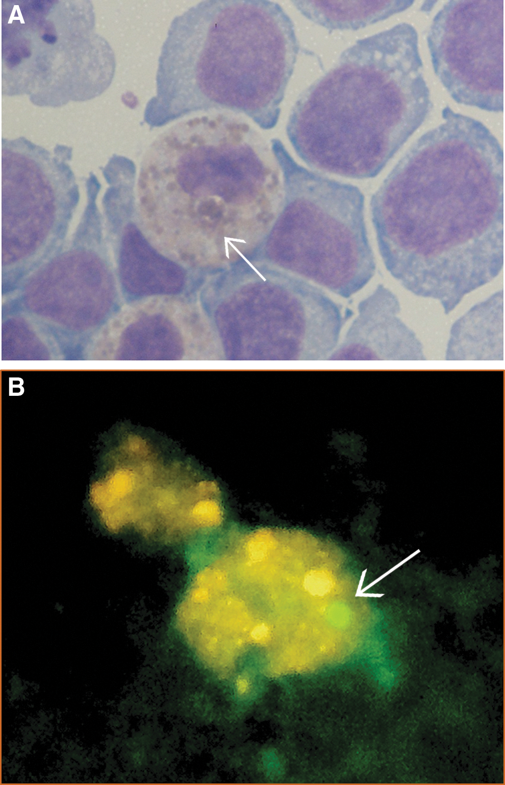
Ehrlichia ewingii morulae visualized in differentiated HL60 cells using Diff-Quik staining
In this study, we present the initial phase of isolation of a previously uncultivated organism, although much remains to be done to optimize propagation of E. ewingii in vitro. Naive goats must be inoculated with cultures to verify the organism's virulence and pathogenicity after in vitro propagation. Differentiation of cells in immortal lines leads to cell types that will eventually cease to replicate, causing culture death. Neutrophils can also release granules into cultures upon apoptosis, creating a toxic environment for replicating cells. Induction of differentiation should be explored further, given the initial promising results, but should be undertaken with careful observation of cells to prevent the rapid loss of cultures. Although this study was not able to produce an optimized protocol for long-term growth of E. ewingii in vitro, we were able to demonstrate the first in vitro propagation of the agent. Given the positive IFA results from the HL60 cell line, the amount of E. ewingii produced is sufficient for limited antigen production—enough, perhaps, to be used to generate IFA control antigens. It is our hope that researchers will be able to use these results to develop a continuous culture for the maintenance and propagation of E. ewingii in vitro and, thereby, expand our diagnostic capabilities and research opportunities for this pathogen.
Footnotes
Acknowledgments
The authors are grateful to Dr. Uli Munderloh for her help, advice, and encouragement throughout the years of trials and disappointments. The authors also thank Dr. S. Karpathy, Dr. C. Paddock, G. Zemtsova, and A. Snellgrove for their contribution.
Author Disclosure Statement
This work was conducted at the Centers for Disease Control and Prevention as part of authors' official duties. The findings and conclusions in this report are those of the authors and do not necessarily represent the official position of the Centers for Disease control and Prevention. No competing financial interests exist.