
Abstract
Anaplasma platys is an uncultivable tick-borne obligatory intracellular bacterium, which is known to infect platelets of dogs. A. platys causes infectious canine cyclic thrombocytopenia in subtropical and tropical regions throughout the world. Several cases of human infection with A. platys infection have also been reported. However, seroprevalence of A. platys exposure and infection has not been determined in most of the regions, in part, due to lack of a simple and reliable assay method. Furthermore, A. platys antigens recognized by dogs are unknown. We previously sequenced gene encoding A. platys major outer membrane proteins P44 and Omp-1X. In the present study, we obtained purified recombinant A. platys P44 and Omp-1X proteins, and using them as antigens in immunoblotting examined seroreactivity in dogs. Of 34 specimens from Venezuela where A. platys infection was previously reported, 25 specimens (73.5%) reacted to rAplP44 and/or rAplOMP-1X. Neither Anaplasma phagocytophilum-seropositive (N = 10) nor A. phagocytophilum-seronegative canine specimens (N = 10) from the geographic regions where A. platys infection has never been reported, reacted rAplP44 or rAplOMP-1X. The result indicates a high A. platys seroprevalence rate in tested dogs from Venezuela and suggests that the immunoblot analysis based on recombinant A. platys major outer membrane proteins can provide a simple and defined tool to enlighten the prevalence of A. platys infection.
Introduction
A
Clinical signs of ICCT are fever, depression, anorexia, and bleeding, which may occur (Harvey et al. 1978, Baker et al. 1987, Baker et al. 1988). In the United States, A. platys infection by itself is a relatively mild disease in dogs, but some strains reported in Greece, Israel, and Chile appear to be more pathogenic (Kontos et al. 1991, Harrus et al. 1997, Abarca et al. 2007). Other Anaplasma species, namely Anaplasma phagocytophilum and Anaplasma marginale, are known to be transmitted by ticks (Rikihisa 2011). Because A. platys DNA has been detected in brown dog tick Rhipicephalus sanguineus and brown ear tick Rhipicephalus bursa, which are commonly found in warmer climates and grassy areas (Inokuma et al. 2000, Motoi et al. 2001, Aktas et al. 2009, Ramos et al. 2014), A. platys transmission to dogs is thought to occur through this tick vector, but this has not been experimentally proven (Simpson et al. 1991).
Clinical cases of ICCT and A. platys infection have been increasingly reported throughout the world, including the United States (Harvey et al. 1978, Mathew et al. 1997, Yabsley et al. 2008, Diniz et al. 2010), Croatia (Dyachenko et al. 2012), Greece (Kontos et al. 1991), France (Beaufils 1985), Sicily (de la Fuente et al. 2006), Japan (Motoi et al. 2001, Unver et al. 2003), Taiwan (Chang and Pan 1996), Spain (Sainz et al. 1999, Aguirre et al. 2006), Southern China (Hua et al. 2000), Italy (Sparagano et al. 2003), Israel (Harrus et al. 1997), Australia (Brown et al. 2001), Chile (Abarca et al. 2007), Thailand (Suksawat et al. 2001), Mexico (Aktas et al. 2015), Turkey (Aktas et al. 2015), Portugal (Cardoso et al. 2010), Algeria (Dahmani et al. 2015), and Venezuela (Suksawat et al. 2001, Huang, 2005).
In addition to dogs, the presence of A. platys-specific DNA fragments has been reported in humans in the United States (Breitschwerdt et al. 2014), Grenada (Maggi et al. 2013), and Venezuela (Arraga-Alvarado et al. 2014), a cat in Brazil (Lima et al. 2010), a sheep in South Africa (Allsopp et al. 1997), and a goat in Cyprus (Chochlakis et al. 2009). A. platys-like inclusions have been observed in stained platelets from HIV patients in Venezuela (Tami and Tami-Maury 2004), a cat in Brazil (Santarém VA et al. 2000), and impalas in South Africa (Du Plessis et al. 1997).
Seroprevalence of A. platys infection of dogs was reported more than 20 years ago in Eastern North Carolina, Florida, and Louisiana in the United States, based on IFA test using infected dog platelets as antigen (French and Harvey 1983, Hoskins et al. 1988, Bradfield et al. 1996). However, since then and in other geographic regions, seroprevalence has not been determined using A. platys antigens due, in part, to the limited availability of A. platys antigen. In Venezuela, based on microscopic observations of blood smear, high prevalence of A. platys infection of dogs has been reported (Arraga-Alvarado et al. 1999, Arraga-Alvarado et al. 2003), and the prevalence rate of A. platys infection in military dogs in Venezuela as determined by PCR is 16% (7/43) (Huang 2005). However, so far no seroprevalence study has been reported in Venezuela. The present study, therefore, examined seroprevalence of A. platys infection in dogs in Venezuela.
We have recently sequenced and annotated the A. platys gene encoding major outer membrane proteins, OMP-1X and P44 (Lai et al. 2011). In the present study, we produced recombinant A. platys OMP-1X and P44 proteins (rAplOMP-1X and rAplP44, respectively) and analyzed sera from dogs in Venezuela. Canine sera from Wisconsin where a granulocyte-tropic Anaplasma species A. phagocytophilum is endemic (Greig et al. 1996), A. platys infection has never been reported, and canine sera from Ohio where neither A. phagocytophilum nor A. platys infection has been reported in dogs were also included in the assay. This is the first report on A. platys seroprevalence and canine immune reactivity to defined A. platys antigens.
Materials and Methods
Canine specimens
Blood samples were collected from 34 mixed-breed dogs living in an animal shelter or belonging to private owners at Caracas, Maracay, and Coro, Venezuela in April, July, and August 2013 (Table 1). Fresh buffy-coat smears were prepared the same day of collection and stained using Diff-Quik (Fisher Scientific, Pittsburgh, PA) or Hemacolor® (Merck KGaA, Darmstadt, Germany). At least 30 microscope fields were observed at 1000 X magnification. Samples were considered positive if more than three platelet inclusion bodies were observed. Ten serum samples collected in 2013 from dogs in Wisconsin were seropositive for A. phagocytophilum by the Canine SNAP® 4Dx® Test Kit (IDEXX Laboratories, Westbrook, ME). Tests were performed according to the manufacturer's instruction. Serum samples collected in Ohio between November 1995 and November 2013 from 10 dogs were seronegative for both A. phagocytophilum and Ehrlichia canis whole bacterial antigens by immunofluorescent antibody tests as previously described (Rikihisa et al. 1992, Rikihisa et al. 1997). All samples were drawn from the cephalic or jugular veins using sterile tubes containing ethylenediaminetetraacetic acid (EDTA) as anticoagulant (Becton Dickinson, Franklin Lakes, NJ) with oral consent from the owners. Blood samples were aliquoted and stored at −20°C until analysis.
F, female; M, male; UN, unknown.
Cloning of A. platys omp-1X and p44
DNA from an A. platys-infected dog from Taiwan, R.O.C. (Lai et al. 2011) was used as a template. DNA fragments encoding full-length omp-1X and p44 without the signal peptide were amplified by PCR. The PCR reaction was performed with Phusion High-Fidelity DNA polymerase (Thermo Scientific, Rockford, IL) according to the manufacturer's instruction with 0.2 μM of both forward and reverse primers at the annealing temperature of 57°C with 1 min/kb for extension for 30 cycles. All primer sequences will be provided upon request. The amplified DNA fragments were purified using GeneJET™ PCR Purification Kit (Thermo Scientific) according to the manufacturer's instruction and digested with restriction enzymes SpeI/XhoI (New England Biolabs, Ipswich, MA) for omp-1X and NheI/XhoI for p44 and ligated into the restriction enzymes NheI/XhoI-digested vector pET33b (Novagen, Madison, WI). Chemically competent PX5-α cells (Protein Express, Cincinnati, OH) were transformed, plasmids were extracted, and the cloned fragments were confirmed by sequencing (GenBank No. KJ155503 and AEH96269). All primer sequences will be provided upon request.
Expression and purification of rAplOMP-1X and rAplP44
rAplOMP-1X and rAplP44 were induced in Escherichia coli BL21/(DE3) with 1 mM isopropyl-1-thio-β-
Immunoblot assay
rAplP44 and rAplOMP-1X (0.1 μg) in Tris-buffered saline (TBS; 50 mM Tris-HCl, 150 mM NaCl, pH 7.4) were individually adsorbed onto nitrocellulose membranes using a slot blot apparatus (Bio-Rad Laboratories, Richmond, CA), blocked for 30 min with TBS containing 5% nonfat dry milk, air dried, and stored at −20°C until use. For immunoassays, the antigen bound to a nitrocellulose strip was incubated with canine serum samples, which were diluted 1:500 in TBS containing 1% nonfat dry milk for 3 h at 4°C. After being washed thrice with TBS containing 0.05% (v/v) Tween 20 (T-TBS), the strip was incubated with horseradish peroxidase-conjugated affinity-purified anti-dog IgG (KPL, Gaithersburg, MD) at a dilution of 1:5000 in TBS containing 1% nonfat dry milk. After being washed with T-TBS, enhanced chemiluminescence (ECL) LumiGLO chemiluminescent reagent (Thermo Scientific) and an LAS3000 image documentation system (FUJIFILM Medical Systems, Stamford, CT) were used to visualize the protein bands. The difference in the densitometry signal for the ECL reaction between the antigen spot and the nonantigen background on the test strip (blank) was analyzed by Multi Gauge software (FUJIFILM Medical Systems).
Results
Venezuelan dog specimens
Tick infestation and health status of dogs from which blood samples were obtained are summarized in Table 1. Majority of dogs were infested with ticks. Although not every tick was examined, they were mostly brown dog tick R. sanguineus. Approximately, one-third of dogs had clinical signs (most frequently cachexia) and the remaining dogs had no obvious clinical signs at the time of blood sample collection. Although it was not independently verified, every blood sample was reported positive for A. platys inclusion bodies when stained blood smears were observed in Venezuela.
Immunoblot assay using rAplP44 and rAplOMP-1X
The recombinant proteins rAplOMP-1X and rAplP44 were expressed and purified to near homogeneity using affinity chromatography (Fig. 1) and used for immunoblot analysis. Molecular sizes of native AplOMP-1X and AplP44 are 34.53 and 43.49 kDa, respectively. Due to some extra amino acid sequences derived from the vector, the predicted rAplOMP-1X and rAplP44 molecular sizes are 36.02 and 45.42 kDa, respectively, although rAplOMP-1X ran slightly slower than the predicted size in the SDS-polyacrylamide gel electrophoresis (Fig. 1). Slot immunoblot analysis showed that based on densitometric analysis, all of 20 canine sera from Ohio and SNAP 4Dx A. phagocytophilum P44–positive dog specimens from Wisconsin reacted with neither rAplP44 nor rAplOMP-1X (relative density value of 0–1) (Fig. 2). Under the assumption of a normal (Gaussian) distribution, the expected true negative rate is 99.9% if the cutoff value selected was equal to the mean of the negative reference serum plus three times the SD (Jacobson 1998). We selected the cutoff value of three to identify positive specimens. Results of the 34 canine sera from Venezuela showed that 25 sera (73.5%) reacted to rAplP44 and 21 sera (61.8%) reacted to rAplOMP-1X (Fig. 2). Majority (21/25) of rP44-positive specimens had strong reactions (relative density value of 15–580). When comparing between rAplOMP-1X and rAplP44, 19 specimens showed stronger reaction to rAplP44 than to rAplOMP-1X, whereas only one specimen (VZ12) showed reverse (Fig. 2).
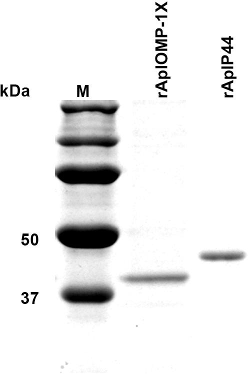
SDS-polyacrylamide gel electrophoresis profiles of the purified recombinant proteins. Affinity-purified recombinant proteins (0.5 μg) were separated in a 12% SDS-polyacrylamide gel and stained with GelCode Blue. Lanes: M (molecular size markers), rAplP44 (45.426 kDa), and rAplOMP-1X (36.02 kDa). The numbers on the left indicate the molecular masses in kilodaltons based on the Precision Plus Protein Standards (Bio-Rad Laboratories, Richmond, CA). SDS, sodium dodecyl sulfate.
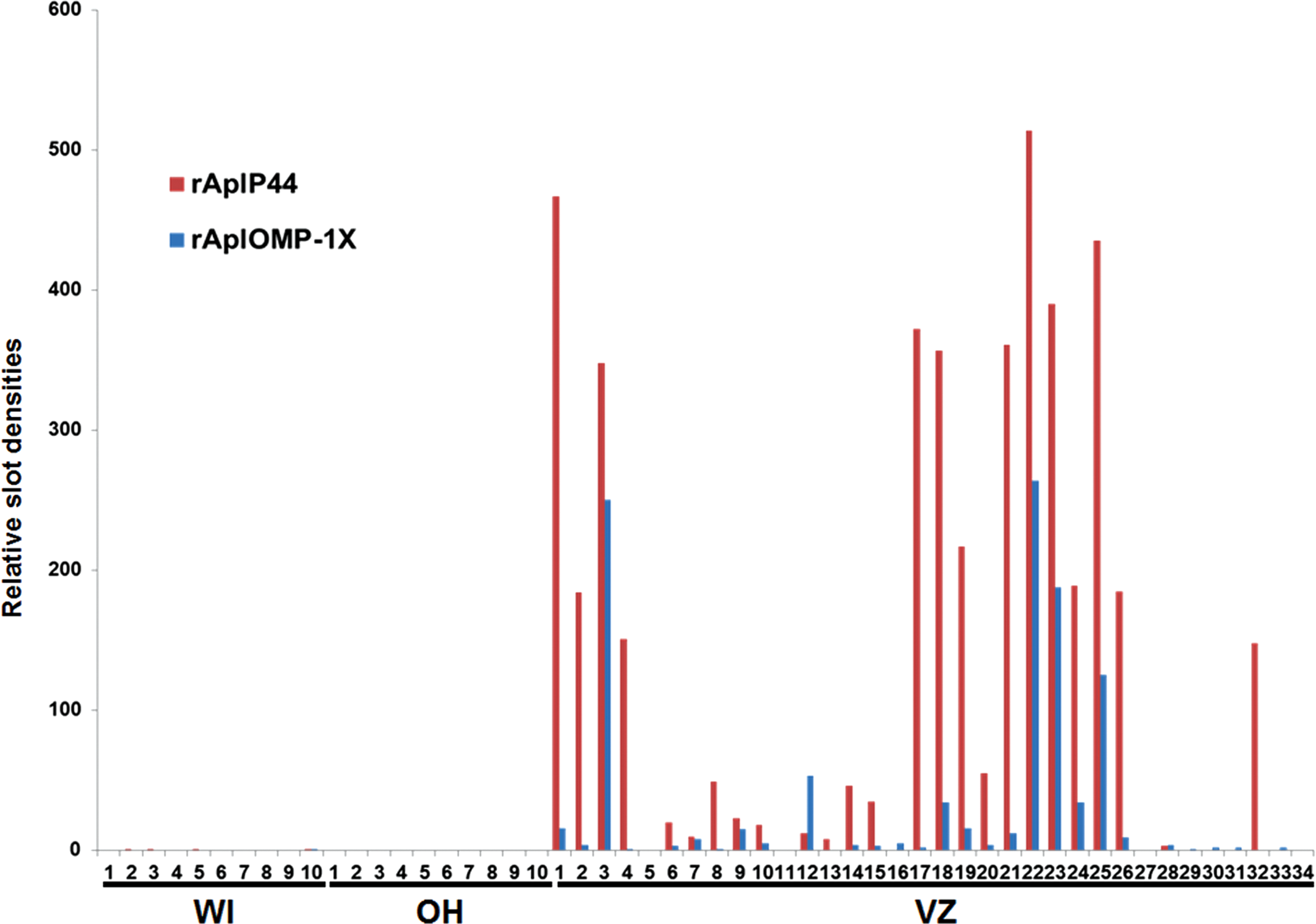
Slot immunoblot analysis of canine serum samples. Nitrocellulose membrane strips were individually loaded (0.1 μg) with rAplOMP-1X (plOMP) or rAplP44 (plP44) and tested with 10 canine sera from Ohio (OH), 10 IDEXX SNAP 4Dx canine anaplasmosis-positive canine sera from Wisconsin (WI), and 34 canine sera from Venezuela (VZ). The difference in ECL reaction signal densities between the antigen band and the nonantigen background on the test strip was analyzed by Multi Gauge software and divided by 104 and is shown in bar. ECL, enhanced chemiluminescence. Color images available online at
Discussion
The present study revealed high seroprevalence rates among tested Venezuelan dogs using two defined A. platys antigens. Among A. platys-positive dog serum samples, 14 of 29 had ≥10-fold higher reactivity to rAplP44 than to rAplOMP-1X, and only one sample had higher reactivity to rAplOMP-1X, suggesting rAplP44 is more readily recognized by immune system of A. platys-infected dogs. It was previously reported that the canine SNAP 4Dx A. phagocytophilum test cross-reacts with A. platys sera (Gaunt et al. 2010, Stillman et al. 2014). However, the reverse, whether canine A. phagocytophilum serum cross-reacts with A. platys antigens, has not been reported. Our current result showed none of canine SNAP 4Dx A. phagocytophilum-reactive sera reacted with our recombinant A. platys antigens. Thus, Venezuelan dogs' antibody reaction to recombinant A. platys antigens is unlikely due to A. phagocytophilum infection. There are reports of A. phagocytophilum infection in horses and dogs in Venezuela using stained buffy-coat films (de Alvarado 1992, de Alvarado et al. 1992). A single A. phagocytophilum PCR-positive dog case had been reported (Suksawat et al. 2001); however, the PCR result was later recanted as false positive (Suksawat et al. 2001, authors' corrections). Our previous study did not find A. phagocytophilum DNA in dog, human, or tick specimens from Venezuela (Huang 2005). Whether A. phagocytophilum infection occurs in Venezuela remains to be investigated.
Finding A. platys within platelets on stained blood films is difficult, because A. platys is a small Gram-negative coccus present at a very low number. Furthermore, false-positive results can occur when other inclusions, granules, particles, or megakaryocyte nuclear remnants are mistaken for A. platys (Simpson and Gaunt 1991, Ferreira et al. 2007, Little 2010). ICCT is a generally chronic disease and at stages between the cyclical bacteremia or at the chronic stage, PCR of blood specimens is often not sensitive enough to detect A. platys infection (Eddlestone et al. 2007), despite PCR testing being sensitive in detecting A. platys infection as early as day 4 postinfection before detection of bacterial inclusions in the platelets on day 7 postinfection (Eddlestone et al. 2007). It is difficult to obtain convenient and consistent serology using A. platys-infected platelet-rich plasma as antigen. Serological assay based on defined rAplOMP-1X and rAplP44 antigens is simple, consistent, objective, and convenient, thus helps generating A. platys prevalence information to raise awareness of ICCT and potential zoonosis.
Furthermore, these recombinant proteins can be applied to develop a simple serodiagnostic test for ICCT in the future. The limitation of the assay is, as in any other serologic assays, false-negative results at early stages of infection and in immunosuppressed individuals. Future steps that would be necessary for the test to become applicable for clinical diagnosis is to determine sensitivity and specificity of the test using a larger number of well-defined canine specimens. For this and understanding the pathogenesis and canine immune responses in ICCT, culture isolation of A. platys is desirable.
In addition to thrombocytopenia and mild clinical signs, dogs with chronic A. platys infection may develop nonregenerative anemia, which results in more severe clinical pathology and poor prognosis (Baker et al. 1988, Little 2010). Majority (21 of 34) of tested dogs did not have obvious clinical signs, suggesting the high rate of subclinical infection and reservoirs of A. platys in Venezuela. In conclusion, our data suggest that the immunoblot assay based on major outer membrane proteins of A. platys is expected to provide a simple tool to enlighten the prevalence and to assess the risk of A. platys infection.
Footnotes
Acknowledgments
The authors appreciate Pedro Aso, Departamento de Biología Celular, Universidad Simón Bolívar, Sartenejas, Caracas, Estado Miranda, Venezuela for his support of the research. The authors thank Jill Mielke at Veterinary Medical Teaching Hospital, University of Wisconsin-Madison, Madison, WI and Catalina Rey at the Universidad Francisco de Miranda, Coro, Estado Falcón, Venezuela and to Miguel Martin, Yuraima Reyes, and Hector Castillo at the Universidad Central de Venezuela, Maracay, Estado Aragua, Venezuela for collecting and shipping samples.
Author Disclosure Statement
No competing financial interests exist.