




Abstract
The presence of tick-borne pathogens and their possible coinfections were evaluated among host-seeking ticks in seven cities from Jiaodong peninsula, Shandong Province, with specific PCR or reverse transcription-PCR tests. Among 2107 ticks collected, four species of three genera were identified with Haemaphysalis longicornis as predominant species, and total of 63 H. longicornis and 10 Rhicephalus microplus were confirmed infected with tick-borne pathogens. These pathogens were consequently identified as severe febrile and thrombocytopenic syndrome virus (SFTSV), Anaplasma capra, Anaplasma phygocytophilum, and Babesia microti, respectively, with high phylogenetic scores on some fragments of species-specific genes. The infection rates of the pathogens in H. longicornis were presented as 1.03%, 0.84%, 0.58%, and 1.66%, respectively, close related to its field density and clump distribution pattern. Furthermore, coinfection of A. capra and SFTSV was also discovered from two female H. longicornis in Pingdu city. These results indicated that the potential human pathogens other than severe febrile and thrombocytopenic syndrome might be transmitted by hard ticks separately or in combination, and more reliable differential diagnosis, proper administrations, rational prevention, and control measures should be developed with the support of precision laboratory tests.
Introduction
T
Jiaodong peninsula, surrounded by the Yellow Sea and the Bohai Sea of China, shares the typical ecological environments characterized by a warm humid climate, mountainous or hilly forested landscape, which provides ticks with more adaptive habitats and hosts to survive and breed (Teng and Jiang 1991, Liu et al. 2014a). In the peninsula, ticks of Haemaphysalis longicornis and Rhicephalus microplus were recognized as predominate tick species and found frequently infested on human and domestic animals (Teng and Jiang 1991, Xing et al. 2015). Although more and more casualties infested with ticks have been documented in the peninsula during the past few years (Meng et al. 2015, Sun et al. 2015, Xing et al. 2015), the potential importance in term of human health caused by these ticks remain to be evaluated. Recently, Jiaodong peninsula was demonstrated as one of three hot spots of SFTS that accounted for most of SFTS cases in China (Liu et al. 2015). The prominent emergence of various tick-borne diseases in Jiaodong peninsula suggested the pivotal urgency and necessity to assess its potential health risk of these pathogens on human populations. The objectives of this study were to determine the prevalence of SFTSV, Anaplasma spp. and Babesia spp. in the quest ticks and to characterize their transmission cycles and potential threat in Jiaodong peninsula as one of sentinel sites around China.
Materials and Methods
Tick collection
Unfed ticks were collected by sweeping flags on vegetation or from cattle in the cow houses in the survey sites (Fig. 1) from April 10, 2010 to October 15, 2015. All ticks were identified by morphological features to the species level according to the keys described by Teng and Jiang (1991).

Origins of ticks in the study. Ticks collected from April 2010 to October 2015 in sites marked on the map with tick symbol.
Pathogen detection
Tick specimens was disinfected in 70% ethanol for 15 min, and then washed in phosphate-buffered saline (pH 7.4), dried, and individually homogenized by the use of lysing matrix tube (MP Biochemicals Germany, Eschwege, Germany). Total DNA and RNA were also prepared from tick samples with the RNeasy Mini Kit (Qiagen Co. Ltd, Hilden, Germany) and the QIAamp DNA Mini Kit (Qiagen Co. Ltd), respectively, according to the manufacturer's protocol. To amplify the coding sequence (CDS) of segment L and S of SFTSV from individual, an aliquot of the total RNA (1.6 or 2 μg) was reverse transcribed using an oligo-dT primer (20 pmol) in a reaction volume of 20 μL according to the manufacturer's instructions using an AMV First-Strand cDNA Synthesis Kit (NEB Co. Ltd.). The synthesized DNA was used as templates to amplify the CDS in segment L and S of SFTSV by PCR with the primer sets Fs-Rs and F1-R1, respectively. Genomic DNA from tick sample was screened for Anaplasma spp. infection by nested PCR targeting gltA gene coding the citric acid dehydrogenase and 16S ribosome RNA of Anaplasma, respectively (details in Table 1). A conventional PCR targeting an 18S rRNA gene fragment was performed for the detection of Babesia spp. with the primer set BJ and BN (Casati et al. 2006). The reaction mixture for PCR contained 2 mM Mg2+, 0.2 mM of deoxynucleoside triphosphate, 1.25 U DNA polymerase (Takara Co. Ltd.), 20 μM of each primer, and 4 ng cDNA or genomic DNA as template in a final 50 μL volume. The primer details and cycling conditions for PCR are also shown in Table 1. The amplified products were visualized under UV light with a ChampGel-3200 Photographic system and purified according to the manufacturer's protocol provided for the QIAquick Gel Extraction Kit (Qiagen Co. Ltd.). To clone target sequences, the purified DNA fragments were ligated into the cloning vector using the pEASY-T1 Simple Cloning Vector Kit (Transgen Co. Ltd.) and then transformed into the competent Escherichia coli strain trans5α (Transgen Co. Ltd.). The purified positive plasmids were sequenced on an automatic DNA sequencer (ABI 3730) with M13 primers by the Sangon Bio-technique Company (Beijing, China).
Nested PCR with Outer-f and Outer-r for the first round amplification and Inner-f and Inner-r for the second one.
SFTSV, severe febrile and thrombocytopenic syndrome virus.
Phylogenetic analysis
Bidirectional sequences obtained were compared with previously published sequences deposited in GenBank (National Institutes of Health) by using Basic Local Alignment Sequence Tool
Results
Among seven sites surveyed, a total of 2107 unfed ticks were harvested and identified as four species of three genera in family Ixodidae. These ticks comprised of H. longicornis (89.9%), R. microplus (9.1%), Haemaphysalis campanulata (0.8%), and Dermacentor silvarum (0.2%). The relative high density of the four tick species appeared in Qingdao, Jiaonan, Pingdu, and Jimo cites among these surveillance sites. H. longicornis appeared to be the predominant species in Jiaodong peninsula. H. campanulata and D. silvarum were only found in Jimo and Qingdao cities, respectively, with lower density.
Among these ticks, neither H. campanulata nor D. silvarum was detected positive for any pathogen described above. From the rest of two tick species, a total of 73 positive ticks were found infected with one or two species of pathogens. Only two female H. longicornis from Pingdu were double infected with Anaplasma capra and SFTSV. No other coinfections were detected. The prevalence of the pathogens in the ticks is listed in Tables 2 and 3. Briefly, total 27 H. longicornis ticks were found positive infected by Anaplasma spp. (1.43%; 95% CI: 0–2.12), which were consequently identified as two species, A. capra and Anaplasma phagocytophilum, with molecular evidences from sequences analysis. No double infection of the Anaplasma agents was recorded in these positive ticks. Among the ticks infected with A. capra, seven females (1.15%; 95% CI: 0–1.23), three males (0.78%; 95% CI: 0–0.91), and six nymphs (0.66%; 95% CI: 0–0.75) were found mainly distributed in Jiaonan, Qingdao, and Pingdu cities (Table 2). The infection rates of A. capra were shown with no significant difference in H. longicornis between sex (χ2 = 0.642; p = 0.216), adult, and immature ones (χ2 = 0.217; p = 0.39). Whereas among those with A. phagocytophilum, five females (0.82%; 95% CI: 0.41–1.3), four males (1.04%; 95% CI: 0.37–2.11), and two nymphs (0.22%; 95% CI: 0–0.58) were harvested from Jiaonan, Qingdao, and Pingdu cities. Differences of the positive rates of A. phagocytophilum in H. longicornis were found without any statistic significance between sex (χ2 = 0.558; p = 0.324), adult, and immature ones (χ2 = 0.425; p = 0.258) (Table 2). With regard to Babesia microti, 22 (12 females, 6 males, and 4 nymph) out of 1894 (1.16%; 95% CI: 0–2.04) H. longicornis ticks and 5 (4 females and 1 male) out of 190 R. microplus (2.63%; 95% CI: 0–6.67) were positive. The positive H. longicornis ticks were mainly collected from Jiaonan, Qingdao, and Pingdu cities and the prevalence rate in males (1.93%; 95% CI: 0.86–3.21) was near to that in females (2.04%; 95% CI: 0.88–2.95) with no significant difference (χ2 = 0.817; p = 0.23). No relationship between the positive R. microplus and their sites of origin were observed (Table 3). In addition, SFTSV infection was also recovered from 16 out of 1549 H. longicornis screened (1.03%; 95% CI: 0.9–2.4) and 5 out of 164 R. microplus (3.04%; 95% CI: 0.9–17.4). The prevalence rates of SFTSV in the seven sites ranged from 0% to 2.11% with the highest in Jiaonan city. In the positive H. longicornis ticks, the infection rate in males (1.7%; 95% CI: 0.86–3.21) was near to that in females (1.7%; 95% CI: 0.88–2.95) with no significant difference (χ2 = 0.317; p = 0.41) (Table 2). Similar to those infected with B. microti, positive R. microplus was also found without any relationship to their origins (Table 3).
Bold indicates positive infection of pathogens.
Bold indicates positive infection of pathogens.
To observe the possible genetic variants of pathogens in these tick samples, all amplicons were bidirectional sequenced and submitted to blast in GenBank. All the sequences obtained were identical to the corresponding fragment of reference strains or isolates as expected. Among 16 H. longicornis infected with A. capra, no mutation was observed in both 792 bp citrate synthase gene (gltA) and 1260 bp 16S ribosomal RNA. Both the genes yielded over 99.99% identity under the 100% coverage with A. capra isolates (KM206274, KJ700627) from patients in Mudanjiang and DNA fragments of Anaplasma spp. from H. longicornis (KR261618∼KR261621; KR261623∼KR261628) in Shandong Province (Sun et al. 2015), China. Another 11 ticks were identified to be infected with an A. phagocytophilum isolate form Apodemus agrarius (GQ412340, GQ412337) in Jilin Province, China with sequence differences distinctive as interspecies level with A. capra. The phylogenetic trees also clustered them into two distinct branches as they deserved, respectively (Fig. 2A, B). Among the ticks with SFTSV, the genes of both segment L and S supported that the virus detected was identical to SFTSV human isolate 2011YSC60 (KF711863, KF711899) with over 99.9% identity and 100% coverage. There was also no mutation observed among the deduced amino acid sequences from different species and sites of origin (Fig. 2C, D). As for the Babesia protozoan infected in H. longicornis and R. microplus, no nucleotide variant was observed among those sequences with 456 bp in length despite the tick species and collection sites. These sequences were identical to B. microti strain HN-Bm (KC147722) with 100% identity and coverage in their phylogenetic tree (Fig. 2E). The two female H. Longicornis, double infected, were demonstrated to carry 2011YSC60 isolate SFTSV and Mudanjiang isolate A. capra based on their sequences data.
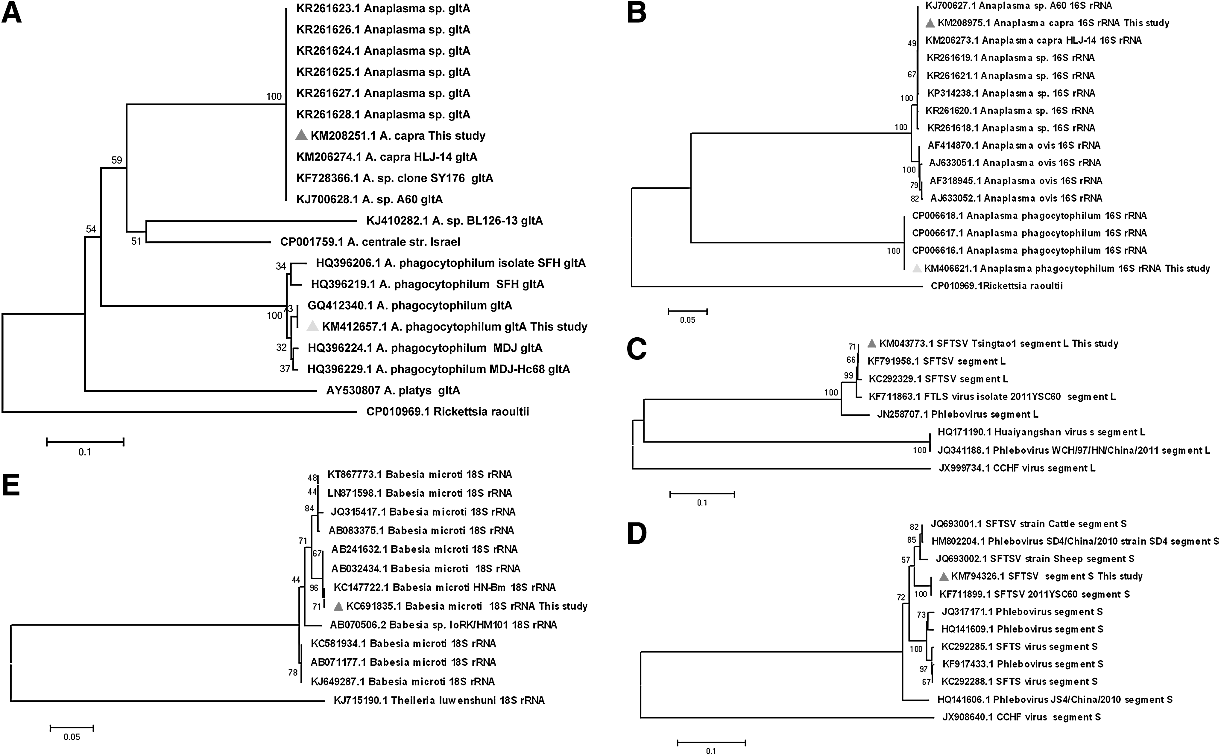
Phylogenetic tree of Anaplasma spp. SFTSV and Babesia microti. Phylogenetic tree of Anaplasma based on variation-deduced amino acid sequences of the citrate synthase (gltA)
Discussion
As an emerged tick-borne disease, SFTS had seriously threatened public health with disparity patterns in Shandong Province, China (Zhao et al. 2012, Cui et al. 2013, Meng et al. 2015, Xing et al. 2015). Jiaodong peninsula, one hot spot with aggregate SFTS cases (Liu et al. 2015), might have some epidemiological clues needed to be elucidated. Our results confirmed the natural infection of the Phlebovirus in the peninsula and the prevalence in its competence vector H. longicornis (Luo et al. 2015, Yun et al. 2016). The infection rates of SFTSV reflect in previously published results with prevalence ranging from 0.1% to 12.8% (Zhao et al. 2012, Cui et al. 2013, Meng et al. 2015), which was shown closely related to the density and the aggregate geographic pattern of H. longicornis. Besides SFTSV, we also successfully detected another three pathogens, A. phagocytophilum, A. capra, and B. microti, from ticks in these areas. Among them, A. phagocytophilum was known to cause anaplasmosis in human and animals. While A. capra was first discovered last year from goats, patients, and taiga ticks Ixodes persulcatus, respectively, in Mudanjiang city, Heilongjiang Province, China (Li et al. 2015). The new Anaplasma species was nominated as A. capra for its origin host Capra aegagrus and recognized as causative agent of an undifferentiated influenza-like human anaplasmosis, with fever, headache, malaise, dizziness, chills, and some gastrointestinal, neurological, or skin symptoms (Li et al. 2015). Since then, A. capra-like DNA fragments were demonstrated in H. longicornis from Shandong Province (Sun et al. 2015) and Beijing (Wang et al. 2016). Our results from Jiaodong peninsula do not merely repeat these investigations with different prevalence rates due to the different sites or seasons, but also suggest the potential vector competence of H. longicornis to transmit A. capra to human and animals. Moreover, B. microti was also demonstrated in both H. longicornis and R. microplus from the peninsula in our survey. The intraerythrocytic sporozoites, the causative agent of human babesioses, might widely be infectious to residencies and animal hosts due to the frequent contacts between the vector ticks with human and animal populations in the peninsula. These results in our surveys pose further questions as to how to make a differential diagnosis of these pathogens and to develop proper therapy, prevention, and control strategies of various tick-borne diseases.
As known to us, clinical symptoms caused by most tick-borne pathogens are not so pathognomonic, but consistent with many other infectious causes, including bacteria, rickettsia, and other viruses. Most clinical physicians make the diagnosis of SFTS with the symptoms of acute fever, thrombocytopenia and leukocytopenia, and exposure history to ticks in patients (Liu et al. 2014b). However, much more victims suffering from the above symptoms might be caused by pathogens other than SFTSV. For instance, the occurrence of Rickettsia or Anaplasma in patients usually present clinical symptoms such as fever, lymphopenia and thrombocytopenia resembling SFTS cases (Dumler et al. 2007, Zhang et al. 2009, Kok and Jin 2011, Dugat et al. 2015). Thus, anaplasmosis and Rickettsioses are difficult to distinguish clinically from many viral infections (Walker et al. 2008), as well as SFTSV (Kok and Jin 2011). In addition, coinfections with different tick-borne pathogens in both human and animals are commonly reported in recent years (Zhao et al. 2013, Moniuszko et al. 2014, Mayne et al. 2015, Liu et al. 2016). The coinfection in human populations, caused by the bites of either single tick coinfected with pathogens or multiple ticks with single pathogen, might have enormous impact on the diagnostic methods and highly relevant to public health (Moutailler et al. 2016). In the present survey, the coinfection in two H. longicornis individuals discovered suggests the possible coinfection of A. capra and SFTSV in human populations in the peninsula due to their concurrence in the same vector ticks H. longicornis and transmission cycles. Since coinfections may enhance the severity and duration of disease, most coinfected patients experienced a greater number of more severity symptoms for a longer duration than those with single pathogen alone (Liu et al. 2016, Moutailler et al. 2016). Therefore, the current diagnostic tools for tick-borne disease in China may not match the range of pathogens carried by vector ticks from all specific geographical areas. The occurrence of Rickettsia or Anaplasma might confuse the clinical diagnosis of SFTS for these bacteria and their possible coinfection with SFTSV (Liu et al. 2016), which might partially explain the presence of detectable antibody to Anaplasma spp. in clinical SFTS-like patients (Li and Zhang 2008, Zhang et al. 2009), and the reasons that quite a few of severe acute febrile illness with tick exposure history were cured by certain antibiotics instead of antiviral therapy (Kok and Jin 2011, Diuk-Wasser et al. 2016). To overcome the limitation of current diagnostic tools, more reliable differential diagnosis techniques and rational prevention and control measures are imperative to be developed with precision laboratory tests.
Footnotes
Acknowledgments
The authors are grateful to Prof. Wuchun Cao and Rongman Xu for their helpful comments and for revising the article. This study was supported by grants from the National Key Technology Research and Development Program of the Ministry of Science and Technology of China (grant no. 2014BAI13B02), and the Key Program of State Key Laboratory of Pathogen and Biosecurity (grant no. SKLPBS1504) of China. Prof. Tongyan Zhao was supported by grant 2012ZX10004219 from China Mega-project for Infectious Diseases sponsored by Chinese Ministry of Science and Technologies.
Y. Sun, PhD, works at the State Key Laboratory of Biosecurity and Microbiology, Beijing Institute of Microbiology and Epidemiology, Beijing, People's Republic of China. His primary research interest is ticks and tick-borne diseases.
Author Disclosure Statement
No competing financial interests exist.