
Abstract
The intraleukocytic parasite, Hepatozoon canis, causes the sometimes fatal tick borne disease canine hepatozoonosis. In this study, dogs from Islamabad, Lahore, and Multan Districts of the Punjab region of Pakistan were surveyed to investigate the presence and prevalence of H. canis infection and to determine the effects of the parasite on hematological parameters. Blood samples were collected from 151 domestic dogs (149 pet, 2 stray) of both sexes and varying ages. Data on sex, age, tick infestation, and clinical factors (body temperature, mucous membrane status, and presence of hematuria and vomiting) were collected. Using PCR, 18 dogs (11.9%) were found positive for the presence of H. canis DNA. Partial sequences of the 18S rRNA gene shared 99–100% similarity with the corresponding H. canis isolates. This epidemiological survey revealed higher prevalence of H. canis in Islamabad (11/49, 22.4%) compared to Lahore (3/52, 5.8%) and Multan (4/50, 8%) in Pakistan. No investigated epidemiological or clinical factors were found to be associated with the presence of H. canis (p > 0.05) in dogs. H. canis positive dogs exhibited higher minimum inhibitory dilution (p = 0.04), mixed inclusion (p = 0.008) and relative distribution width of red blood cells (p = 0.02), and lower hematocrit (p = 0.03) and mean hemoglobin content (p = 0.03) than did dogs in which H. canis was not detected. We are recommending this PCR-based protocol to the veterinary practitioners for the detection and/or confirmation of H. canis in dogs suspected for hepatozoonosis to improve their health status.
Introduction
H
Little published information is available regarding the prevalence of H. canis in dogs in Pakistan. This study was designed to investigate the presence and prevalence of H. canis in domestic dogs from Lahore, Islamabad, and Multan districts in Punjab and aimed to compare the studied hematological parameters and selective clinical findings of parasite-positive and negative dogs. For the first time, to the best of our knowledge, 18S rRNA gene of H. canis was sequenced in dogs from Pakistan and phylogenetic analysis of the parasite was also conducted.
Materials and Methods
Study area and sample collection
One hundred fifty-one domestic dog blood samples were collected from three districts of Punjab from Pakistan [Islamabad (n = 49), Lahore (n = 52), and Multan (n = 50)] from December 2014 to May 2015 (Fig. 1). With the exception of two strays, all dogs included in the study were pets, and informed consent was obtained from owners before their inclusion in the study. Sampled dogs included healthy animals, as well as those with clinical symptoms, including fever (diagnosed with thermometer), pale mucous membranes, hematuria, and vomiting (physical observation). The dogs were divided into two age groups with those aged 6 months to 1 year designated as young and those from 1 to 7 years considered adults. A questionnaire was completed at the sampling site to gather data on sex, age, tick infestation, and clinical factors (noted above) associated with canine hepatozoonosis. Blood samples were taken from the cephalic vein into a tube coated with ethylenediaminetetra-acetic acid.
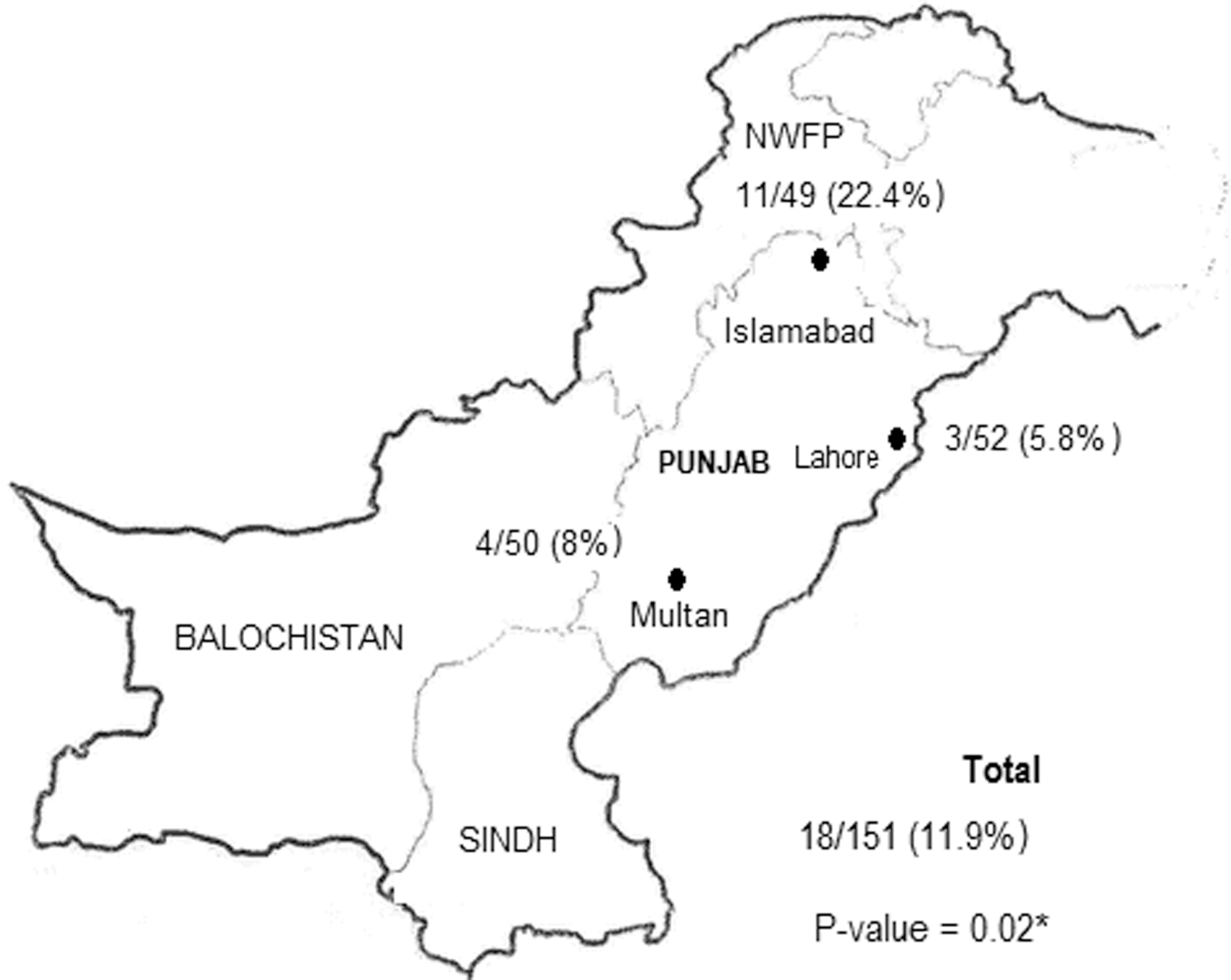
Prevalence of Hepatozoon infection in dogs from three districts of Punjab, Pakistan using PCR. p Value represents the results of one-way ANOVA calculated for Hepatozoon canis prevalence at three sampling sites.
The body of each dog, with special attention to the ears, was examined for the presence of ticks. If present, they were removed with forceps and placed in bottles with moistened cotton wool and transferred to the laboratory for identification.
DNA extraction and PCR
An inorganic method of DNA extraction was used as previously described elsewhere (Saeed et al. 2015). Briefly, blood samples were suspended in 500 μL of lysis buffer (20 mM Tris-HCl, 1 mM EDTA, 30 mM DTT, and 0.5% SDS) supplemented with 0.4 mg/mL proteinase K (Fermentas). The samples were kept overnight in a heating block set at 55°C. After the lysis, samples were heated at 95°C for 10 min. Then equal volume of phenol: chloroform: isoamyl alcohol (25:24:1 v/v/v), was added, vortexed for 30 s, and centrifuged at 12,000 × g for 10 min. The aqueous phase was transferred to new clean tube, and equal volume of ice cold isopropanol was added. The DNA was pelleted by centrifugation at 12,000 × g for 15 min; the pellet washed with 70% ethanol and dried at 65°C for 5 min. The DNA was finally resuspended in 50 μL sterile distilled water. The quality of the DNA obtained was assessed through optical density counts at 260–280 nm and submerged gel electrophoresis to determine purity. For the detection of H. canis, a PCR assay was performed as previously described by Otranto et al. (2011). DNA amplification was carried out in a DNA thermal cycler (GeneAmp® PCR system 2700 Applied Biosystems, Inc., United Kingdom). The thermo-profile of PCR consisted of an initial denaturation at 95°C for 5 min followed by 40 cycles of denaturation at 95°C for 30 s, annealing at 57°C for 30 s, elongation at 72°C for 1 min and 30 s, and a final extension at 72°C for 10 min (Karagenc et al. 2006). PCR products were held at 4°C until separated by electrophoresis on a 1.5% agarose gel and visualized under a UV Trans illuminator (Biostep, Germany).
Sequencing and phylogenetic analysis
To confirm PCR results, four representative positive PCR products (two from Islamabad, one Lahore, and one Multan) were randomly chosen for sequencing. The generated DNA fragments were purified with a PCR Purification Kit (GF-1 96-well PCR Clean-up Kit; Vivantis, United Kingdom) and sent to a commercial laboratory (Macrogen, Korea) for sequencing. DNA sequences obtained were evaluated with Chromas Lite software, version 2.1.1 (Technelysium Pty Ltd., Australia) and compared for similarity to registered sequences in GenBank (
A phylogenetic analysis was carried out using the software MEGA (v.6;
Hematological analysis
To compare hematological parameters of H. canis positive and negative dogs, complete blood counts were obtained using an automated cell counter (Abbott Cell-Dyn 3700).
Statistical analysis
Data are expressed as mean ± standard deviation. The statistical package Minitab (v.16) was used for analysis of the results. The prevalence of H. canis in the surveyed districts was compared by one-way analysis of variance (ANOVA). The prevalence (%) and 95% binomial exact confidence intervals were calculated for the PCR results for H. canis using Sourceforge (
Ethics approval
All animal handling procedures and laboratory protocols were approved by the Ethics Committee of the Institute of Molecular Biology and Biotechnology, Bahauddin Zakariya University, Multan, Pakistan.
Results
Prevalence of H. canis
Eighteen of the 151 (11.9%) blood samples amplified a 666-bp amplicon specific for 18S rRNA gene of the parasite. H. canis infections were found in all the three investigated districts of Punjab. The highest prevalence of parasite was observed in Islamabad (11/49, 22.4%), followed by Multan (4/50, 8.0%) and Lahore (3/52, 5.8%). Statistical analysis revealed that prevalence of H. canis in the dogs varied significantly among the three study sites (p = 0.02; Fig. 1).
Sequencing and phylogenetic analysis
Four sequences of the 18S rRNA gene of H. canis were obtained and submitted to the EMBL/GenBank database under the Accession numbers KU535868 (dog from Lahore district), KU535869 (dog from Lahore district), KU535870 (dog from Rawalpindi district), and KU535871 (dog from Multan district). A BLAST search indicated that the sequences were 99–100% identical to the H. canis isolates registered in GenBank.
Phylogenetic analyses revealed that the obtained H. canis sequences clustered in the group with H. canis sequences obtained from dogs and foxes in India, Brazil, Turkey, and Thailand, and sequences were clearly distinct from Hepatozoon americanum, Hepatozoon felis, and Hepatozoon catesbianae. However, separate clusters were present within H. canis isolates, which may represent a strain variant of H. canis (Fig. 2).

Phylogenetic relationship of H. canis detected during present study to H. canis reported from other countries based on the partial sequence of the 18S rRNA gene. The evolutionary history was inferred by the maximum likelihood method based on the Kimura three-parameter model. Toxoplasma gondii was used as out-group. Bootstrap analysis, used to estimate the node reliability of the trees, was conducted with 1000 replicates as implemented in MEGA6. Hepatozoon spp., host species, country of origin from where these sequences were derived, and the GenBank accession numbers are included for each sequence.
Analysis of epidemiological factors
When prevalence of H. canis was compared between various breeds of dogs, it was observed that H. canis was not restricted to a particular breed (p > 0.05). Stray dogs and Spitzer were among the highly infected breeds while minimum incidence of H. canis was recorded in Labrador breed. H. canis was not detected in Boxer, Pug, Russian, Shih Tzu, and Spanial breeds (Table 1).
p > 0.05, nonsignificant.
All dogs were examined for the presence of ixodid ticks. Eleven of 151 dogs (7.3%) were infested with adult and nymphal R. sanguineus. Forty-eight ticks (29 nymphs, 19 adults) were removed from dogs (Table 2). Analysis of epidemiological factor data revealed that all the studied parameters were not associated (p > 0.05) with the prevalence of H. canis in collected dog samples (Table 2).
p Values represent the results of Fisher's exact test calculated for each studied parameter. p > 0.05, nonsignificant.
Comparison of hematological parameters
H. canis positive dogs showed higher minimum inhibitory dilution (MID) (p = 0.04), mixed inclusion (MI) (p = 0.008), and relative distribution width of red blood cells (p = 0.02) with lower hematocrit (p = 0.03) and mean hemoglobin content (p = 0.03) than did dogs in which H. canis was not detected (Table 3).
Data are expressed as mean ± SD. p Value indicates the results of two sample tests calculated for each parameter. p > 0.05 = nonsignificant; p < 0.05 = least significant *; p < 0.01 = significant (**). GR%, relative (%) content of granulocytes; GRA, granulocytes; HCT, hematocrit; HGB, hemoglobin; LY%, relative (%) content of lymphocytes; LYM, lymphocyte; MCH, mean content of hemoglobin; MCHC, mean concentration of hemoglobin in erythrocytes; MCV, mean volume of erythrocytes in cubic micrometers; MI%, mixed inclusion; MID, minimum inhibitory dilution; MPV, mean platelet volume; PCT, plateletcrit; PDWc, platelets distribution width; PLT, platelets; RBC, red blood cells; RDWc, the relative distribution width of red blood cells; SD, standard deviation; WBCs, white blood cells.
Discussion
Canine hepatozoonosis is a tick borne disease of increasing importance in dogs and has become an active area of research in recent years, although it has been unexplored in Pakistan. In this study, H. canis infections were found in the three surveyed districts, with overall prevalence of 11.9%, suggesting that H. canis is present throughout Punjab. The prevalence of Hepatozoon infection revealed by PCR was similar to a previous report in stray dogs and cats in Bangkok, Thailand (11.4%) (Jittapalapong et al. 2006), but lower than that observed in India (30%) (Rani et al. 2011), Turkey (22.3%) (Aktas et al. 2015), and Italy (57.8%) (Otranto et al. 2011). This variation in the prevalence of H. canis could be due to many factors, including the distribution and population density of the vector (Otranto et al. 2011), the sampling methodology, and the characteristics of the targeted dog population (Gomes et al. 2010, De Miranda et al. 2014).
None of the other studied clinical factors (body temperature, mucous membrane status, hematuria, and vomiting) were shown to be correlated with the presence of H. canis infection in the tested dogs (Table 1). These observations are in agreement with the previous findings that H. canis usually causes chronic infection with relatively mild or no clinical manifestation in its host (Little et al. 2009). Although the prevalence was higher in adult dogs (19%) than in young (10%), age differences were not significant in the present study (p = 0.14). The finding conflicts with previous reports of significantly higher prevalence of H. canis in adult dogs (Gomes et al. 2010, Aktas et al. 2015). However, Rojas et al. (2014) had also concluded that the differences in prevalence of H. canis infection in adult and young dogs were not significant (Rojas et al. 2014). In this survey, sex differences were also not significant (p = 1), confirming previous studies reporting that there is no correlation of sex with presence of H. canis infection in dogs (O'Dwyer et al. 2001, Forlano et al. 2005, Gomes et al. 2010, Rani et al. 2011, Aktas et al. 2013, 2015).
During present study, we did not find a correlation between presence of H. canis DNA with the presence of R. sanguineus in dogs (p = 0.12). The finding is not consistent with previous studies reporting Hepatozoon infection in dogs to be positively correlated with the presence of ticks (O'Dwyer et al. 2001, Forlano et al. 2005, Aktas et al. 2013). Most of the animals sampled in this study were pets and groomed by the owners, and ticks were only found on 11 of 151 (7.2%) sampled dogs. Good care and management of dogs by owners could be the reason that most dogs were not found infested with ticks, and hence, we have not seen a positive correlation between presence of ticks and prevalence of H. canis in blood of enrolled dogs (Table 1).
MID and MI values were significantly higher in H. canis positive samples compared to negative samples (p = 0.04, p = 0.008, respectively), whereas the mean hemoglobin concentration and hematocrit were low in parasite positive samples (p = 0.03), indicating that H. canis attacks leukocytes (Table 2). MID cells include less frequently occurring and rare cells correlating to monocytes, eosinophils, basophils, blasts, and other precursor white cells that fall in a particular size range (Saeed et al. 2015). Total leukocyte count is presented in three parts in our data; lymphocyte count, neutrophil count, and MI that refers to monocyte, eosinophil, and basophil counts. All these leukocytes are an important part of the immune system, hence an increase in MID and/or MI (%) indicates an infectious state in H. canis infected dogs. Reduced mean hemoglobin concentration and hematocrit in H. canis infected dogs are indicating the developing anemia; one of the major symptom of hepatozoonosis in dogs (Shaw et al. 2001, Otranto et al. 2011).
In conclusion, we have used a PCR protocol for the detection of H. canis in dogs by amplifying their 18S rRNA gene and reporting their partial gene sequence along with the phylogenetic analysis. We are recommending the use of PCR to the veterinary practitioners and pet owners for the detection and/or confirmation of H. canis in dogs suspected for hepatozoonosis to improve their health status.
Footnotes
Acknowledgments
Authors are grateful to pet owners and veterinarians who aided in sampling and epidemiological data collection. Positive control DNA used in this study was kindly donated by Prof. Dr. Chengming Wang (College of Veterinary Medicine, Yangzhou University, Yangzhou, China).
Author's Contributions
R.S.S. designed the study and M.Q., M.I.M., Q.U.A., and M.L. collected the samples. M.Q. and M.I.M. conducted the laboratory investigations. M.A. performed the phylogenetic analysis. M.A. and F.I. analyzed the data and prepared the article.
Author Disclosure Statement
No competing financial interests exist.