
Abstract
Bats are reservoir hosts for many paramyxoviruses, some of which cause human and zoonotic diseases of public health importance. We developed a Nipah virus nucleoprotein enzyme-linked immunosorbent assay to detect cross-reactive antibodies in serum samples from several bat species in Brazil. Our results warrant further investigation of henipa-like virus reservoirs in the Western hemisphere.
P
Bats were sampled for blood and swabs (oral/fecal) according to protocols approved by the Ethics Committee on Animal Experimentation of the Institute of Biomedical Sciences, University of São Paulo and with the permission of the Brazilian Institute of Environment and Renewable Natural Resources (IBAMA, process number 16575-3), and were released on completion of sample collection. All samples were stored in liquid nitrogen in the field and maintained at −80°C. RNA extracted from all swab samples tested negative for the presence of NiV N gene RNA by quantitative reverse-transcription polymerase chain reaction (RT-PCR) (Lo et al. 2012). Aliquoted serum samples were shipped on dry ice to the U.S. Centers for Disease Control and Prevention and were inactivated by gamma-irradiation (5 × 10⁶ rad) before being tested.
We developed an enzyme-linked immunosorbent assay (ELISA) using recombinant NiV nucleoprotein (N) antigen from human embryonic kidney cell lysate (HEK293T) transfected with a eukaryotic expression plasmid expressing NiV N gene (pC-NiVN). Our negative control antigen was cell lysate from HEK293T cells transfected with empty vector plasmid (pC-293T). We validated our NiV N ELISA using both NiV seropositive human and bat sera (Fig. 1A, B), and also confirmed the specificity of our NiV N ELISA by showing a lack of significant cross-reactivity against NiV N antigen by antisera specific for other paramyxoviruses (Fig. 1C).
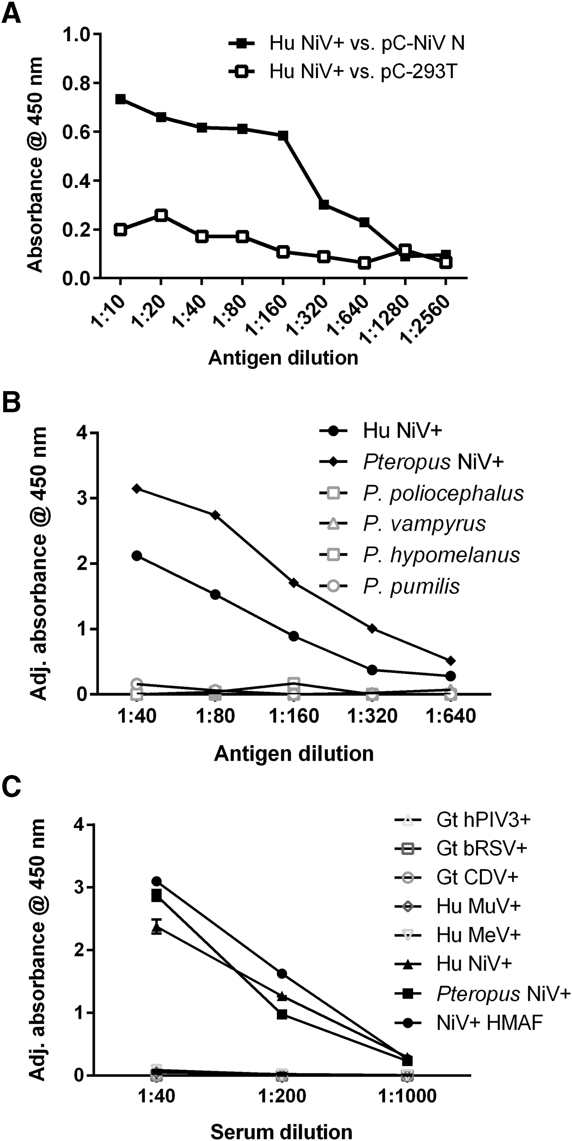
Development and validation of NiV N ELISA.
We then used the ELISA to query our collection of Brazilian bat sera. Bat serum was considered positive if its adjusted absorbance ([absorbance against pC-NiVN]–[absorbance against pC-293T]) readout was higher than the cutoff value, which was defined as the average absorbance value of the NiV-positive control human serum against negative control antigen (performed in triplicate) plus three times the standard deviation of the average absorbance value. Of the 11 species of bats tested, 5 species tested positive by NiV N ELISA, with 9 of 76 total serum samples testing positive (∼12%) (Table 1). To confirm our results, we assayed these sera by indirect immunofluorescence assay (IFA) on formalin-fixed NiV-infected African green monkey kidney (Vero) cells (Fig. 2 and Supplementary Fig. S1; Supplementary Data are available online at

Immunofluorescence assay. NiV seropositive pooled Pteropus serum, Glossophaginae sp3 serum (1:40 final dilution), and NiV seronegative Pteropus poliocephalus serum were incubated with both mock-infected and NiV-infected African green monkey kidney (Vero) cells. A goat anti-bat antibody and a donkey anti-goat antibody conjugated with DyLight 550 (Bethyl) were used, respectively, to detect bat serum reactivity against NiV antigen. DAPI was used as a nuclear counterstain. Representative images were taken at 40× magnification using a Nikon Eclipse Ti inverted fluorescence microscope. Scale bar indicates length of 50 μm. DAPI, diamidino-2-phenylindole dye.
ELISA, enzyme-linked immunosorbent assay; IFA, immunofluorescence assay.
In summary, 13 of 76 total serum samples (17%) tested positive by IFA. These positive sera were from Glossophaginae sp3, (n = 6), Artibeus planirostris (n = 2), Carollia perspicillata (n = 2), Glossophaginae sp1 (n = 1), Artibeus lituratus (n = 1), and Desmodus rotundus (n = 1). Our study provides serological evidence of henipa-like virus infection in five Brazilian bat species (Glossophaginae sp1 and sp3 are combined as one species due to the inability to identify to species level). The overall henipa-like virus seroprevalence in our study falls within the range documented from prior bat henipavirus seroprevalence studies (from 2% to up to 63%) (Iehle et al. 2007, Jonathan et al. 2008, Li et al. 2008, Andrew et al. 2010, Peel et al. 2012, Hume et al. 2013).
Although our study lacked molecular evidence of henipa-like virus infection, our results support existing molecular evidence available for bat henipa-like virus infection in the Western hemisphere (Drexler et al. 2012). While only pteropodidae bats have been identified as reservoirs of henipaviruses to date, all of the bat species tested in this study were from the diverse family Phyllostomidae. Hollow trees, fruit groves, caves, as well as man-made buildings and tunnels can all serve as dwellings for phyllostomid bats. Although the majority of bats sampled in this study were frugivorous and/or insectivorous, there also were two common vampire (D. rotundus) bats sampled, one of which tested positive by both NiV N ELISA and IFA.
The bats captured for this study were at a location directly adjacent to human development, which highlights the potential of human–bat interactions leading to disease spillover events by zoonotic pathogens due to increasing human encroachment into undeveloped areas. Similar to a study that provided evidence of anti-henipavirus antibodies in insectivorous bats in China (Li et al. 2008), the results of our study expand the host range for henipa-like viruses and provide impetus for future efforts to identify new henipa-like viruses as well as their potential host reservoirs in the Western hemisphere.
Footnotes
Acknowledgments
Financial support for this research came from CDC core funding and grant sponsor: Fundação de Amparo a Pesquisa do Estado de São Paulo-FAPESP, grant number: 2006/00572-0. We thank Marcel Soares for identification of the bats. This research has been approved by the animal subjects research review board at University of Sao Paulo.
Disclaimer
The findings and conclusions in this article are those of the authors and do not necessarily represent the views of the CDC, U.S. Department of Health and Human Services.
Author Disclosure Statement
No competing financial interests exist.
References
Supplementary Material
Please find the following supplemental material available below.
For Open Access articles published under a Creative Commons License, all supplemental material carries the same license as the article it is associated with.
For non-Open Access articles published, all supplemental material carries a non-exclusive license, and permission requests for re-use of supplemental material or any part of supplemental material shall be sent directly to the copyright owner as specified in the copyright notice associated with the article.