
Abstract
Cryptosporidium is a parasitic protozoon with a wide range of vertebrate hosts. Cryptosporidiosis has been reported from numerous countries, including Iran. Molecular identification can be applied to characterize Cryptosporidium, of which there are over 30 species and 50 genotypes. Herein, we report the genetic diversity of Cryptosporidium spp. in Iranian dogs for the first time based on 18S ribosomal RNA gene sequencing. One hundred forty fecal samples of herd dogs were collected from Isfahan, central Iran. The samples were concentrated using sucrose flotation and subjected to Kinyoun staining. DNA extraction of positive samples was performed, and molecular diagnosis was carried out using highly specific seminested PCR for the characterization of Cryptosporidium species. Finally, sequencing and DNA analysis were performed to identify Cryptosporidium species. A total of 2.14% of herd dogs were positive for cryptosporidiosis in both microscopy and molecular methods. In all cases, the causative agent was identified as Cryptosporidium parvum. Dogs associated with positive samples had been in close relationship with livestock. Cryptosporidiosis in the herd dogs in Isfahan could be due to their close contact with animals, particularly cattle and sheep. Given that dogs with cryptosporidiosis lack clinical symptoms, they are a potential source of zoonotic transmission of this disease as they are companion animals for humans. Dogs with cryptosporidiosis are a potential source of the zoonotic transmission of this disease.
Introduction
C
As a zoonotic intestinal parasite, Cryptosporidium includes over 30 species (Ranjbar et al. 2016) and at least 50 genotypes, of which Cryptosporidium parvum is the most common pathogenic species, infesting a wide variety of mammals. Infection with or parasitic attachment by C. parvum has been reported in more than 150 countries in the world (Abe et al. 2002, Xiao and Fayer 2008, Koompapong et al. 2014).
Although dogs are commonly infected with species-specific Cryptosporidium canis, the occurrence of zoonotic C. parvum in dogs has raised a concern that these animals may serve as a potential reservoir for human transmission (Tangtrongsup et al. 2017). Although studies have shown that most infections in dogs are caused by C. canis, other species, including C. parvum, Cryptosporidium muris, and Cryptosporidium meleagridis, have also been identified (Fayer 2004).
Several epidemiological studies in Iran have demonstrated that the prevalence of Cryptosporidium spp. in dogs is different in various provinces; a prevalence of 5% in Khorasan-Razavi (Beiromvand et al. 2013), 7.14% in Ilam (Kakekhani et al. 2011), 3.1% in northeast of Iran (Arzamani et al. 2016), and 3% in Kerman (Mirzaei and Fooladi 2013) provinces has been reported.
Small subunit rRNA, as a molecular target to identify Cryptosporidium species, has been used in more than 80% of studies published in the past 2 years (Xiao 2010, Gil et al. 2017). In Thailand C. canis was identified in 2 of 95 temple dogs in central Thailand using PCR, amplifying an 830-bp fragment of small subunit ribosomal RNA (rRNA) gene (rDNA) (Tangtrongsup et al. 2017). In one study in Japan, C. canis and C. parvum were identified using nested PCR and sequencing methods. In addition, a study in Spain by using the same gene showed that C. canis and Cryptosporidium hominis were found in dogs (Gil et al. 2017).
To our best knowledge, there has been no previous research concerning Cryptosporidium spp. genotypes in dogs in Iran. Since this protozoal infection is a potential public health concern, determining the prevalence and genotypes of these organisms in dogs living in close proximity to humans and other animals is essential (Tavalla et al. 2017). Thus, the aim of this study was to determine the prevalence of Cryptosporidium species in dogs in Isfahan, Iran, using sequencing-based molecular approach.
Materials and Methods
Sample collection and fecal preparation
One hundred forty fecal samples of herd dogs were obtained from ranches that traditionally kept sheep and cows (Fig. 1). Around 10 g of each fecal sample was put in a sterile container and stored at 4°C at the parasitology laboratory at Isfahan University of Medical Sciences until further analysis. A 0.5 g aliquot of each sample was mixed with phosphate-buffered saline (PBS) to make homogeneous suspension, passed through double-layered gauze, and centrifuged at 500 × g for 3 min. Three milliliters of the obtained pellet was concentrated using cold flotation method (5 mL of 4 M sucrose; 1280 g of sugar/liter) by centrifugation for 10 min in 500 × g. The cloud layer was carefully separated and washed twice with PBS for remove the remaining sucrose. From the resulting sediment, slides were prepared for Kinyoun staining for the detection of Cryptosporidium spp.
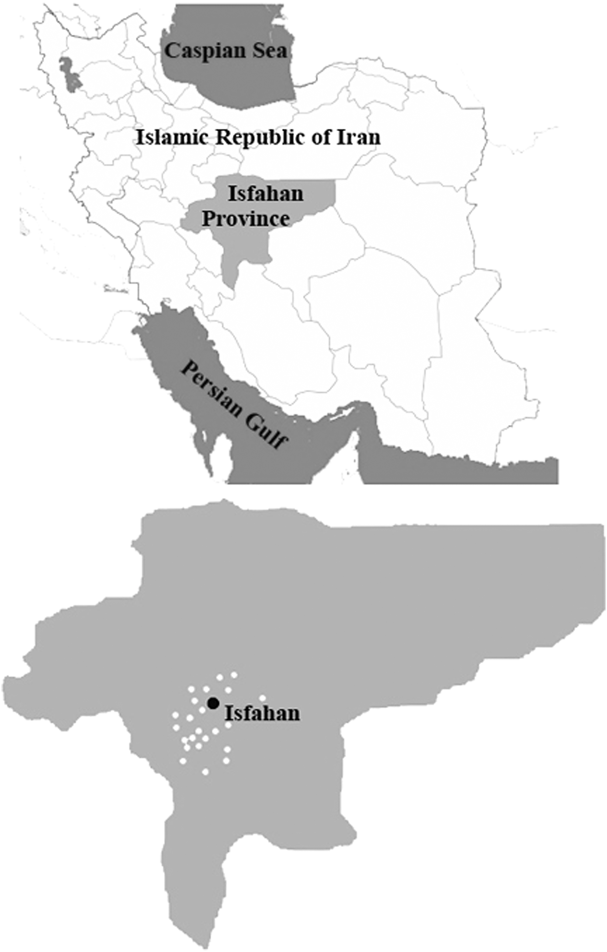
Locations of herd dog fecal samples collected from Isfahan, Iran.
DNA extraction
The samples positive in Kinyoun staining were subjected to DNA extraction. For complete destruction of Cryptosporidium oocysts, 300 μL of lysis buffer (10 mM Tris, 1 mM EDTA pH 8, 100 mM NaCl, 2% Triton X-100, 10% SDS), 300 mg of glass beads (425–600 μm in diameter, G9268; Sigma-Aldrich), and 100 μL of pretreatment suspension obtained by the flotation method were added to a 1.5-mL tube and highly homogenized at 4500 rpm (Micro Smash ms-100R) for 2 min. Subsequently, DNA was extracted using the phenol–chloroform method (Mirhendi et al. 2006). The extracted DNA was stored at −20°C until being used for PCR.
PCR amplification and sequencing
To amplify a fragment of the 18S rRNA gene from various Cryptosporidium species, a highly specific seminested PCR protocol was developed using primers complementary to conserved regions of published 18S rDNA nucleotide sequences of Cryptosporidium species (downloaded from GenBank), using Geneious 9.1.4 software (Fig. 2). The outer forward primer was 5′-GGA AGG GTT GTA TTT ATT AGA TAA AG-3′, which was previously described by Xiao et al. (1999), and the common reverse primer was Cr550 5′-TGA AGG AGT AAG GAA CAA CCT CC-3′; they amplified an ∼835-bp fragment containing a variable region that allows for identification of different Cryptosporidium species. The second step of nested-PCR was performed with the inner forward primer Cr250 5′-GGA ATG AGT KRA GTA TAA ACC CC-3′ and the common reverse primer (Cr550) with an amplicon of ∼530 bp.

Seminested PCR (for identification of Cryptosporidium spp.) primer positions have been shown on C. canis 18S rRNA gene (GenBank acc. no. AF112576.1).
The PCR mixture with a volume of 25 μL for each of the two steps of seminested PCR contained 1 × PCR buffer, 1.5 mM MgCl2, 0.4 mM dNTP, 2.5 U Taq (Ampliqon Taq DNA polymerase), 2 μL of DNA template (1 μL of PCR product for second step), and 5 pmol/μL of each primer. A total of 30 cycles, each consisting of 95°C for 30 s, 55°C for 30 s, and 72°C for 30 s, as well as an initial preheat step at 95°C for 5 min and a final extension step at 72°C for 5 min, were considered for the first PCR step. For the second step of seminested PCR, an initial denaturation step at 95°C for 3 min was followed by 25 cycles of denaturation at 94°C for 30 s, annealing at 59°C for 30 s, and extension at 72°C for 30 s.
PCR products were analyzed on a 1.5% agarose gel, which was visualized by KBC Power Load (Kawsar Biotech Co., Iran). PCR products were sequenced using the Cr550 primer, and the obtained nucleotide sequences were subjected to BLAST analysis in comparison with the reference sequences in GenBank.
Results
Average age of studied dogs was 27 months, with the frequency of 51 females and 89 males. Using morphological criteria, the overall rate of infection with Cryptosporidium was 2.14% (3/140) (Fig. 3), all of them were positive in seminested PCR (Fig. 4). No clinical signs were apparent in any of the Cryptosporidium-positive dogs at the time of specimen collection. In addition, no significant relationship was observed between sex or age and cryptosporidiosis (p > 0.05). Dogs infected with Cryptosporidium had close life relationship with sheep or cattle. Overall, 72.1% of the herding dogs lived indoors in sheepfolds with sheep and cattle. The remaining 27.9% of herding dogs lived outdoors and did not have close relationship with livestock. A total of 87.4% of dogs were associated with sheep and 12.6% of dogs were associated with cattle (Table 1). Moreover, two infected dogs lived indoors and had daily relationship with sheep and one infected dog lived with cattle inside sheepfold.

Cryptosporidium oocyst identified in a fecal sample of herd dog using Kinyoun staining that is shown by the arrow.
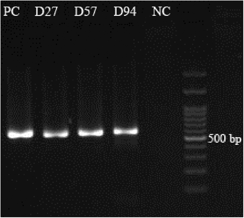
Seminested PCR products of herd dog fecal samples collected from Isfahan, Iran. PC, D27 isolate, D57 isolate, D94 isolate, 100-bp DNA ladder, NC. NC, negative control; PC, positive control.
BLAST analysis of the sequences confirmed the species of Cryptosporidium strains, which had already been identified by seminested PCR. All of these sequences had high similarity with C. parvum (GenBank acc. no. KU200956.1).
Discussion
By molecular characterization, C. hominis and C. parvum have been identified as the predominant species of Cryptosporidium, isolated from hosts, including humans (Xiao and Fayer 2008). Dogs have been suggested to be a significant source of human cryptosporidiosis (Xiao and Feng 2008). Although many studies were performed on the prevalence of cryptosporidiosis in dogs, few reports have investigated the genetic diversity of the isolates (Yoshiuchi et al. 2010). In the present study, Cryptosporidium strains were detected in 3 out of 140 (2.14%) herding dogs, all recognized as C. parvum. Previous reports on cryptosporidiosis of dogs in Iran were based on morphological criteria. In the field, infection of dogs with Cryptosporidium spp. has been reported in Chenaran, Kerman, and Hamedan, with infection rates of 5%, 4.08%, and 3.8%, respectively (Mirzaei 2010, Beiromvand et al. 2013, Sardarian et al. 2015).
Molecular identification of Cryptosporidium species showed that the most common species in dogs is C. canis (Brinklov et al. 2009). Various studies have confirmed this issue globally, including the work by Soriano et al. (2010) with a prevalence of 1% in Argentina, Yoshiuchi et al. (2010) with a prevalence of 3.9% in Japan, Wang et al. (2012) with a prevalence of 2.3% in the United States, and several reports with frequencies of 3.8%, 2.2%, and 8% from China (Jian et al. 2014, Li et al. 2015, Xu et al. 2016). Based on analysis of the 18S rRNA gene, other Cryptosporidium species have also been found around the world, such as C. meleagridis and C. parvum in the Czech Republic (Hajdusek et al. 2004), and C. parvum with a prevalence of 1 of 89 in Germany (Sotiriadou et al. 2013). A molecular epidemiological study of cryptosporidiosis in humans and animals in Isfahan, Iran, showed that C. parvum had the highest incidence (Azami et al. 2007).
Cryptosporidiosis in the herd dogs in Isfahan could be due to their close relationship with animals particularly cattle and sheep. Given that dogs with cryptosporidiosis lack clinical symptoms (Abe et al. 2002), they are a potential source of zoonotic transmission of this disease (Smith et al. 2009) as they are companion animals for humans. The existence of C. parvum in stool specimens of three herd dogs in this study can probably be due to the close relationship between two dogs with sheep and another one with cattle, and then, these relationships are likely to be a potential risk factor for infecting dogs.
The collection of stool samples from herd dogs and the danger of approaching them were a limitation in our study. Likewise, setting up the molecular methods for the detection of Cryptosporidium spp. in feces was problematic mainly due to the presence of several types of microorganisms, including parasites, fungi, bacteria, and many other cells in dog stools.
Conclusion
Given that only one oocyst is sufficient for Cryptosporidium infection in a host (Ramirez et al. 2004), the fecal shedding of dogs and livestock in one place and the possible contamination of their food and drinking water is notably important. Moreover, given the fact that traditional animal husbandry is often in the vicinity of farmland and herd dogs freely move in agricultural crops and sometimes their stools are discarded into these places, the importance of the issue in the field of human health is also clarified.
Footnotes
Acknowledgments
We thank the “Central Laboratory of School of Medicine, Isfahan University of Medical Sciences” and the “Clinical Research Development Center of Baqiyatallah hospital” for their kind cooperation.
Author Disclosure Statement
No competing financial interests exist.