




Abstract
Q fever is a disease caused by Coxiella burnetii. It is a disease of public health concern in many parts of the world. In this study, we described the seroprevalence of Q fever among selected populations of Orang Asli (OA), indigenous people, many of whom live within the forest fringe areas of Peninsular Malaysia. Serum samples were obtained from 887 OA participants from selected villages. Samples were analyzed for the presence of IgG antibodies reactive against C. burnetii by enzyme-linked immunosorbent assay. Statistical methods were used to identify possible associations between seropositivity for C. burnetii and a number of demographic variables obtained from the questionnaires. In total, 9.6% (n = 85/887) of the serum samples were reactive to C. burnetii. Statistical results suggest that elderly male OA residing in OA village, Bukit Payung, were most likely to be tested seropositive for C. burnetii. This study suggests that OA are at a significant risk of contracting C. burnetii infection, and both demographic and geographic factors are important contributors to this risk. Further prospective studies are needed to establish the true burden of C. burnetii infection within the indigenous population as well as within Peninsular Malaysia as a whole.
Introduction
C
Q fever typically presents with nonspecific symptoms of fever, sweats, headache, joint pain, cough, shortness of breath, and fatigue (van Woerden et al. 2004, Amitai et al. 2010, Dijkstra et al. 2012). Such nonspecific symptoms make accurate clinical diagnoses challenging and likely results in the underreporting of C. burnetii infections. Although rare, in severe cases of human infection, C. burnetii can spread to the liver, lungs, spleen, bone marrow, and female genital tract (Maurin and Raoult 1999). Osteomyelitis, meningitis, meningoencephalitis, pericarditis, or myocarditis can also occur (Raoult et al. 2000, Merhej et al. 2012). Infection of the female genital tract in humans during pregnancy may result in spontaneous abortion, intrauterine growth retardation, oligoamnios, stillbirth, and premature delivery (Carcopino et al. 2009).
Transmission of Q fever occurs through inhalation or ingestion of C. burnetii particles present in milk, urine, feces, birth fluids, and placenta of infected animals (Fishbein and Raoult 1992, Guatteo et al. 2006, Reusken et al. 2011). Human-to-human transmission has also been described, but such transmissions are considered rare (Milazzo et al. 2001). Direct transmission through ticks to humans has never been reported despite evidence of C. burnetii in saliva secreted by ticks (Rehácek and Brezina 1968).
Q fever has been documented in Southeast Asia countries (Marchette 1966, Chomel et al. 1993, Tay et al. 1998, Suttinont et al. 2006, Rai et al. 2011). However, only a single study describes human infection in Peninsular Malaysia, and this study was limited to three neighboring farms in mainland Penang (Rai et al. 2011). One of the participants was initially hospitalized with a provisional diagnosis of brucellosis, which was subsequently laboratory confirmed to be Q fever. Owing to its nonspecific symptomatic profile, the limited use of diagnostics to test for infection, and the absence of epidemiological studies, the true burden of disease caused by C. burnetii remains unknown.
The indigenous people of Peninsular Malaysia, known as the Orang Asli (OA), live largely within the forest-fringe areas. Members of this population often work in the oil palm or rubber plantations, participate in self-sustaining small-scale animal husbandry, and many still forage in the forests for food and gather forest products for sale (Rahim et al. 2010, Tay et al. 2013). Many OA live in close contact with wildlife and domestic animals, which include cats, dogs, poultry, cattle, sheep, and goats. Constant exposure of the OA to nearby forested areas, wildlife, and domesticated animals, and any ticks these animals may harbor may pose an increased risk of contracting C. burnetii infection and Q fever. As part of the “Comprehensive study on tropical disease among indigenous people of Malaysia,” this study aims to elucidate the risk of C. burnetii infection among this unique community and identify demographic variables and factors that may influence this risk.
Materials and Methods
Ethics statement
The study protocol was approved by the Medical Ethics Committee of the University of Malaya Medical Center, Malaysia (MEC ref. no. 824.11). Permission was also obtained from the Department of Orang Asli Development (JAKOA) [ref. no. JHEOA.PP.30.052 Jld. 6 (19)], a department under the Malaysian Ministry of Rural and Regional Development entrusted to oversee the affairs of OA. Subjects or legal guardians of the subjects provided written consent for the study before enrollment.
Study area and sample collection
The 16 OA villages, Lubuk Legong (LL), Sungai Perah (SP), Tumboh Hangat (TH), Jekjok (JJ), Sangwai (SW), Paya Pelong (PP), Paya Sendayan (PS), Donglai Baru (DB), Paya Lebar (PL), Dusun Kubur (DK), Ulu Kelaka (UK), Bukit Payung (BP), Bukit Sebang (BS), Pasir Intan (PI), Semanggar (SM), and Sungai Selangi (SS), selected for this study were located throughout 8 of the states in Peninsular Malaysia (Fig. 1). A total of 945 blood samples were collected from healthy OA ≥5 years of age during the period from July 2012 to June 2013. At least 5–10 mL of blood was collected from each participant using blood tubes with clot activator (BD, Franklin Lakes). The samples were centrifuged at 1000 g for 10 min. Collected serum was aliquoted into tubes and stored at −80°C freezer until analysis. OA participants also completed a given questionnaire detailing their basic personal data (marital status, education level, occupation, monthly household income, and household size).
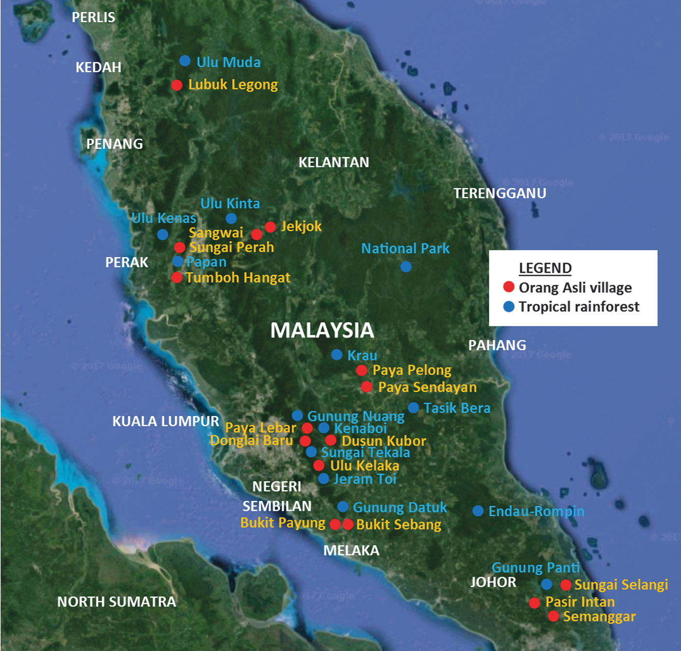
Participating OA villages and surrounding tropical rain forests. OA, Orang Asli.
Detection of C. burnetii IgG
The presence of C. burnetii IgG in serum samples was determined using a commercial Enzyme-Linked Immunosorbent Assay (ELISA) Kit (Coxiella burnetii ELISA IgG; Vircell, Granada, Spain). The assays were performed strictly following the protocol provided by the manufacturer.
Statistical analysis
Statistical analysis of ELISA results and questionnaire data were conducted using the Statistical Package for the Social Science (SPSS) v24 (IBM Corp., NY). Binary Logistic Regression was used to test the association between age, education level, gender, monthly household income, household size, marital status, type of house, and village of residence (independent variables) with C. burnetii seropositivity as dependant variable. Samples with equivocal results were excluded to generate a binary variable. OA villages with fewer than 10 participants were excluded due to low participation rates.
Results
A total of 945 OA from 16 villages volunteered to participate in the study. Out of the 16 villages sampled, 2 villages, BS and SS, comprising 11 participants in all, were excluded due to low levels of participation. Another 47 individuals were excluded due to the following reasons; incomplete demographic data, insufficient volume of serum sample available or having equivocal results. The remaining 887 individuals from 14 villages were included in this study. Of those individuals considered, 519 (57.3%) were female. The mean age of all of those enrolled was 23.1 years of age (standard deviation = ± 16.0, range = 5–83) and median was 18.0. Analysis of the 887 blood samples revealed that 9.6% were positive for C. burnetii IgG (mean age = 31.2, range = 6–69) (Table 1). Among the 887 participating OAs, 386 provided responses to the administered questionnaire, which were included in the statistical analysis below.
Education level and annual household income are both categorical variables. Categories with highest percentage in each OA village are displayed.
Not included in the analysis due to low number of respondents (n = 4).
The percentage of OA with annual household income of “<500” and “500–1000” are the same (46.2%).
BP, Bukit Payung; DB, Donglai Baru; DK, Dusun Kubur; JJ, Jekjok; LL, Lubuk Legong; OA, Orang Asli; PI, Pasir Intan; PL, Paya Lebar; PP, Paya Pelong; PS, Paya Sendayan; MYR; Malaysian Ringgit; SD, standard deviation; SM, Semanggar; SP, Sungai Perah; SW, Sangwai; TH, Tumboh Hangat; UK, Ulu Kelaka.
Univariate analysis was performed using binary logistic regression to test the association between the dependent variable (i.e., seropositivity) and the independent variables. Age, gender, and village of residence were found to be significant predictors of the presence of C. burnetii-specific IgG (p < 0.05) (Table 2). Five variables with p value of <0.2 were included in multivariate analysis using binary logistic regression. The multivariate analysis performed suggested that age, gender, and village of residence are significant predictors (p < 0.05) for C. burnetii seropositivity. A single unit change in age (1 year older) increased the risk for C. burnetii infection, as evidenced by the presence of specific IgG antibodies, by 1.03 times. Male participants were 2.14 times more likely to test positive for C. burnetii IgG in comparison to female participants (p < 0.04). Among OA villages, the seropositivity rate of questionnaire respondents from DK (14.0%) was similar to the overall seropositivity rate of participants who responded to questionnaire (14.0%), hence they were used as reference for comparison among the OA villages. Participants residing at BP and UK were 13.35 (p < 0.001) and 5.65 (p < 0.011) times more likely to test positive for C. burnetii IgG when compared with participants residing in DK. Education level, monthly household income, household size, marital status, and type of house were found not to be significant predictors of seropositivity in the univariate analyses.
Variables with p < 0.20 will be included in multivariate analysis.
Binary logistic regression was performed with OA village, DK as reference.
CI, confidence interval; OR, odds ratio.
Significant p-value < 0.05 are bolded.
Out of the 386 OA who answered the questionnaire, 54 (14.0%) respondents did not give a specific occupation. The remaining respondents (n = 332; 86.0%) stated their occupation as housewife (n = 175), plantation worker (n = 66), odd-job worker (n = 20), student (n = 17), forager of natural resources (n = 14), retired (or unemployed) (n = 7), landscaper (n = 7), cleaner (n = 6), and others (n = 20) (Fig. 2). Occupation however, was not a significant predictor for seropositivity by binary logistic regression.

OA occupation by village. Occupation of questionnaire respondents was used to form 100% stacked bar graph. Occupations denoted by Others consists of factory worker, educator, construction worker, fisherman, bus driver, sales clerk, and self-employed.
Discussion
In this study, we attempted to establish the baseline prevalence and distribution of C. burnetii by determining the seroprevalence of C. burnetii IgG in serum samples obtained from consenting OA. Serological testing showed that 9.6% of the participating OA were positive for C. burnetii IgG with the seroprevalence within each of 14 OA villages ranging from 0% to 47.4%. The two OA villages with highest seroprevalence, BP (47.4%), and UK (24.5%) were located at the periphery of two adjoining forests, Hutan Lipur Jeram Toi and Hutan Lipur Gunung Datuk, whereas other villages were located elsewhere (Fig. 1). This suggests that the degree of C. burnetii dissemination in local terrain or geographical factors may influence the seroprevalence. However, data collected for this study were insufficient to examine this possibility.
Findings from our study among the OA of Peninsular Malaysia suggest higher C. burnetii exposure rates in comparison to earlier studies from Indonesia (Bali) and Greece that reported a rate of only <1.0% (Chomel et al. 1993, Maltezou et al. 2004). With a focus on children and teenagers in their studies, the lower seroprevalence in the Indonesian and Greek studies may be due, in part, to the relatively young populations they targeted, as age was a significant predictor of seropositivity in our study as well as other published reports (Raoult et al. 2005, McCaughey et al. 2008). Our results are comparable, however, to previous results from studies on high-risk individuals conducted in the United States, Northern Ireland, Gambia, and Zambia in which the seroprevalence ranged from 3.1% to 12.8% (Okabayashi et al. 1999, McCaughey et al. 2008, Anderson et al. 2009, Bok et al. 2017).
Physical contact with farm animals, the principal risk factor for Q fever, is an important factor influencing seroprevalence (de Rooij et al. 2012). While animal husbandry was not mentioned as an occupation among those queried in our study, one possible route of exposure may involve ticks. As stated previously, the OA often live and work along forest fringe areas where domestic animals, wildlife, and ticks are present. The presence of C. burnetii in ticks removed from wild animals caught in Malaysian forest; and domestic animals visiting the veterinary clinic were recently confirmed by molecular detection (Watanabe et al. 2015, Khoo et al. 2016). Taken together with the seroprevalence rates from our study, these findings highlight domestic animals and ticks as potential sources of C. burnetii exposure among OA. However, unlike disease transmission by mosquitoes, transmission of pathogens through tick bite requires a moderate duration of attachment and feeding (Piesman et al. 1987), and may not be as efficient in infecting OA villagers. On the other hand, domesticated animals from OA villages and wild animals infected through tick bite may act as amplifying hosts and shed large amounts of infectious particles in bodily secretions. Pathogen amplification and distribution through the subsequent aerosolization of these secretions may provide a more efficient, alternative transmission route of infection (Brooks et al. 1986, Astobiza et al. 2011b). Furthermore, intensive interactions with C. burnetii-infected animals, such as the hunting and butchering in which the OA are known to engage (Rahim et al. 2010, Tay et al. 2013), may further facilitate transmission (Buhariwalla et al. 1996, Astobiza et al. 2011a). The hunting of wild animals to supplement household food supply and exposure to bodily fluid from domestic animals were not captured in our questionnaire. This may explain the lack of association between occupation and seropositivity in this study.
Earlier studies, however, have reported possible serological crossreactions among C. burnetii and Coxiella-like bacteria (CLB), Bartonella quintana, Bartonella henselae, and Legionella micdadei (La Scola and Raoult 1996, Musso and Raoult 1997, Angelakis et al. 2016). CLB are endosymbionts with high prevalence in ticks (Khoo et al. 2016). Transmission of CLB to host through tick bites resulted in sensitization of host and production of anti-CLB antibodies. Crossreaction of antibodies induced by CLB with C. burnetii antigen remains unverified; however, anti-C. burnetii antibodies were reported to crossreact with CLB-derived antigens (Angelakis et al. 2016). CLB, Bartonella and Legionella spp. are found in Malaysia, raising the possibility that the 9.6% seropositivity we identified could be an overestimation due to possible crossreactivity of the assay (Yong et al. 2010, Mokhtar and Tay 2011, Khoo et al. 2016). Improved diagnostics and a future study involving symptomatic patients are, therefore, needed to obtain a more accurate estimate of C. burnetii infection.
Conclusions
Although Q fever is an infectious disease of significant importance, information on it is severely lacking in most Southeast Asian countries, including Malaysia. This study described the presence of IgG antibodies reactive to C. burnetii among the OA of Malaysia with the estimated seropositivity at 9.6%. Age, gender, and village location were significant predictors for C. burnetii seropositivity with elderly males residing in the OA village, BP most likely to be seropositive for C. burnetii. The importance of geographic locations and contact with infected wildlife needs further investigation.
Footnotes
Acknowledgments
The authors would like to thank all volunteers, JAKOA, and Orang Asli village chiefs for their cooperation and assistance in sample collection activities. This study is funded in parts by the U.S. Naval Medical Research Center—Asia (Work Unit no. D-1101) and the U.S. Department of State, Biosecurity Engagement Program (NAMRU: J-55025-75053), University of Malaya Research Grant (UMRG: FL001-13HTM), University Research Grant 2015, Center of Excellence (COE) Programs (RU016-2015) and Ministry of Higher Education (MOHE), and High Impact Research (HIR)-MOHE Grant (E000013-20001).
Author Disclosure Statement
The authors declare that they have no competing interests. B.L.P. is a U.S. military service member. This work was prepared as part of his official duties. The opinions and assertions contained herein are those of the author and are not to be construed as official or reflecting the views of the Department of the Navy, Department of Defense, or the U.S. Government. Furthermore, Title 17 U.S.C. §105 provides that “Copyright protection under this title is not available for any work of the United States Government.” Title 17 U.S.C. §101 defines a “U.S. Government work as a work prepared by a military service member or employee of the U.S. Government as part of that person's official duties.”