
Abstract
Rickettsia spp. has been detected in dog fleas in Bangkok, Thailand. With the intent of collecting evidence to confirm the presence of rickettsioses in dogs and to assess the level of associated potential for accidental human infection, human buffy coat from patients with fever of unknown origin (n = 168), whole blood samples from dogs (n = 353), and 19 flea groups from our dog sample population were studied during the 2012 to 2014 study period. The presence of Rickettsia was investigated by molecular detection of 23S rRNA gene of Rickettsia genus, citrate synthase (gltA) gene, and 17-kDa outer membrane gene. All positive samples were confirmed by DNA sequence analysis. Using phylogenetic analysis, three groups of Rickettsia were detected, as follows: Rickettsia felis in 8 patients and 8 dogs; R. felis-like sp. in 2 patients, 5 dogs, and 11 flea samples; and Rickettsia typhi in 3 patients. In addition to confirming the presence of R. felis in Thai patients, the findings of this study suggest that R. felis-like sp. isolated from fleas that were symbiotically coexisting with dogs that we evaluated in this study can transmit and cause disease in dogs and humans in Bangkok.
Introduction
T
Spotted fever rickettsioses are emerging infections that are caused by various species of Rickettsia. In Thailand, spotted fever rickettsioses were first recorded by positive antibody in three patients in 1994 (Sirisanthana et al. 1994). In 2003, Parola et al. reported on eight patients who tested positive by antigen detection, including one R. felis, two Rickettsia conorii, and five Rickettsia helvetica infections in a district on the Thai–Myanmar border. They also found Rickettsia RF31 and Rickettsia RF2125 (GenBank acc. nos.: AF516331 and AF516333, respectively), both of which were in the R. felis group in fleas from 3 of 90 dogs by PCR detection of specific rickettsial DNA citrate synthase (gltA) gene and DNA outer-membrane protein (ompB) gene (Parola et al. 2003).
In a previous study, we reported the finding of rickettsial DNA (Rickettsia sp. BKK2007; GenBank acc. no.: JF511461) in 67.4% of flea samples (n = 152) from dogs in Bangkok, Thailand (Foongladda et al. 2011). This finding triggered some concern, because there was no evidence of rickettsial DNA in fleas harvested from cats (n = 54) or in ticks collected from dogs (n = 304). In addition, our rickettsial DNA finding was similar to Rickettsia sp. cf1and5 and Rickettsia sp. SE313 (Parola et al. 2003), and was found to be related to R. felis (96.6% similarity). However, it was suspected of being endosymbiotic in fleas. Accordingly, association between domestic dogs and this type of Rickettsia infection may be an indicator of the pathogenicity of the organism. Moreover, humans may become exposed to the organism and disease may develop in some people. The aim of this study was to further investigate Rickettsia DNA in dog blood samples, in flea samples collected from studied dogs, and in blood samples of patients who had suspected murine typhus, scrub typhus, or leptospirosis.
Materials and Methods
Samples and DNA preparation
A total of 353 leftover dog EDTA blood samples (100–200 μL) from sick dogs and flea samples from the same set of 353 dogs (19 pairs, 7–10 fleas/dog) were collected from a veterinary clinical laboratory (Vet Central Lab, Bangkok, Thailand). These samples were evaluated for Rickettsia during the 2012 to 2014 study period.
Buffy coat (n = 168) from human patients who developed fever with unknown cause, but suspected murine typhus, scrub typhus, or leptospirosis, were retrospectively investigated for rickettsial DNA detection. They were stored at −20°C before DNA extraction. The protocol for this study was approved by the Siriraj Institutional Review Board (SIRB), Faculty of Medicine Siriraj Hospital, Mahidol University, Bangkok, Thailand.
DNA was extracted using the High-Pure PCR Template Preparation Kit (Roche Applied Science, Penzberg, Germany), according to the manufacturer's instructions. DNA samples were stored at −20°C until use in real-time PCR and/or DNA sequencing.
Real-time PCR detection of Rickettsia spp.
DNA was tested by real-time PCR and DNA sequencing to detect the citrate synthase (gltA) gene, 23S rRNA, and 17 kDa antigen gene of Rickettsia. Samples found to be positive for rickettsial DNA by real-time PCR were further subjected to DNA sequencing analysis.
Real-time PCR with probes was used to amplify the 167 bp fragment of the citrate synthase (gltA) gene and the 180 bp fragment of the 23S rRNA gene for screening detection of SFG rickettsial DNA. A PCR was in 20 μL of 2× concentrated iTaq™ Universal Probes Supermix (Bio-Rad Laboratories, Hercules, CA) with 1 μM each of primers and probes (Table 1) and 5 μL of DNA sample. The reactions were performed on a CFX96™ Real-Time PCR Detection System (Bio-Rad Laboratories). Cycling conditions for gltA gene detection included an initial phase at 95°C for 8 min, followed by 45 cycles of amplification procedure delineated into 5 s at 95°C and 20 s at 55°C. For 23S rRNA, PanR8 assay was used as described by Kato et al. (2013). PCR conditions were 95°C for 8 min, followed by 45 cycles each of 5 s at 95°C and 30 s at 60°C. Nuclease-free water replaced DNA template as a negative control, while R. typhi DNA was used for positive control.
Positive DNA samples were then used as a template for the 17 kDa antigen gene. The 320 bp of the 17 kDa antigen gene were amplified by semi-nested PCR with primers Rr17.61p, Rr17.492n, and F-Jari-17-FDW. The amplification conditions were previously described (Schriefer et al. 1994, Foongladda et al. 2011).
PCR product analysis
PCR products were resolved by agarose gel electrophoresis and were purified by using the QIAquick PCR Purification Kit (Qiagen, Hilden, Germany). Purified PCR products were sent to First BASE Laboratories Sdn Bhd (Selangor, Malaysia) for sequencing. Nucleotide sequencing data were edited and compared with data in the GenBank DNA database using BLAST sequence analysis software (
Results
Positive detection of Rickettsia genes in samples
Results of the three Rickettsia gene (gltA, 23S rRNA, and 17 kDa antigen) detection are presented in Table 2. A total of 26 of 168 (15.48%) human buffy coat samples showed positive. However, only 8 of 26 (4.76%) samples were positive by gltA and 23S rRNA (4 samples), 23S rRNA and 17 kDa antigen (3 samples), and all three regions (1 sample) (Table 2).
+, positive detection; −, negative detection.
Rickettsia DNA was detected in 15 of 353 (4.25%) dog blood samples and 11 of 19 (57.89%) flea specimens. Six (1.7%) dog blood samples were positive for at least two genes detected, including one that were positive for 17 kDa antigen gene. Among 19 flea samples, 11 (57.89%) were positive for Rickettsia DNA and 8 of those showed positive for all three regions (Table 2). Only one pair of dog and flea showed positive for gltA gene detection.
R. felis and R. felis-like sp. results in sequence analysis
All of the positive PCR results, including gltA gene in 34 samples, 23S rRNA gene in 36 samples, and 17 kDa antigen gene in 15 samples, were subjected to nucleotide sequence analysis. The phylogenetic relationships of partial gltA gene and the 17 kDa antigen protein gene are shown in Figures 1 and 2, respectively. Phylogenetic trees show the following three groups of DNA detected in human samples: R. felis, Rickettsia sp. Rf31/RF2125 (R. felis-like sp.), and R. typhi. R. felis and Rickettsia sp. Rf31 and RF2125 were found in dog and flea samples, but no R. typhi was found.
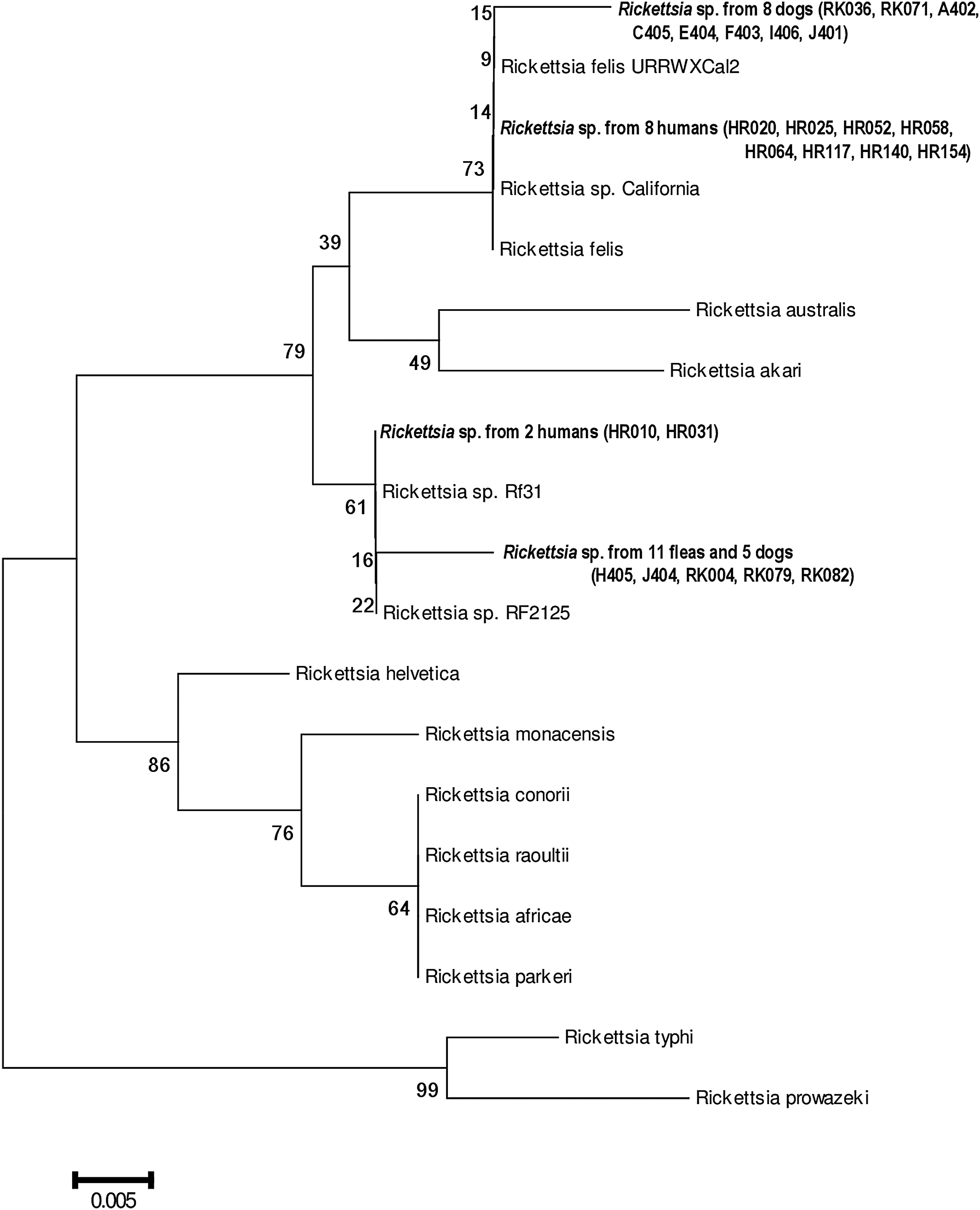
Phylogenetic relationships among 135 bases of gltA gene of Rickettsia DNA from 10 human blood (buffy coat) samples (HR020, HR025, HR052, HR058, HR064, HR117, HR140, HR154, HR010, and HR031; deposited in GenBank under the acc. nos.: MG264722–MG264731, respectively) 13 dog blood samples (RK036, RK071, A402, C405, E404, F403, I406, J401, H405, J404, RK004, RK079, and RK082; deposited in GenBank under the acc. nos.: MG264714–MG264721, MG264732–MG264736, respectively) 11 flea-group samples from Bangkok (deposited in GenBank under the acc. nos. MG264737), and 15 corresponding Rickettsia from GenBank under the following acc. nos.: Rickettsia felis URRWXCal2 CP000053.1, Rickettsia africae KJ645939.1, Rickettsia akari CP000847.1, Rickettsia australis U59718.1, Rickettsia raoultii KM288494.1, Rickettsia sp. California 2 AF210692.1, Rickettsia helvetica KF859959.1, Rickettsia conorii KT345977.1, Rickettsia monacensis GI 749495816, Rickettsia sp. Rf31 AF516331.1, Rickettsia australis CP003338.1, Rickettsia sp. RF2125 AF516333.1, Rickettsia typhi CP003398.1, and Rickettsia prowazekii CP004889.1 Phylogenetic analyses were constructed by neighbor-joining method using MEGA6 sequencing software (

Phylogenetic relationships between 267 bases of 17 kDa antigen protein gene of Rickettsia DNA from human blood samples HR052, HR072, HR103, and HR124 (deposited in GenBank under the acc. nos. MG264710, MG264711, MG264712, and MG264713, respectively), a dog blood sample A402 (deposited in GenBank under the acc. no. MG264709), and from 7 flea-group samples from Bangkok (deposited in GenBank under the acc. nos. MG452137–MG452143) and 15 corresponding Rickettsia from GenBank under the following acc. nos.: Rickettsia felis URRWXCal2 CP000053.1, Rickettsia rickettsii GU723476, Rickettsia rhipicephali DQ865207, Rickettsia hoogstraalii FJ767736, Rickettsia australis M74042, Rickettsia raoultii JX885457, Rickettsia cooleyi AF031534, Rickettsia helvetica JQ711257, Rickettsia monacensis GU292312, Rickettsia sp. SE313 DQ166937, Rickettsia sp. cf1and5 AY953286, Rickettsia australis M74042, Rickettsia endosymbiont of Ctenocephalides felis isolate F144 JF511462, Rickettsia typhi CP003398, and Rickettsia prowazekii AY730679. Phylogenetic analyses were constructed by neighbor-joining method using MEGA6 sequencing software (
Regarding PanR8 assay for 23S rRNA gene detection, 33 sequence results (82 bp) were identical to those of SFG Rickettsia in the GenBank database, including Rickettsia rickettsii CP006010, Rickettsia massiliae CP000683, Rickettsia raoultii CP010969, Rickettsia rhipicephali CP013133, and Rickettsia monacensis LN794217. The other three sequences were from human samples (HR072, HR103, and HR124) and all three were similar to R. typhi CP003397 in the GenBank database. These three samples also showed 17 kDa antigen positive, with a sequence similar to R. typhi (Fig. 2).
There were 13 dog blood samples that were gltA positive. Eight of those samples (RK036, RK071, A402, C405, E404, F403, I406, and J401) were 99% similar (131/132 bp) to R. felis URRWXCal2 (Fig. 1). One dog sample (A402) in this group also showed positive for the 17 kDa antigen gene that also had a sequence identical to R. felis (Fig. 2). The gltA gene from five dog samples (H405, J404, RK004, RK079, and RK082) was identical to Rickettsia sp. RF2125 and Rickettsia sp. Rf13, as shown in the same branch in Figure 1.
The PCR product of Rickettsia gltA gene found in 11 fleas were from the R. felis-like sp., Rickettsia sp. RF2125, or Rickettsia sp. Rf13 groups (Fig. 1). In addition, 8 of 11 showed 17 kDa antigen sequences similar to Rickettsia sp. cf1and5 17 kDa antigen gene (GenBank acc. no.: AY953286.1) and Rickettsia in Ctenocephalides canis 17 kDa antigen gene (GenBank acc. no.: JF511461), similar to and as described in our previous report (Foongladda et al. 2011).
Regarding pairs of dog blood and flea samples, only one pair was positive by gltA gene detection. That pair was in the same group of Rickettsia sp. RF2125 and Rickettsia sp. Rf13.
Analysis results of all 26 positive human samples revealed that 8 gltA gene positive samples were identical to the gltA gene of R. felis URRWXCal2 (GenBank acc. no.: CP000053). In this group, one sample (HR052) was also positive by 17 kDa antigen PCR and showed a sequence identical to the 17 kDa antigen gene of R. felis URRWXCal2 (Figs. 1 and 2). Therefore, these findings confirm R. felis infection in Thai patients, as shown in the phylogenetic analysis in Figure 1. The other two gltA gene-positive human samples (HR010, HR031) presented 99% similarity (133 and 134 bp, respectively) to Rickettsia sp. RF2125 (GenBank acc. no.: AF516333) and Rickettsia sp. Rf31 (GenBank acc. no.: AY953289) (Fig. 1).
Discussion
Rickettsia is a fastidious, obligate, intracellular bacteria that cannot be grown on simple culture media. Although, PCR technique was developed to detect the organism, false negative could be presented given the nature of intraendothelial cells to low-level copy in blood circulation (Williams et al. 1992, Walker et al. 1996) and the possibility of missampling during low-severity phase of intermittent bacteremia. In this study, we used real-time PCR assay with probe detection 23sRNA gene fragment (PanR8 assay), which has proven high sensitivity for detection of Rickettsia spp. in blood samples (Kato et al. 2013). Additionally, we detected the gltA gene fragment by real-time PCR with probe and the 17 kDa antigen by nested PCR for species analysis of amplicon sequences. In these detections, no false positives were observed in the negative control. By phylogenetic analysis, three groups of Rickettsia gene were detected, such as R. felis, R. felis-like sp., and R. typhi.
According to a previous finding of R. felis-like sp. in fleas from dog only, not from cat (Foonglaadda 2011), this study confirmed the organism in five dogs. The gltA gene sequences found in these dogs were similar to those found in fleas in previous studies (Parola et al. 2003, Foongladda et al. 2011). Interestingly, all fleas in this study, 57.89% containing R. felis-like sp., were collected from dogs with fever, anemia, and petechial skin, but only one of the pair dog showed positive results. This may be due to missampling during bacteremia or other pathogens.
Eight dogs had gltA gene of R. felis and 17 kDa antigen sequence could additionally validate in only one dog. However, this was enough evidence to reveal R. felis infection in dogs in Thailand. Both importantly and regrettably, we were unable to analyze fleas known with certainty to have come from that validated dog for pathogen investigation. R. felis has been reported as an emerging vector-borne pathogen with worldwide distribution in cats, dogs, and humans, with cat fleas (Azad et al. 1992, Raoult et al. 2001). However, fleas and ticks from dogs and cats in Bangkok showed no positive detection of R. felis (Foongladda et al. 2011.). Other vectors, such as tick, mosquito, lice, and mites, have been reported (Parola et al. 2005, Mediannikov et al. 2013, Sekeyová et al. 2013, Khrouf et al. 2014). Therefore, these other arthropods are probable vectors of transmission that should be further studied.
In human blood, we found R. felis in eight patients, R. felis-like sp. in two patients, and R. typhi in three patients. One of eight R. felis patients was positive by 17 kDa antigen gene detection. R. typhi in three patients were positive by both 23S rRNA and 17 kDa antigen gene detection. These were corrected gltA gene negative detection due to the SFG-specific group of the primers. These three patients had positive antibody by indirect immunofluorescence to R. typhi. This confirmed results that they were not cross-reacted with SFG rickettsiae.
Thirteen human and two dog samples were positive by 23S rRNA, but negative by the other genes for species analysis, which may be due to variations in DNA in this genus (Fournier and Raoult 2007, Fournier and Raoult 2009, Merhej et al. 2014).
This study has some mentionable limitations. In this study, Rickettsia DNA was found in 4.25% of dog blood samples, which was lower than the 15.47% prevalence found in patient blood samples. The high proportion of positive results in human patients has several possible explanations. Blood samples were taken from patients with suspected scrub typhus, murine typhus, or leptospirosis, whereas dog blood samples were taken from any dog with nonspecific symptoms. Moreover, we used buffy coat samples from patients and whole blood samples from dogs. The predominance of white blood cells in buffy coat samples may yield more Rickettsia than the whole blood samples from dogs. In addition, it is possible that bacteremia was intermittent and perhaps missed due to low level of severity during the sampling period. Moreover, our use of detection assays was limited by the fact that no internal control was used. Consequently, specimens with PCR inhibitors could have been falsely reported as negative.
This study was the first to report R. felis-like sp. DNA in patient buffy coat samples and dog blood samples in Bangkok. It is possible that R. felis-like sp. may have a preference for living and replicating as an endosymbiont in fleas. It is then and therefore possible that dogs and humans may be accidentally infected through the bite of an infected flea. Accordingly, differential diagnosis of this disease and disease control measures should be established as components of a public health strategy to thwart this disease.
Footnotes
Acknowledgments
The authors gratefully acknowledge the assistance of Kevin P. Jones for editing the article. This research project was supported by Mahidol University.
Author Disclosure Statement
No conflicting financial interests exist.