
Abstract
Bluetongue is a re-emergent arthropod-transmitted viral disease that affects all wild and domestic ruminant species, reducing herd productivity. The epidemiology of bluetongue virus (BTV) infection is poorly defined in much of the world, including extensive portions of Asia and the Middle East. In the Republic of Korea (ROK), scarce information is available on the status of BTV infection. Despite evidence of BTV in neighboring countries, such as the People's Republic of China and Japan, and the presence of Culicoides species that have recently been reported as possible vectors for BTV transmission, no serological data on BTV infection or circulation have been reported in the ROK. The objective of this study was to investigate the seroprevalence of BTV antibodies in the domestic goat population to glean further insights into BTV epidemiology in ROK. The results of this study indicate that in 2012–2013, on a countrywide level, the estimated seroprevalence rate of BTV antibodies was 15.7% (50/318) in domestic goat flocks and 11.7% (94/802) in individual animals. To our knowledge, this is the first serological evidence of antibodies against circulating BTV in the domestic goat population in the ROK.
Introduction
B
This vector-borne viral disease has high socioeconomic and sanitary consequences. The subsequent imposition of strict guidelines on the movement of animals, embryos, semen, and other animal products from BTV endemic regions has caused substantial economic damage to BTV-affected areas, exceeding even the direct economic losses caused by BTV-induced morbidity and mortality (MacLachlan and Osburn 2006). Worldwide, BTV has been estimated to cause direct (disease) and indirect (trade, vaccines, etc.) losses of more than $3 billion per year (Tabachnick 1996, Gethmann et al. 2015, Rushton and Lyons 2015, Grewar 2016). It is therefore listed as a “notifiable disease” by the Office International des Epizooties (OIE) (Maclachlan 2011, Rushton and Lyons 2015).
Because small ruminants, including goats and sheep, can be regarded as indicators of BTV circulation, many previous studies conducted in many worldwide countries have used goats to monitor circulating BTV or for surveillance of BTV antibodies, for example, in Australia (St George 1985a, 1985b), the Netherlands (Elbers et al. 2008), Poland (Czopowicz et al. 2010), Albania (Lelli et al. 2004), Croatia (Listes et al. 2009), Kazakhstan (Lundervold et al. 2004), Iran (Najarnezhad and Rajae 2013), Iraq (Hafez et al. 1978), Saudi Arabia (Hafez and Taylor 1985, Abu Elzein et al. 1998), Turkey (Taylor and Mellor 1994), India (De et al. 2009), Taiwan (Lee et al. 2010), Nigeria (Taylor and McCausland 1976), Sudan (Eisa et al. 1979), the Caribbean and Florida (Gibbs and Greiner 1985), and some countries in the Caribbean region and South America (Gibbs et al. 1983, Gumm et al. 1984).
BTV occurs throughout most tropical and temperate regions worldwide (Osburn 1994, Maan et al. 2007). By and large, this distribution is a reflection of the distribution of its Culicoides vectors and the temperature required for BTV replication in and transmission by vectors. In the field, BTV is transmitted between its vertebrate hosts almost entirely through the bites of various species of the Culicoides biting midge (Standfast and Muller 1989, Mellor 1990, Schwartz-Cornil et al. 2008, Maclachlan et al. 2009).
The epidemiology of BTV infection is poorly defined in much of the world, including extensive portions of Asia and the Middle East. In the Republic of Korea (ROK), BTV was first identified in Chungnam Province in 2014 (Seo et al. 2015), but scarce information is available concerning the status of BTV infection in the ROK to date. Despite the evidence for BTV in neighboring countries in the Far East, such as the People's Republic of China and Japan (Nianzu and Kirkland 1998, Kirkland et al. 2002, Sugiyama et al. 2009), and the presence of Culicoides species that have been recently reported as being possible vectors for BTV transmission (Kim et al. 2012, Oem et al. 2013), no serological information on BTV infection or circulation has been reported in the ROK. Therefore, the objective of this study was to investigate the seroprevalence of BTV antibodies in domestic goat populations to gain further insights into the epidemiology of BTV in the ROK. As a serological indicator of BTV circulation, this study targeted goats distributed countrywide in the ROK because the sheep population was too small to be investigated, although bluetongue is an especially economically important infectious disease in this species (ROK goat 10,291 flock 242,787 individual animals vs. sheep 103 flock 2522 individual animals in 2013) (Livestock Management Division 2015).
Materials and Methods
Field samples for serological surveys
The seroprevalence rates of BTV antibodies were estimated at the herd and animal levels in goats. A serological survey was conducted between 2012 and 2013 on domestic goats for specific antibodies to BTV. The serum samples from goats were obtained from two separate sources: (1) from the serum bank of the National Foot-and-Mouth Disease (FMD) Surveillance Program maintained by the Foreign Animal Diseases Division of the National Veterinary Research and Quarantine Service (Anyang, Republic of Korea) and (2) from goats sampled in close collaboration with local veterinary practitioners and/or government veterinary officers. In the samples obtained from the serum bank, the sera used in this study were initially tested for FMD according to the monitoring measures issued and conducted by the Ministry of Agriculture, Food, and Rural Affairs between 2012 and 2013, and only the sera that were confirmed to be negative for the FMD virus were analyzed for bluetongue-specific antibodies in this study. To collect the serum samples only for BTV serosurveillance in this study, the farm selection and blood sampling were conducted in close collaboration with local veterinary practitioners and/or government veterinary officers. As a result, a total of 802 goat blood samples collected from 318 flocks in 11 provinces in the ROK (33°06′ N–39°25′ N, 124°36′ E–131°52′ W) were obtained. Signs typical of bluetongue disease were not observed in any of these animals. The number of samples from each province is shown in Table 1. The blood and serum separated from the blood samples were stored at −20°C until further analysis. The animals were classified into three age groups based on tooth replacement and livestock owner questionnaires: juveniles (between 6 months and 1 year of age), subadults (between 1 and 2 years of age), and adults (>2 years of age). Unfortunately, the ages of eight animals could not be recorded. The study population included 82 animals that aborted or showed reproductive problems during 2012–2013 and 720 that did not. We investigated the association between the seroprevalence and local seropositivity risk factors. These included the population sizes of flocks, stocking animal density, adult/juvenile ratios of flocks, availability of a vector habitat in the environment (whether the herd was located in a rural area or near an urban area), age, and reproductive problems, including abortion. Statistical analyses were performed with SPSS software (SPSS for Windows, version 11.5; SPSS, Inc., Chicago, IL) using the Chi-squared test, Fisher's exact test, and Student's t-test as appropriate.
Serotypes identified by SNTs.
Number of seropositive herds or animals (individuals).
BTV serotype identified by RT-PCR for blood sample.
AP, apparent (estimated) prevalence; BTV, bluetongue virus; ND, not detected; SNT, serum neutralization test; TP ±95% CI, 95% confidence interval for the true prevalence.
Field samples for the retrospective study
Goat serum samples from the serum bank of the National Animal Disease Surveillance Program maintained by the Foreign Animal Diseases Division of the National Veterinary Research and Quarantine Service were used for the retrospective serological investigation. In total, 2911 goat serum samples collected between 2003 and 2008 were analyzed for the presence of bluetongue-specific antibodies (Table 2).
NA indicates samples not available.
Bluetongue-specific antibody testing
It has previously been reported that specific serodiagnostic techniques, such as the competitive enzyme-linked immunosorbent assay (cELISA) or BTV neutralization tests, should be used for bluetongue surveillance in Ibaraki virus (IBV)-endemic areas because IBV-positive serum samples may result in false-positive bluetongue agar gel immunodiffusion test reactions (Shimizu et al. 2004). Because the ROK is an IBV-endemic region, two commercially available commercial cELISA kits based on a VP7-specific monoclonal antibody, namely, the BTV Antibody Test Kit (VMRD cELISA; Veterinary Medical Research and Development, Inc., Pullman, WA) and the BTV VP7 Antibody Test Kit (IDEXX cELISA; IDEXX Laboratories, Inc., Institute Pourquier, Montpellier, France), were used to identify the presence of antibodies against BTV in the serum samples. BTV VP7 is the major structural protein of the inner core and contains group-specific antigenic determinants (Mertens et al. 1984). Therefore, most diagnostic methods for BTV-specific antibody detection have been based on BTV VP7 (Cloete et al. 1994, Zhou et al. 2001, Mecham and Wilson 2004, Chand et al. 2009, 2017, Yang et al. 2010, Yin et al. 2011, Venkatesan et al. 2015, Xu et al. 2015). The assays were performed according to the manufacturer's instructions. The specificity and sensitivity of the VMRD cELISA Kit are 99.3% and 69.5%, respectively, as stated by the manufacturer, and those of the IDEXX cELISA kit are 100% and 82.8%, respectively (Niedbalski 2011). To maximize the test specificity of this study, all samples were tested using both cELISA kits, and a serum sample was defined as “positive” only when it yielded positive results from both tests. A herd was defined as “positive” when at least one seropositive sample was present. The seroprevalence rates and the 95% confidence intervals were calculated using the program “Survey Toolbox for Livestock Diseases” (Ausvet).
The serum neutralization test (SNT) was used to confirm the positive ELISA results. Positive and negative controls for the SNT were obtained from the Institute for Animal Health, Pirbright, United Kingdom. The microtiter neutralization method was used in this study according to the Manual of Diagnostic Tests and Vaccines for Terrestrial Animals (OIE-World Organization for Animal Health 2008). Only serum samples collected from goats between 2012 and 2013 were available for SNT, whereas field samples for the retrospective study could not be used due to an insufficient volume and quality. Briefly, ∼100 TCID50 (50% tissue culture infective dose) of the standard or untyped virus was added in 50 μL volumes to test wells of a flat-bottomed microtiter plate and mixed with an equal volume of standard antiserum that had been serially diluted in tissue culture medium. Approximately 104 cells were added per well in a volume of 100 μL and assessed after incubation for 4–6 days using an inverted microscope. Wells were scored for the degree of cytopathic effects observed.
RT polymerase chain reaction
A total of 802 blood samples were tested for the presence of BTV RNA by means of a nonserotype-specific reverse transcription–polymerase chain reaction (PCR) assay targeting a conserved region within the VP3 gene of the BTV genome according to the method described by Yeh et al. (2011). It is known that RNA segment 3 of BTV encodes a serogroup-reactive protein, VP3, and that VP3 sequences are often used for the genetic characterization of BTV serotypes worldwide (Huismans and Erasmus 1981, Gould and Pritchard 1991, McColl and Gould 1991). Briefly, total nucleic acids were extracted from 400 μL of whole blood. Automated extraction was performed using a BioRobot M48 workstation apparatus (Qiagen GmbH, Hilden, Germany) with the MagAttract Virus Mini M48 Kit (Qiagen GmbH). Nucleic acids were recovered in 50 μL of elution buffer. The reverse transcription-polymerase chain reaction (RT-PCR) was performed using the One-Step RT-PCR Kit (Qiagen GmbH). The reactions were prepared in a volume of 25 μL containing 2 μL of RNA, 1× buffer [Tris-Cl, KCl, (NH4)2SO4], 2.5 mM MgCl2, 0.2 mM deoxynucleoside triphosphates, 0.4 μM aliquots of each of the specific primers, 5 U of RNase inhibitor (Intron Biotechnology), and 1 μL of enzyme mix (Omniscript and Sensiscript RTs, HotStartTaq DNA polymerase; Qiagen GmbH). Sequences of oligonucleotide primers for the dual priming oligonucleotide system used in this study are shown in Table 3. Reverse transcription amplification was accomplished in one step with the following optimized incubation program: 30 min at 50°C; 15 min at 95°C; 40 cycles of 94°C for 30 s, 61°C for 90 s, and 72°C for 50 s; and 1 min at 72°C. RT-PCR amplifications were performed using an Eppendorf MasterCycler gradient thermal cycler (Eppendorf). RT-PCR amplification products (5 μL) were analyzed by gel electrophoresis on a 3% agarose gel containing 0.5 μg of ethidium bromide/mL. For identification and differentiation of bluetongue virus (BTV) serotypes, previously published serotype-specific RT-PCR (Maan et al. 2012) was performed using BTV RNA-positive samples identified by RT-PCR for the detection of pan-BTV. The positive control for the BTV serotype RNAs was prepared according to a method previously described by Yeh et al. (2011). The RT-PCR assays were not performed on the retrospective goat serum samples from 2003 to 2008 because of an insufficient sample volume.
Abbreviation for mixed base code. Y = Pyrimidine (C or T); H = A, C, T (not G); R = Purine (A or G).
Statistical analysis
Statistical significance was set at the 5% level, and two-sided p-values were calculated for analysis of the correlation between the data by age and the studied area categories, respectively. The statistical analysis was performed through the paired t-test, and a p-value <0.05 was accepted as significant. All data were analyzed using GraphPad PRISM software (version 6.07 for Windows; GraphPad Software, Inc.).
Results
In 2012–2013, at a countrywide level, the estimated seroprevalence rate of BTV antibodies was 15.7% (95% confidence interval: 12.1–20.1%) for domestic goat flocks (50/318) and 11.7% (95% confidence interval: 9.7–14.1%) for individual goats (94/802) (Table 1). For reference, true prevalence (TP) is the proportion of animals that present the event of interest in the population at a given point in time. In general, the true value is practically impossible to determine, but it can be estimated from the apparent prevalence (AP). AP is the proportion of animals from a representative sample of the population that are positive using a diagnostic method. In this study, TP estimates were calculated as described by Rogan and Gladen (1978). The prevalence of BTV antibodies in each province ranged from 0% to 33.3% for goat flocks and from 0% to 40.0% for individual goats.
Table 1 shows the prevalence of antibody-positive serum samples collected in 2012–2013 from goats of mixed ages in the different municipalities. The municipality survey results suggested that BTV may have been widely distributed across goat populations in the ROK.
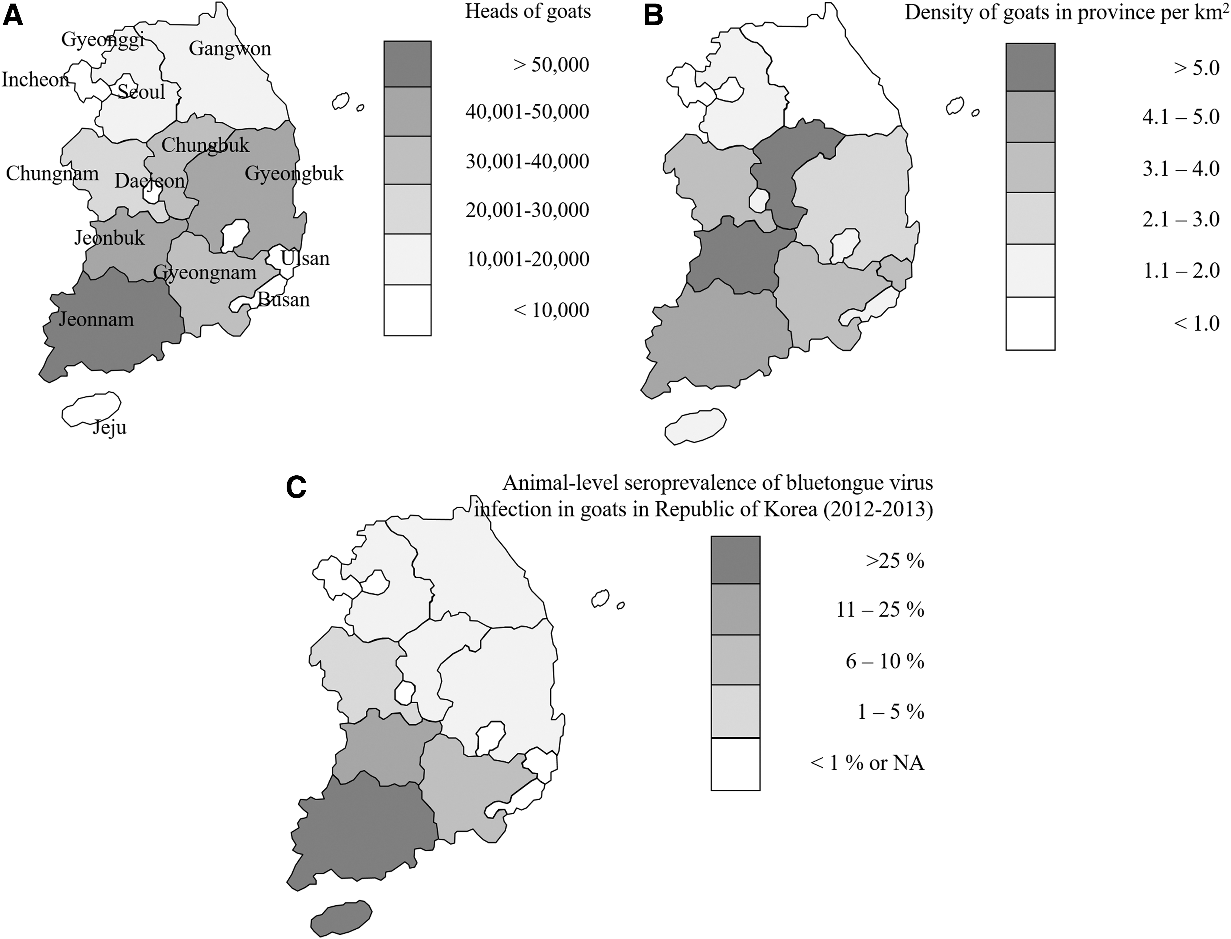
The risk of being seropositive for BTV was associated with age and geographic zone. The seroprevalence was higher in adults (>2 years) than in subadult (1–2 years) and juvenile (6 months to 1 year) goats (Table 4). The risk of being a seropositive animal was 3.5 and 5.5 times higher in the adult population than in subadults and juveniles, respectively. The seroprevalence increased progressively from northern to southern zones, without clinical evidence of BTV infection throughout the country. A significantly higher seropositivity was also found in western provinces than in eastern provinces (Fig. 1), and the risk of being a seropositive animal was 3.7 and 3.2 times higher in the southern and western provinces than in the northern and eastern provinces, respectively. Of the positive goat flocks, 76.0% (38/50) had a seroprevalence either <20% or >81%, suggesting a bimodal frequency distribution (Fig. 2).

Frequency distribution of the seroprevalence rates of BTV infection in goats in the ROK (2012–2013).
The results in this study revealed a significant association between age and BTV seroprevalence (p < 0.05) and showed that more seropositive flocks were located in the western and southern areas, indicating that the risk of BTV exposure was significantly higher than that in the eastern and northern areas (p < 0.005).
Number of seropositive herds or animals (individual animals).
Adults, >2 years of age; subadults, 1–2 years of age; juveniles, 6 months to 1 year of age; age information of some animals in this study was not available.
In this study, the northern half of the ROK includes Incheon, Gyeonggi, Gangwon, Chungbuk, and Chungnam, whereas the southern half includes Ulsan, Jeonbuk, Jeonnam, Gyeongbuk, Gyeongnam, and Jeju (divided along latitude 36°N). The western half of South Korea includes Incheon, Gyeonggi, Chungnam, Jeonbuk, Jeonnam, and Jeju, whereas the eastern half includes Ulsan, Gangwon, Gyeongbuk, and Gyeongnam (divided along longitude 127°E). ROK, Republic of Korea.
Attempts to establish a correlation between seropositive rates and (1) the population sizes of flocks, (2) the adult/juvenile ratios of flocks, and (3) the availability of a vector habitat in the environment (whether the herd was located in a rural area or near an urban area) were inconclusive (data not shown). In addition, although there were substantial regional differences in seroprevalence within the ROK (Fig. 1), there was no significant correlation between serological status and reproductive problems, including recent abortion, as 10/82 (12.2%) of recently aborted goats and 48/412 (11.6%) of female goats that delivered normally were seropositive for BTV.
Although no clinical cases of bluetongue have been reported, the serological results showed that the virus has been widely distributed in goats in the ROK since 2005 (Table 2), with seropositive rates ranging from 0% to 9.1% between 2003 and 2008. Of the 94 ELISA-positive samples, only 41 were positive by SNT (Table 1) and neutralized 1 or more BTV serotypes: 1 (21 serum samples), 2 (17 serum samples), 3 (14 serum samples), 4 (11 serum samples), 15 (8 serum samples), and 16 (9 serum samples). In contrast, three goat serum samples failed to neutralize any known BTV serotype. In addition, the RNA of the BTV-1, -2, -3, and -4, 16 serotypes was detected in 10 serologically positive blood samples by RT-PCR, indicating that BTV was circulating in the goat populations in the studied area. Further phylogenetic analysis and virus isolation for these blood samples could not be performed using these samples because of an insufficient blood volume and quality of the positive samples.
Discussion
The widespread occurrence of infection with BTV in the ROK has been demonstrated by ELISA, SNT, and RT-PCR. Our serological results showed that approximately one in seven goat flocks and one in nine goats were infected (Table 1). To understand the results of the present study, it is important to note that the ROK has no vaccination program for bluetongue. Thus, considering that all the sera evaluated in this study were collected from domestic animals and not from imported animals from countries that are BTV endemic or that vaccinate for BTV, the high seroprevalence of BTV infection can be assumed to reflect natural infection of domestic goats in the ROK.
Our results revealed a significant association between age and BTV seroprevalence in the ROK (p < 0.05), similar to previous reports by several groups (Ward et al. 1994, Linden et al. 2010, Garcia-Bocanegra et al. 2011). The higher seroprevalence in adult animals was probably due to a greater exposure of this age group to circulating endemic virus over time (Falconi et al. 2011, Garcia-Bocanegra et al. 2011).
Our analysis showed that location was a significant factor, which indicates that the univariate results were confounded by the location variable because more seropositive flocks were located in the western and southern area of the ROK. Thus, the risk of BTV exposure in these areas was significantly higher than that in the eastern and northern areas (p < 0.005).
The immunological analysis demonstrated that BTV was widely distributed, although bluetongue has been clinically uncommon in goats in the ROK, probably because clinical signs in infected goats are far less obvious than in sheep, allowing BTV to circulate undetected. Similarly, no clinically affected goats were reported during the 2006 epidemic in Northwest Europe (Elbers et al. 2008).
A retrospective study found BTV antibodies in the ROK goat population as early as 2005, although the ROK was believed to be officially BTV free. The present study showed that since 2005, the geographic distribution of antibodies against BTV has remained stable, but the overall seroprevalence has increased, which is indicative of ongoing virus transmission. Together with the serological evidence, RNA detection of BTV-1, -2, -3, and -4, 16 serotypes in 10 antibody-positive blood samples collected from animals without obvious clinical signs of disease indicates a circulation of BTV in goat populations in the studied area. Further studies might include virological and serological investigations of BTV-15 circulating in the ROK because neutralizing antibodies against BTV-15 have been detected in goat blood samples from Chungnam and Jeonnam provinces. However, none of the samples in the present study showed virological evidence of BTV-15. Collectively, the significance of these serological findings has been discussed, and planning for further studies to isolate local BTV strains from sentinel flocks of small ruminants, including goats has been proposed. Future surveillance programs for BTV should be extended to include other susceptible animals such as sheep, cattle, other ruminants, and biting Culicoides midges.
Conclusion
To our knowledge, this is the first evidence of antibodies against circulating BTV in the ROK. Nevertheless, an outbreak of BTV among native sheep in the ROK has yet to be officially reported; and BTV insect vectors and specific virus serotypes circulating in the ROK remain to be identified. Further investigation is required to characterize the serotypes of BTV present in the ROK. In addition, surveillance studies should be implemented to monitor the spread of BTV and the emergence of new serotypes. Future surveillance programs for BTV should be extended to include other susceptible animals, such as sheep, which are more likely to suffer the effects of BTV infection and therefore lead to economic losses. In addition, the high-risk animal species (native sheep) and the distribution of Culicoides vectors in the ROK should also be monitored to facilitate the prediction and response to a possible BTV outbreak in the ROK.
Footnotes
Acknowledgments
The authors thank all of the farmers who volunteered to participate in the study. The authors would also like to acknowledge the individuals who helped to recruit farms to the study and the farmers who allowed us to collect serum samples at their farms. The authors are very grateful to the local government veterinary officers and veterinary practitioners for their help in collecting blood samples from goats throughout the country. They are grateful to two anonymous reviewers who reviewed the first draft of the article and for their critical comments and suggestions. Bluetongue-specific antibody testing, SNT, and RT-PCR were performed at the National Veterinary Research and Quarantine Service. Literature review, data analysis, statistical analysis, and the writing of the article were conducted at Incheon National University (Incheon, Republic of Korea). This work was supported by a 2014 Incheon National University Convergence Research Grant to Jung-Yong Yeh.
Author Disclosure Statement
No competing financial interests exist.