
Abstract
The open reading frame of the nucleocapsid protein (NP) of Rift Valley fever virus (RVFV) strain MP12 was cloned and expressed in Vero E6 cells. The recombinant NP (rNP)-expressing cells were used as antigens for an indirect immunofluorescent antibody assay (IFA). The rNP-based IFA and RVFV-infected Vero E6 cell (authentic antigen)-based IFA showed similar IFA profiles with immune rabbit serum, which was prepared by immunization with rNP expressed using a baculovirus vector. A total of 942 traditional cattle sera obtained in five districts in Central, Southern, and Western provinces of Zambia were screened for anti-RVFV antibodies by the authentic antigen-based and rNP-based IFAs. Significant agreement was obtained between the two IFAs. The findings show that the rNP-based IFA is a safe and useful diagnostic tool as an alternative to the authentic antigen-based IFA. The antibody titers given by the rNP-based IFA were higher than those by the authentic antigen-based IFA. Therefore, the rNP-based IFA might be useful for serosurveillance of RVFV infection among cattle. Antibody prevalence rates in the five districts were 1.3% to 13.5% in the authentic antigen-based IFA and 6.0% to 21.4% in the rNP-based IFA. The results indicated that despite no reports of active cases of RVF in these provinces of Zambia, the virus is circulating among cattle herds.
Introduction
R
In Zambia, serological evidence of RVF was first reported in 1974 after an epizootic of disease among cattle and sheep in Chisamba (Central Province) and Mazabuka (Southern Province) districts, and some parts of Copperbelt Province (Hussein et al. 1987, Davies et al. 1992). However, currently, there have been limited reports on the geographical distribution of RVFV in Zambia, even though the presence of the virus in neighboring countries has been reported (Morita 1988, Hasebe et al. 1989, Dautu et al. 2011). The existence of wildlife and livestock interfaces in Zambia provides a conducive environment for maintenance and spread of the virus (LaBeaud et al. 2011). Comprehensive surveillance of the disease in cattle would clarify the extent of virus distribution and whether the virus is circulating in livestock in the country. To understand the epidemiology of RVF in Zambia, a simple, rapid, and safe diagnostic and surveillance tool is required.
Saijo et al. (2002, 2005) reported that the recombinant nucleocapsid protein (rNP)-based indirect immunofluorescent antibody assay (IFA) system for diagnosis of Crimean-Congo hemorrhagic fever virus infection among suspected patients was a safe, sensitive, and specific diagnostic method.
In this study, rNP of the RVFV strain MP12 was expressed in mammalian cells and used as an antigen for IFAs in cattle sera. The usefulness of the rNP-based IFA for serological screening of cattle sera was evaluated by comparison with the reactivity to the strain MP12-infected cell-based IFA (authentic IFA antigen).
Materials and Methods
Preparation of an authentic indirect IFA antigen
RVFV strain MP12 was used to prepare an authentic antigen for IFA as previously described (Fukuma et al. 2016). Briefly, Vero E6 cells were infected with RVFV. Briefly, Vero E6 cells were infected with RVFV at a multiplicity of infection of 1.0 or mock-infected and cultured overnight. After overnight incubation, almost all of the cells were infected with the virus. The cells were trypsinized and washed with phosphate-buffered saline (PBS). Mixture of virus-infected and mock-infected cells at ratio 1:3 were spotted onto 14-well slides and fixed with acetone at room temperature for 5 min. Mixing with uninfected cells makes it easy to distinguish the positive fluorescent staining from nonspecific staining in IFA. The slides were stored at −80°C until use.
Preparation of an rNP-based IFA antigen
Total RNA was obtained from RVFV strain MP12-inoculated Vero E6 cells as previously described (Fukushi et al. 2012). The NP gene was amplified from total RNA by one-step reverse transcription PCR using a QIAGEN OneStep RT-PCR kit (QIAGEN) and the primer pair SacI-MP12-NP1F (5′-CCGAGCTCATGGACAACTATCAAGAGCTTGCG-3′) and SphI-MP12-NP738R (5′-GCGCATGCTTATTAGGCTGCTGTCTTGTAAGCC-3′). The amplified NP gene tagged with SacI and SphI recognition sites was cloned into a mammalian expression plasmid vector, pCAGGSMCS, following SacI and SphI treatment of the NP amplicons and pCAGGSMCS. Vero E6 cells were grown in Dulbecco's minimum essential medium (DMEM) containing 10% heat-inactivated fetal bovine serum, penicillin, and streptomycin.
The Vero E6 cells were transfected with pCAGGSMCS containing the coding region of RVFV NP using X-tremeGENE HP DNA transfection reagent (Sigma-Aldrich) according to the manufacturer's instructions. The transfected cells expressing RVFV rNP were trypsinized and cultivated on 24-well glass slides overnight at 37°C in 5% CO2. Cell sheets on the wells were washed with PBS and then fixed with acetone at room temperature for 10 min and rinsed with distilled water. After drying, the slides were stored at −80°C until use. Cells with high transfection efficiency such as HEK293T cells were also examined to prepare an IFA slide antigen, but Vero E6 cells were chosen because of their tight adhesion to a glass slide (data not shown). Transfection efficiency of Vero E6 cells was estimated to be about 5–10% because of the ratio of rNP-expressing cells detected by immune rabbit serum as described below. Because 90% of the cells did not express rNP antigens, untransfected cells were not mixed with transfected cells as carried out in preparation of an authentic antigen slide.
Immune rabbit serum
Rabbit immune serum against rNP of RVFV was prepared as previously described (Fukushi et al. 2012). Briefly, rNP of RVFV strain MP12 was expressed by a baculovirus vector as a six histidine-tagged protein. The rNP was purified by nickel chelate column and the purified rNP was used for immunization.
Cattle serum
A total of 942 serum samples were collected in 2014 from traditional cattle herds in five districts of three provinces of Zambia: Central Province (Mumbwa), Southern Province (Mazabuka and Kazungula), and Western Province (Mwandi and Sesheke) (Fig. 1). The samples were collected for routine surveillance. Also, serum samples were humanely collected in collaboration with the Ministry of Fisheries and Livestock, Zambia, and with the informed consent from cattle owners. The districts were chosen on the basis of accessibility, cooperation by the owners or managers, and availability of animal health and other relevant records. The sera were transported to the laboratory of the University of Zambia and stored at −80°C until analysis.
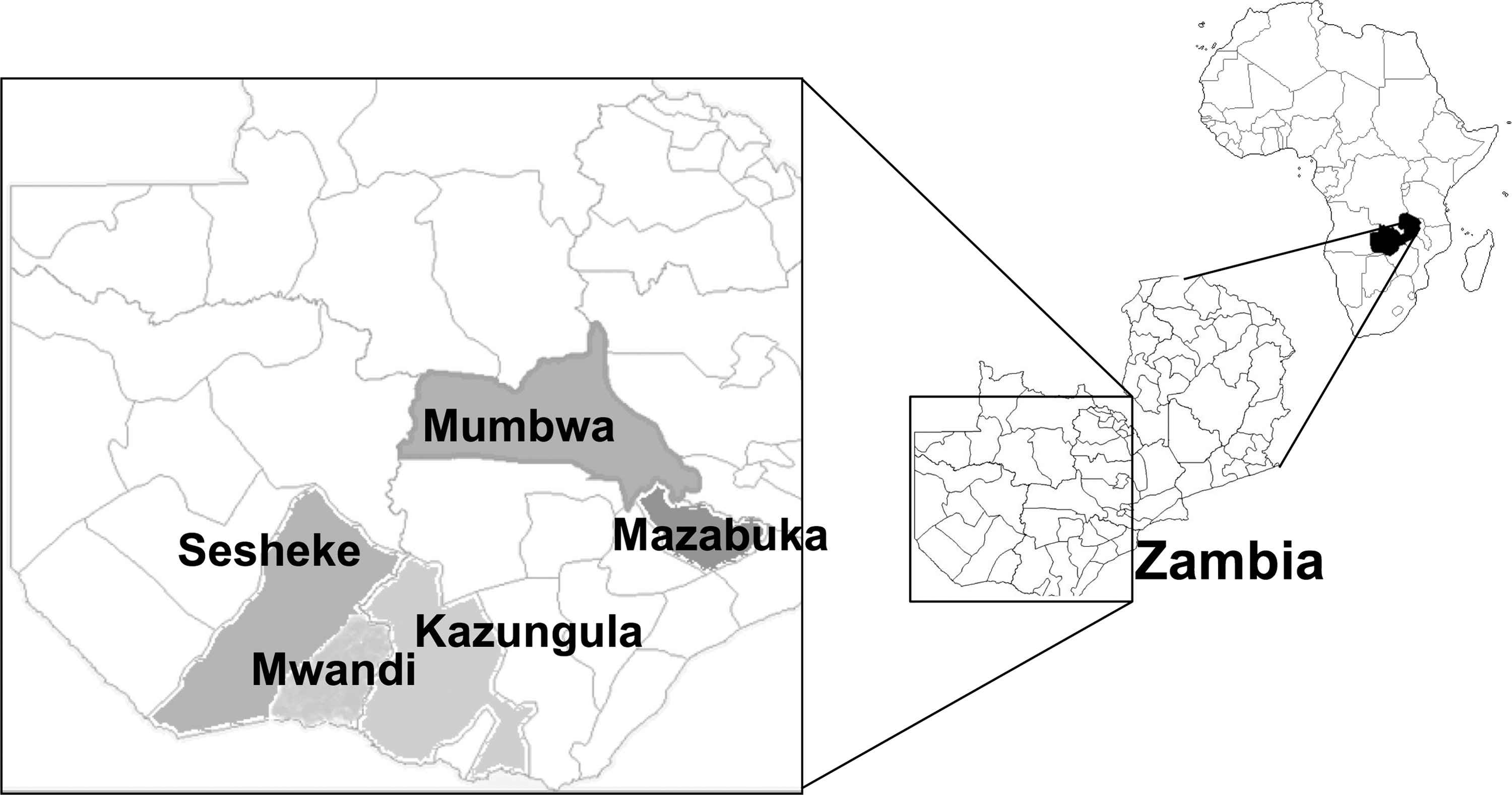
Map of Zambia showing the locations of sampling sites. Sera were collected from traditional cattle herds in five districts of three provinces: Central Province (Mumbwa), Southern Province (Mazabuka and Kazungula), and Western Province (Mwandi and Sesheke).
Indirect IFA
The IFA was performed as basically the same method as described previously (Fukushi et al. 2012) with Alexa Fluor 488®-conjugated goat anti-rabbit IgG antibody (Thermo Fisher Scientific) and FITC-conjugated goat anti-bovine IgG antibody (Kirkegaard & Perry Laboratories, Inc.) as secondary antibodies. Immune rabbit sera against rNP were diluted at 1:160 and cattle sera were diluted at 1:100 with PBS. Twenty microliters of the diluted sera was applied on wells of IFA slides. After a 1-h incubation at room temperature, the slides were soaked in PBS to wash their wells thrice for 5 min each time. Secondary antibodies at 1:1000 dilutions with PBS were applied on the slide at 500 μL per slide. After incubation for 1 h at room temperature, the slides were washed with PBS as described above. Finally, each slide was covered with 50% glycerol in PBS and a cover glass. Cattle serum showing clear immunofluorescence in the cell cytoplasm was regarded as positive by the IFA, and a cattle serum specimen not showing a specific fluorescent profile was regarded as negative. Judgment of the IFA test was carried out by two to four examiners. A serum specimen for which judgment varied among the examiners was regarded as “intermediate.”
Determination of IFA antibody titers of cattle sera
After screening of bovine sera at 1:100 dilution, IFA antibody-positive or IFA antibody-intermediate sera were selected and then their IFA antibody titers were determined. Ultimately, 48 cattle sera were chosen and diluted with PBS at 1:20. Then, twofold serial dilutions of them were applied on wells of an IFA antigen slide. The IFA antibody titers were expressed as the reciprocals of serum dilution that caused clear immunofluorescence in the cell cytoplasm.
Statistical analysis
(1) Agreement between results of the two IFA tests with authentic antigen and rNP in cattle sera shown in Table 1 was statistically analyzed by Kappa test of vcd package of R ver.3.4.0 (
IFA, immunofluorescent antibody assay; rNP, recombinant nucleocapsid protein.
(2) A total of 48 sera were selected from positive and/or intermediate reactions in both IFA tests and their IFA antibody titers were determined. The correlation of antibody titers from the two IFA tests was statistically analyzed by the function “CORREL” and regression line of Microsoft Excel ver. 15.28.
Results
Expression of rNP
IFA profiles of authentic (Fig. 2A) and rNP-based (Fig. 2B) antigens against immune rabbit serum at a dilution 1:160 were almost the same with specific immunofluorescence in the cytoplasm. No specific staining with the immune rabbit serum was observed in cells without transfection (data not shown). In addition, negative control rabbit serum that was obtained from a rabbit before immunization exhibited no specific florescence in both authentic and rNP-based IFA antigens (data not shown). These results confirmed that the rNP of RVFV was expressed in Vero E6 cells.

IFA profiles of authentic and rNP antigens. Authentic antigen-
Reactivities of cattle sera to authentic and rNP antigens in IFA
One cattle serum that was positive by the authentic antigen-based IFA was selected (Fig. 2C) and used for comparison with the reactivity to the rNP-based IFA. The positive serum showed a clear IFA staining pattern in rNP-expressing cells (Fig. 2D), which was the same as that with immune rabbit serum (Fig. 2B). No specific staining was observed in cells without transfection (data not shown) with staining with positive control rabbit serum and negative cattle sera. Furthermore, negative cattle sera showed no specific staining in both authentic and rNP IFA antigens (data not shown). A total of 942 cattle sera were screened for RVFV antibodies in IFAs with authentic and rNP antigens (Table 1). A total of 46 sera were positive by both IFAs and 826 sera were negative by both IFAs. The overall agreement ratio was 872/942 (92.5%). The Kappa intertest reliability agreement of the rNP and authentic protein was high (Kappa = 0.626). Therefore, results of both IFA tests significantly agreed. Most of the sera that were positive to the authentic antigen were also positive to the rNP antigen. However, the number of sera that were positive to the rNP antigen was about twice more than the number of sera that were positive to the authentic antigen. Twenty-one sera could not be diagnosed with the authentic antigen as they showed IFA patterns with a high background and were thus tentatively categorized as intermediate. On the other hand, there was no intermediate result with the rNP antigen due to the relatively low background staining.
Comparison of antibody titers between IFAs with authentic and rNP antigens
To compare antibody titers between two IFAs, IFA titers of sera showing positive reaction in the screening assay were determined. Forty-eight sera, including 42 sera that were positive to both IFA antigens and 6 sera that showed an intermediate reaction to the authentic antigen, but a positive reaction to the recombinant antigen, were selected depending on the availability of the serum samples. All results for IFA antibody titers against authentic and rNP antigens were plotted in Figure 3. As shown in Figure 3, IFA titers to the two antigens were correlated (r = 0.71). However, the regression line was tilted to the right and the section of the y-axis (rNP antigen) of the regression line was calculated to be 10 × 23.43 (the value was about 107.8). This indicates that antibody titers to the rNP antigen were ∼10 times higher in specimens with low antibody titers.

Comparison of antibody titers between authentic and rNP-based IFAs. A total of 48 sera were selected and their IFA titers were plotted. IFA titers to the two antigens were significantly correlated (r = 0.706). Regression line y = 0.558x + 3.43 and R 2 = 0.498.
Seroprevalence of RVFV among cattle in Zambia detected by IFAs
As shown in Table 2, RVFV antibodies were detected in cattle sera collected in all districts with seroprevalence rates of 1.3–13.5% in the authentic antigen-based IFA and 6.0– 21.4% in the rNP-based IFA. While the positive rates varied among the districts, samples collected in Mumbwa and Mazabuka districts showed high positive rates in both of the authentic antigen- and rNP-based IFAs. In the other districts, positive rates were relatively lower (less than 10%) for both antigens.
+, positive; ±, intermediate; −, negative.
Discussion
In this study, the NP gene of RVFV strain MP12 was cloned and expressed in Vero E6 cells. Since the IFA staining patterns of Vero E6 cells with an authentic antigen and an rNP against anti-rNP immune rabbit serum are the same, the rNP-expressed Vero E6 cells were useful for the IFA antigen (Fukushi et al. 2012). Although BSL-2 facilities are required to handle the infectious RVFV strain MP12, such a containment facility is not needed to prepare an rNP-based IFA antigen. This is an important advantage of the use of a recombinant antigen. Enzyme-linked immunosorbent assay (ELISA) is a simple, rapid, and safe diagnostic method. We tried to offer a different choice from commercial ELISA by establishing IFA. Although preparation of IFA slides requires cell culture facilities, IFA slides can be stored for at least 1 week in a cool box and easily carried to remote area. Also, IFA slides are useful for screening a small number of sera. Furthermore, reliability of diagnosis may increase by examining with multiple diagnostic methods.
Generally, sensitivities and specificities of serological diagnoses are determined by comparison of the results with a sufficient number of true-negative and true-positive sera. However, in the case of livestock animals and wild animals, it is difficult to obtain a large number of such negative/positive control specimens. Therefore, as an alternative, we screened a large number of field samples in comparison to an IFA with an authentic antigen for evaluation of the rNP-based IFA. The results for 942 cattle sera showed a relatively high overall agreement ratio (872/942, 92.5%) between the authentic antigen- and rNP-based IFAs. Statistical analysis also showed that the results of the two IFAs significantly agreed. These results indicated that the rNP-based IFA has sensitivity and specificity comparable to those of the authentic antigen-based IFA.
RVFV strain MP12-infected cells were used for the IFA (Billecocq et al. 1996). The authentic antigen-based IFA gave intermediate (±) results representing high background immunofluorescence. On the other hand, the rNP-based IFA caused relatively low background immunofluorescence and, as a consequence, no intermediate results were obtained. In addition, antibody titers to the rNP antigen tended to be higher, especially in specimens with low antibody titers. Therefore, the rNP-based IFA might be more sensitive for detecting a specific antibody with a low titer. The difference in antibody titers is considered to be due to differences in the quantities of antigens in cells. IFA antibody titers of positive sera were about two- to four-times higher in the rNP-based IFA than in the authentic antigen-based IFA. The total number of positive sera was about two-times higher in the rNP-based IFA than in the authentic antigen-based IFA. Thus, the rNP-based IFA might be useful for serological surveillance to detect RVFV-specific antibodies in cattle sera.
A previous study on RVF in one farm in Chisamba showed a prevalence of 2.5% (Hussein et al. 1987). Other previous studies have also shown evidence of seroconversion of sentinel cattle to RVFV-infected cell IFA antigens in Mumbwa district of Central province (Davies et al. 1992). Our study further showed high RVFV antibody prevalence in Mumbwa, indicating that RVFV has been maintained among cattle in Zambia. Since these areas are adjacent to Kafue National Park, the role of wildlife such as buffalo as a source of RVFV should be considered.
In summary, the rNP-based IFA is a safe and sensitive method for serosurveillance of RVFV infection in cattle. Continuous surveillance for RVFV infection of livestock and humans using this novel tool will help to understand the epidemiology of RVF in Zambia.
Footnotes
Acknowledgments
This work was supported by the Japan Initiative for Global Research Network on Infectious Diseases (J-GRID) and the Japan Agency for Medical Research and Development (AMED)/Japan International Cooperation Agency (JICA) within the framework of the Science and Technology Research Partnership for Sustainable Development (SATREPS). There is no conflict of interest to declare.
Author Disclosure Statement
No competing financial interests exist.