
Abstract
Leishmaniases are classified as tegumentary leishmaniasis (TL) and visceral leishmaniasis (VL). Brazil is among the countries with the highest number of TL and VL cases. This study was undertaken to standardize the multiplex polymerase chain reaction (PCR) for the detection of the genus Leishmania in sandflies of endemic regions, on islands in the Upper Paraná River, northwestern Paraná. The sandflies were collected on 10 islands, from November 2012 to November 2014, with Falcão light traps, identified and conserved in tubes containing isopropanol, for subsequent DNA extraction. Two pairs of primers were used for multiplex PCR: A1/A2 and 5Llcac/3Llcac. Nyssomyia neivai was the predominant species of the collected specimens. A total of 3870 samples of female sandflies were analyzed and submitted to multiplex PCR, for the validation of the technique. All pools showed the 220 bp fragment for sandfly DNA detection, but no ∼120 bp fragment of Leishmania DNA was found. Although no natural infection of Ny. neivai by Leishmania was found in this study, the interaction of sandflies with Leishmania and its natural reservoirs continues in these Paraná River islands, despite the low diversity of the sandfly fauna. Some of these islands have permanent residents and are frequented by tourists.
Introduction
L
In studies of natural sandfly infection rates, polymerase chain reaction (PCR) has been widely used to detect Leishmania DNA because of its high sensitivity and specificity (Michalsky et al. 2002). Considering TL is endemic in several municipalities in Paraná and VL has been reported in the states neighboring Paraná, this study was undertaken to standardize the multiplex PCR technique for the detection of the genus Leishmania in sandflies of endemic regions, on islands in the Upper Paraná River, northwestern Paraná, Brazil.
Materials and Methods
Sandflies were collected on Mutum, Bandeira, Chapéu Velho, Japonesa, Santa Rosa, and Carioca islands. These islands are located close together, in the municipality of Porto Rico (22°46′S and 53°16′W). Sandflies were also collected on Fina and Floresta islands in the municipality of Querência do Norte (23°05′S and 53°29′W) and on São José Catarino and Cruzeiro islands in the municipality of São Pedro do Paraná (22°49′S and 53°13′W). These 10 islands are situated in the middle stretch of the Upper Paraná River in northwestern Paraná, within the Environmental Protection Area of Islands and Lowlands of the Paraná River (Fig. 1). The number of residences varies from island to island; some of them are occupied only part of the year because of the environmental protection regulations.
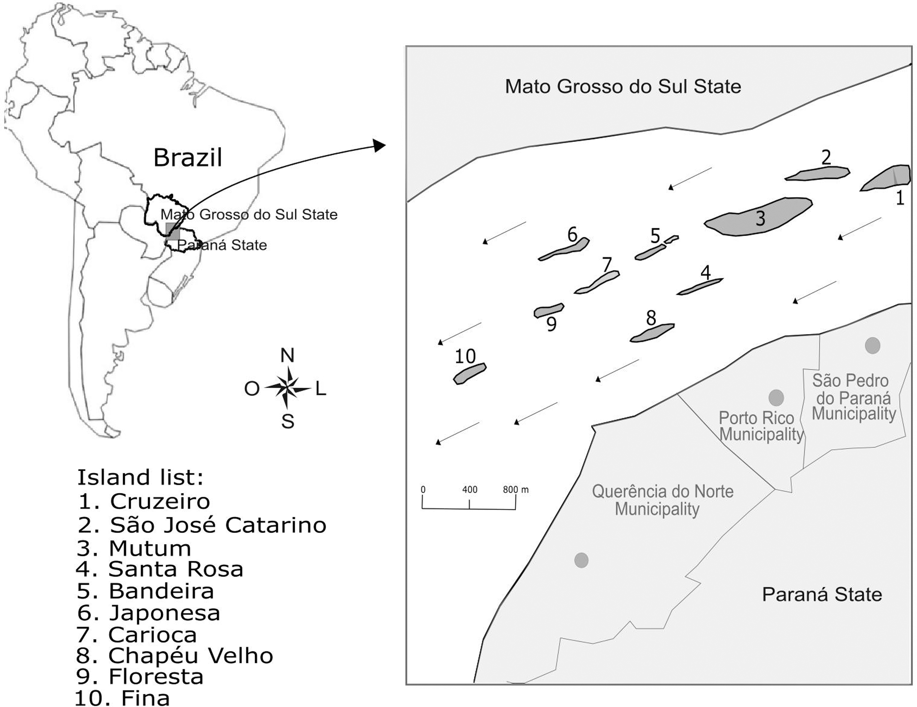
Islands of Paraná River where sandflies were collected, the municipalities of São Pedro de Paraná, Porto Rico and Querência do Norte, state of Paraná, southern Brazil.
The sandflies were collected with Falcão light traps at night, in five different ecotopes on the islands: interior of the forest, forest edge, uninhabited residence, inhabited residence, domestic-animal shelter, and abandoned domestic-animal shelter. The collections were performed from November 2012 to November 2014. On Mutum Island, the collections totaled 156 h per trap. On most of the other islands, the collections totaled 48 h in each ecotope. The total trap-hours differed on three islands: Carioca Island, a total of 5 h per trap; on Fina Island, 12 h per trap; and on Bandeira Island, 29 h per trap. Samples of the insects collected on each island were randomly chosen for multiplex PCR.
The insects were processed according to Oliveira et al. (2011) and identified at the Leishmaniases Laboratory in the Department of Clinical Analyses and Biomedicine of the Universidade Estadual de Maringá (Paraná State, Brazil). The collected sandflies were killed with chloroform and conserved in tubes containing isopropanol 80% for identification and subsequent DNA extraction (Santos et al. 2016). The nomenclature follows Galati (2003), and abbreviations follow Marcondes (2007).
DNA was extracted according to Loxdale and Lushai (1998), with modifications. The isopropanol-preserved insects were macerated with a plastic pestle in a microtube containing 200 μL of a solution of 5% Chelex resin. For every 22 samples extracted, a positive control was used [male sandflies plus 105 promastigotes of Leishmania (Viannia) braziliensis] and a negative control (male sandflies). The DNA was stored at 4°C until use.
For the multiplex PCR, two pairs of primers were used for DNA amplification: A1 [5′-(G/C)(G/C)(C/G) CC (A/C) CTA T (A/T) T TAC ACC AAC CCC] and A2 (5′-GGG GAG GGG CGT TCT GCG AA), which amplify a fragment of ∼120 bp of the conserved region of DNA from the minicircle of the kinetoplast (kDNA) of genus Leishmania (Romero et al. 2001); and 5Llcac (5′-GTG GCC GAA CAT AAT GTT AG-3′) and 3Llcac (5′-CCA CGA ACA AGT TCA ACA TC-3′), which amplify a fragment of 220 bp from the IVS6 cacophony gene region of insects of genus Lutzomyia (Lins et al. 2002). The PCR mixture (final volume 25 μL) was composed of 0.5 μM of each primer (Invitrogen), 0.24 mM dNTP (Invitrogen), 1 IU Taq DNA Polymerase (Invitrogen), 1.5 mM MgCl2, 1 × enzyme buffer, and 2 μL DNA template. The amplification was carried out in a G96G & G96GEN cycler (Biosystems) at 95°C for 5 min for initial denaturation, followed by 35 cycles, each divided into three stages: denaturation (30 s at 95°C), annealing (30 s at 55°C), and polymerization (30 s at 72°C). Next, the extension was continued for a further 10 min at 72°C, and the tubes were kept at 4°C until analysis. Positive controls [L. (V.) braziliensis, Leishmania (Leishmania) amazonensis, and Leishmania (Leishmania) infantum DNA] and a negative control (reaction mixture plus water) were added for each amplification set/round.
The amplification products were submitted to electrophoresis in 3% agarose gel, stained with 0.1 μg/mL ethidium bromide, at 10–15 V/cm. The presence of bands was observed in a transilluminator (Lo MacroVue™ UV-20; Hoefer).
For the multiplex PCR standardization, 0.20 and 0.24 mM of dNTP used in the reaction mixture was tested. The annealing temperatures tested were 50°C, 51°C, 52°C, 53°C, 54°C, and 55°C. The number of cycles tested were 25, 26, 30, and 35.
Samples of DNA from L. (V.) braziliensis (MHOM/BR/1987/M11272), L. (L.) amazonensis (IFLA/BR/1967/PH8 Isolate IOCL 0575), and L. (L.) infantum (MHOM/BR/2002/LPC-RPV isolate IOCL 2906) were used as controls.
To determine the limit of detection of the multiplex PCR technique for Leishmania DNA, different concentrations of Leishmania DNA were added to 200 pg of male sandfly DNA and analyzed. The concentrations of L. (V.) braziliensis and L. (L.) infantum were 1 pg, 100, 10, 1, 0.1 fg, and of L. (L.) amazonensis were 100, 10, 1, 0.1, 0.01 fg. The multiplex PCR technique was already tested for detecting DNA from Trypanosoma cruzi (1256 DTU II).
Results and Discussion
Initially, the multiplex PCR was standardized. The best results were obtained using 0.24 mM dNTP, an annealing temperature of 55°C for 30 s, and 35 cycles. In these conditions, a band of 220 bp was obtained for the IVS6 cacophony gene region, and a band of ∼120 bp was obtained for L. (V.) braziliensis, L. (L.) amazonensis, and L. (L.) infantum (Fig. 2). To determine the Leishmania species detected by multiplex PCR, another PCR specific to Leishmania species was necessary.
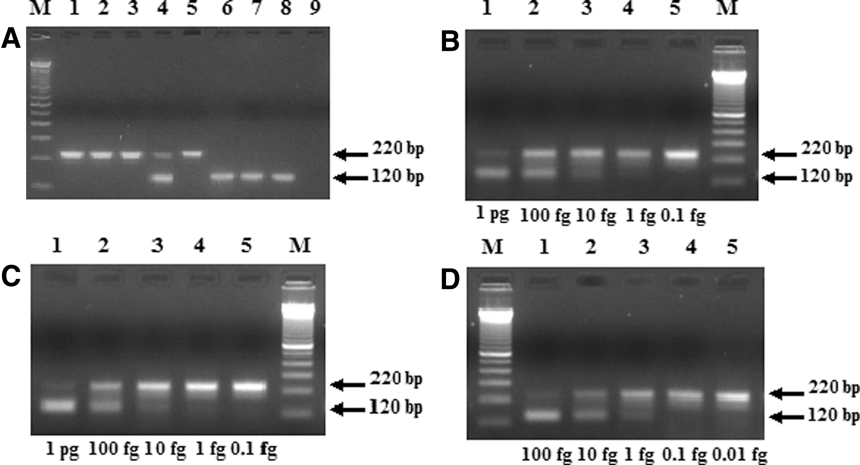
Multiplex polymerase chain reaction (PCR) in 3% agarose gel showing fragments of 120 and 220 bp. The fragment of 120 bp from the kDNA mini circle region of genus Leishmania were amplified with A1 and A2 primers. The fragment of 220 bp from the IVS6 cacophony gene region of the insects of genus Lutzomyia were amplified with 5Llcac and 3Llcac primers. (
A total of 76,145 sandflies were collected on the islands. An amount of 3870 samples of female sandflies were randomly separated and submitted to multiplex PCR. All were Nyssomyia neivai (Pinto). A 220 bp fragment from the IVS6 cacophony gene region of the sandflies was detected in the 387 pools of sandflies tested, each pool with 10 females, demonstrating the absence of Taq DNA polymerase inhibitors. No ∼120 bp fragment of Leishmania DNA was found (Fig. 2A). Therefore, no natural infection was detected in the sandfly samples analyzed.
The limit of detection of the multiplex PCR using A1–A2 and 5Llcac/3Llcac primers was 10 fg of L. (V.) braziliensis DNA (Fig. 2B) and L. (L.) infantum DNA (Fig. 2C) and 1 fg of L. (L.) amazonensis DNA (Fig. 2D). The DNA of T. cruzi was not detected.
No natural infection by Leishmania sp. was observed in the Ny. neivai specimens analyzed. However, in a previous study conducted on Paraná River islands, Santos et al. (2016) detected Leishmania in Ny. neivai, and three human cases of TL (two of these in permanent residents of the island) and eight canine cases of TL were reported from Mutum Island in 2008 (E.C.C and N.A. Membrive, pers. commun.). In addition, Ny. neivai naturally infected by Leishmania were also found in Paraná state by Oliveira et al. (2011) and Neitzke-Abreu et al. (2014). Marcondes et al. (2009), Pita-Pereira et al. (2009), and Córdoba-Lanús et al. (2006) found natural infections in this species in the states of Santa Catarina and Rio Grande do Sul and in Argentina, respectively.
Ny. neivai, Nyssomyia whitmani (Antunes and Coutinho), (França), and Pintomyia fischeri (Pinto) are frequently reported in TL endemic areas in Brazil (Teodoro et al. 2006, Reinhold-Castro et al. 2013). Nevertheless, sandflies of only one species, Ny. neivai, were collected. The predominance of this species has been reported previously. Santos et al. (2016) found 99.9% predominance of Ny. neivai, two specimens of Ny. whitmani, and one of Psathyromyia shannoni (Dyar) on 4/10 islands studied here. Gasparotto et al. (unpublished data) found a similar predominance of Ny. neivai on Mutum Island. Studies in Doutor Camargo municipality, Paraná state, have shown that this species is well adapted to modified environments and forest edges (Reinhold-Castro et al. 2013).
Ny. whitmani and Ny. neivai are vectors of L. braziliensis (Córdoba-Lanús et al. 2006, Marcondes et al. 2009, Pita-Pereira et al. 2009, Oliveira et al. 2011, Neitzke-Abreu et al. 2014). However, in Minas Gerais, Saraiva et al. (2010) found that these sandfly species were infected with Leishmania infantum, the causative agent of VL, a potentially fatal disease. Santos et al. (2012) reported the first find of Lutzomyia longipalpis, the main vector of L. infantum in Foz do Iguaçu, Paraná state. It is worth remembering the increase in the number of VL cases in the states of Mato Grosso do Sul and São Paulo, which border Paraná, especially in the last 20 years (Almeida et al. 2013, Ministério da Saúde Brasil 2015). In addition, Paraná state is considered endemic for L. braziliensis caused TL in humans. However, in Cambé municipality in the same state, Hoffmann et al. (2012) reported a dog with leishmaniasis caused by Leishmania amazonensis, and Silveira et al. 1990) reported a case of TL caused by L. amazonensis in a patient from Maringá who frequently fished in the Ribeirão Pinguin River, Floresta municipality.
The above findings indicate the importance of using PCR testing for the genus Leishmania in sandflies. If the parasite is detected, the particular species of Leishmania can be determined using specific primers. It is important to determine the geographical distribution of these protozoa for epidemiological surveillance of the disease, since human-caused environmental changes can lead to changes in the sandfly fauna, with a consequent redistribution of Leishmania species in different areas.
Therefore, the multiplex PCR was standardized, enabling determination of the infection rate of sandflies by members of the genus Leishmania in a single analysis, with certainty that the material analyzed is suitable for PCR. Moreover, the multiplex PCR is particularly useful when a large number of sandflies has to be analyzed, as in this research. Although no natural infection of Ny. neivai by Leishmania was found in this study, the interaction of sandflies with Leishmania and its natural reservoirs continues in these Paraná River islands, despite the low diversity of the sandfly fauna. Some of these islands have permanent residents and are frequented by tourists who also require attention from the health surveillance authorities.
Footnotes
Acknowledgments
The authors offer thanks to José Luiz Filho, Valmir Ortiz da Silva and José do Porto dos Santos from the Núcleo de Entomologia de Porto Rico for their assistance in collecting sandflies, and to Professor Doctor Mônica Lúcia Gomes from Laboratório de Doença de Chagas (Universidade Estadual de Maringá) for the donation of the T. cruzi epimastigote DNA. This work was supported by the Brazilian agency “Coordenação de Aperfeiçoamento de Pessoal de Nível Superior (CAPES).”
Author Disclosure Statement
No competing financial interests exist.