
Abstract
Cases of wart-like lesions in humans and dromedary camels occurred in eastern Sudan in 2015 were described. Involvement of papillomavirus (PV) in causing these cases was affirmed by PCR and immunoperoxidase test. Mostly, the lesions were observed on the skin of the chest and forearms in addition to lips and mandible. Sequence analysis revealed Camelus dromedarius PV types 1 and 2 genotypes as the causative genotypes. We also observed cases of wart-like lesions on hands and legs of two herders attending the infected camel herd. Partial genome sequencing revealed human PV type 2 in one of the two human samples providing no indications for interspecies transmission of camel PVs, yet provides, for the first time evidence of active circulation of this virus in eastern Sudan.
Introduction
W
The first report of cutaneous papillomatosis associated with PV in dromedaries was published in 1990 (Munz et al. 1990) and showed that dromedary camels in central Somalia 6 months to 2 years of age were primarily affected. Later, cases of papillomatosis in young dromedary camels have also been reported from Kenya (Dioli and Stimmelmayr 1992), United Arab Emirates (Wernery and Kaaden 1995), Sudan (Khalafalla et al. 1998, Ure et al. 2011), and Saudi Arabia (Barakat et al. 2013, Khalafalla et al. 2017). Additionally, PVs were found associated with a 2-kg wart-like growth on the right fetlock joint of a dromedary camel in India (Sadana et al. 1980) and a corneal papilloma mass in the left eye of a 15-year-old dromedary male with a history of chronic severe keratoconjunctivitis (Kilic et al. 2010). Papillomatosis has also been shown in South American camelids (llamas and alpacas; Schulman et al. 2003). The majorities of the described camel papillomatosis cases are usually seen in young animals and occur in the late rainy season, coinciding with outbreaks of camel contagious ecthyma and camel pox (Munz et al. 1990, Khalafalla et al. 1998).
Currently, the genomes of two Camelus dromedarius PV types (type 1, CdPV1, and type 2, CdPV2) have been fully characterized and both are genetically grouped within the genus Deltapapillomavirus (Ure et al. 2011). The two genotypes were isolated from a cauliflower-like nodule and a round oval raised nodule, respectively, observed in 3- and 7-month-old dromedary camels in Sudan. In a recent publication, we investigated outbreaks of camel papillomatosis in Saudi Arabia. Sequencing of eight DNA samples revealed the presence of both CdPV1 and CdPV2, previously identified in infected dromedaries in Sudan (Khalafalla et al. 2017).
During field visits conducted by our team in eastern Sudan, several cases of skin lesions suggestive of warts were observed in camel and also in camel herders who believe it as a zoonotic infection transmitted from the camels. The anxiety from camel-transmitted zoonotic diseases are right now getting extensive consideration in the wake of demonstrating the role played by camels in transmitting the Middle East respiratory syndrome Coronavirus infections and the recent zoonotic cases caused by camelpox virus (Bera et al. 2011, Khalafalla and Abdelazim 2017).
In this study, we collected skin samples from camels and their herders in an attempt to diagnose these cases, molecularly characterize the causative virus, and explore conceivable zoonotic transmission.
Materials and Methods
This study was approved by the University of Khartoum Research Board and written or oral informed consents were obtained from all individuals to collect skin samples.
Cases and sampling
Cases of skin lesions suggestive of warts in camels and their human herders were observed during field visits to eastern Sudan in July–October 2015. A migratory herd of dromedary camels consisting of 87 animals of different age groups was found affected with papillomatosis. Affected animals were restrained and the skin over the entire body was inspected for wart-like lesions. Lesions were described, photographed, and whole biopsies from four affected animals were excised surgically using a local anesthetic. Wart-like lesions were also observed in two herders accompanying the affected herd. Lesions were described and photographed and biopsies were taken from the two subjects with warts under local anesthesia by the staff of Showak Public Hospital. Each sample was divided into two parts, one part was stored at −40°C for DNA extraction and the other part fixed in 10% neutral buffered formalin.
Immunohistochemistry
Wart sample was trimmed, serially dehydrated, paraffin embedded, and sectioned at 5 mm. The major capsid protein (L1) of HPV was detected in paraffin-embedded wart sections, using peroxidase–antiperoxidase (PAP) method. The procedure includes deparaffinization of sections, rehydration, and endogenous peroxidase inactivation by incubation in 0.5% H2O2 for 30 min. For antigen unmasking, slides were incubated in Citrate buffer at 96°C for 25 min. Mouse monoclonal antibodies [BPV-1/1H8+CAMVIR] against HPV were used as primary antibodies (Abcam plc; 330 Cambridge Science Park, Cambridge, CB4 0FLm, United Kingdom). Slides were incubated with secondary antibodies for 30 min, and then horseradish–PAP complex from the mouse was applied. Slides were stained with diaminobenzidine and finally counterstained and examined under light microscope.
Rolling circle amplification
Multiply primed rolling-circle amplification (RCA) was performed with the Illustra TempliPhi Amplification Kit (GE Healthcare Life Sciences) following a protocol that was optimized for the amplification of papillomaviral complete genomic DNA (Rector et al., 2004b, Stevens et al., 2010). To investigate whether papillomaviral DNA was amplified, 2 μL of the RCA product was digested with a restriction enzyme panel consisting of BamHI, EcoRI, SalI, HindIII, and HincII. The digestion products were run on a 0.8% agarose gel to check for the presence of a DNA band consistent with full-length PV DNA (∼8 kb), or multiple bands with sizes adding up to this length.
Degenerate primer PCR and sequencing
Degenerate PV-specific primers were used to screen RCA products that possibly contained amplified PV DNA. The following primer pairs were used: FAP59/FAP64 (Forslund et al., 1999), AR-L1F1/AR-L1R3 (Rector et al. 2004a), and AR-E1F2/AR-E1R3 (Rector et al. 2004b). Previously unpublished primer pairs AR-L1F11/AR-L1R10 and AR-E1F14/AR-E1R12, developed in conserved L1 and E1 regions of ungulate PVs, were also tested (Table 1). Amplification was performed with the Qiagen OneStep RT-PCR Kit following the manufacturer's instructions, using a concentration of 2.4 μM of forward and reverse primer, and 1 μL of RCA product as sample in a final reaction volume of 25 μL. PCR conditions comprised of an initial PCR activation step of 15 min at 95°C, followed by 40 cycles of 30 s denaturation at 94°C, 30 s annealing at 55°C and 1 min elongation at 72°C. After a final elongation step of 10 min at 72°C, PCR products were cooled to 4°C. PCR products were checked by PAGE, and when specific amplification products were detected, these were sequenced with the same degenerate primers that were used for PCR, on an ABI Prism 3100 Genetic Analyzer (Applied Biosystems, Life Technologies). Sequences were analyzed by similarity searches using NCBI BLAST.
Results
The clinical disease
A number of five young camels (<3 years of age) of both sexes and one adult female camel (7 years of age) showed wart-like lesions on different parts of the body. The wart-like lesion was mostly cauliflower-like, measuring from 0.3 cm up to 4.5 cm, when more than one wart coalesces together and were found on the skin of the chest and forearm (n = 2 animals; Fig. 1A), the forearm (n = 3 animals; Fig. 1B), and on the lips and mandible (2 animals; Fig. 1C). The nodular type of lesion was observed only in one of the affected camels. The number of wart-like lesion per animal ranged from 3 to 38 lesions.
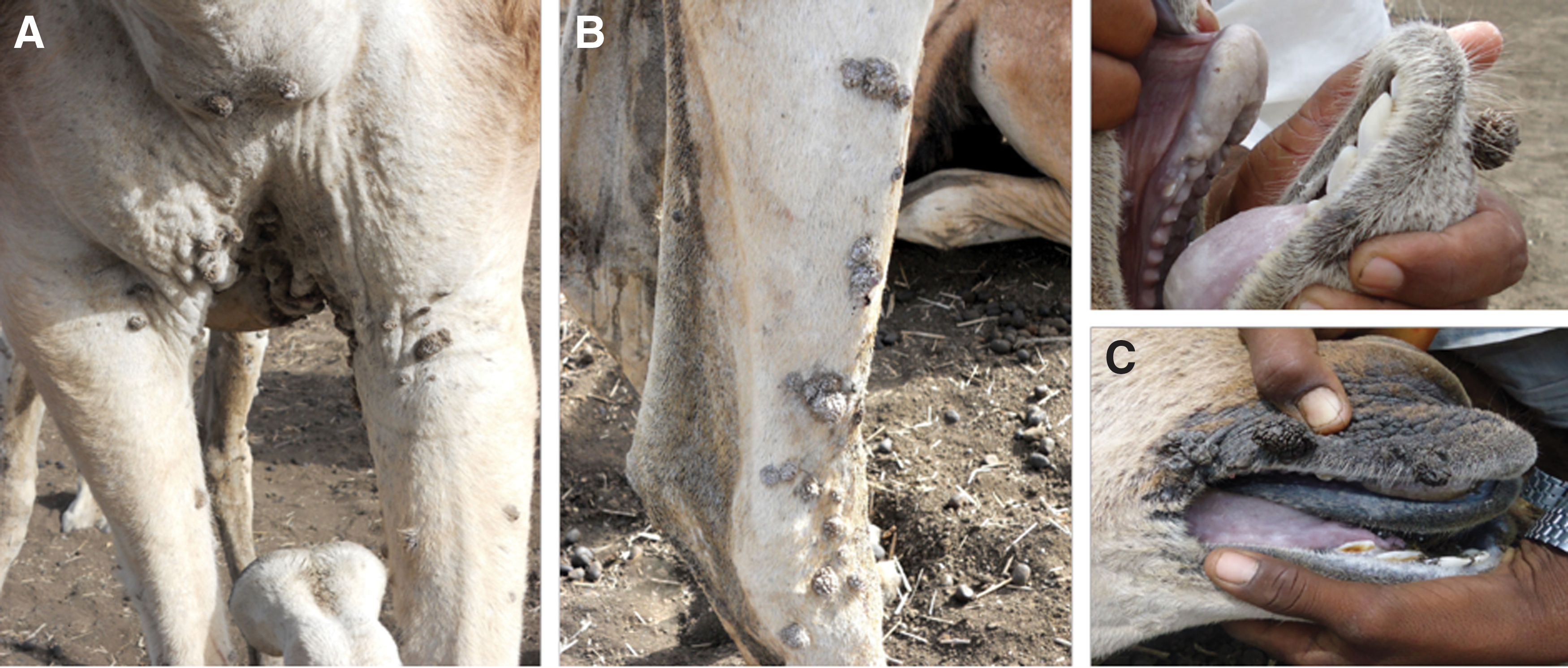
Papillomatosis in dromedary camels in eastern Sudan. Cauliflower-like single and coalesced nodular papillomas on the chest
Two herders who are the shepherds of the same affected camel herd showed wart-like skin lesions. Both men are accompanying the affected herd for 5 years. Case 1: A 45-year-old male who showed a nodular cauliflower-like growth on the index finger (middle phalanx) of the right hand of the size 0.4 × 0.5 cm in addition to two smaller nodules one attached to the cauliflower-like growth (Fig. 2A).

Wart-like lesions on index finger
Case 2: A 59-year-old male who showed wart-like growth on the sole of the foot of size 0.6 × 0.6 cm (Fig. 2B). The growth was sessile with verrucous projections on the surface and was firm in consistency and present for the last 2 months with gradual increase in size.
PCR diagnosis
All the six samples collected from the affected camels and the two camel herders were subjected to PCR using the pan PV primers (FAP 59 and FAP64). PCR amplification was positive in five camel DNA samples and the two samples collected from herders. Samples positive include animals M1–M5 and human X1 and X2 (Table 2).
Immunohistochemistry
Immunohistochemistry examination showed severe localization of PV antigens in the nuclei of stratum granulosum after staining with antibodies (Fig. 3).
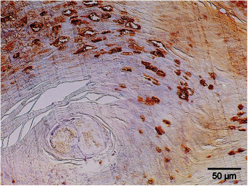
Immunohistochemical reaction of BPV-1 antibodies showing numerous positive cells in the stratum spinosum. A biopsy collected from an adult man (PAP method). PAP, peroxidase–antiperoxidase. Color images available online at
RCA, sequencing, and genotyping
Possible PV genomic amplification by RCA was observed for samples M2 (camel, chest skin lesion), M3 (camel, forearm lesion), and X1 (human). These RCA products were submitted to degenerate primer PCR, and PV-specific sequences could be amplified for all these samples. An overview of the amplicons and sequencing results that were obtained for the different samples, with indication of the most similar established PV type (as identified by BLAST search) and the percentage nucleotide identity of the amplicon sequence to this most similar type, is provided in Table 2.
Discussion
In the present study, we described cases of wart-like lesions in humans and dromedary camels. Involvement of PV in causing these cases was confirmed by PCR and immunoperoxidase test. We observed significant differences in lesion characteristics of the PV infection in terms of location and size, which is not the same as the past depiction of the lesions. In all previous reports on camel PV infection, the wart-like lesions (0.2 to 2.5 cm) were found prevalently on the head, particularly the lips, eyelids, nares, and mandibles (Munz et al. 1990, Khalafalla et al. 1998, Ure et al. 2011). A different clinical picture has been accounted for in the present study that is an occurrence of wart lesions mostly on the chest and forearm areas and the lesion size in some cases was approximately 5 cm due to coalition of more than one wart. One would assume that a virus genotype different from the previously molecularly characterized genotypes with a predilection to skin of the chest and forearm is responsible. However, we found that CdPV1 and CdPV2 genotypes are the causative viruses. These genotypes were detected in camels that exhibited lesions on the head (lips, eyelids, nares, and mandibles), but not the chest or forearms (Ure et al. 2011, Khalafalla et al. 2017).
A new PV type is defined as a cloned full-length papilloma viral genome, whose L1 nucleotide sequence is at least 10% dissimilar from that of any other PV type (de Villiers et al. 2004, Bernard et al. 2010). Although no complete L1 open reading frames (ORFs) were sequenced in this study, the partial L1 sequences that were retrieved from the camel samples showed at least 96% identity to CdPV1 or CdPV2, indicating the presence of subtypes or variants thereof, rather than new PV types. CdPV1 or CdPV2 were previously detected in wart-like lesions collected from dromedary camels in Sudan (Ure et al., 2011) and Saudi Arabia (Khalafalla et al., 2017), therefore, our findings further confirm the circulation of these two genotypes, yet points to the circulation of this neglected cutaneous papilloma virus infection in people in eastern Sudan.
During our regular visits to eastern Sudan, particularly to Showak area, where University of Khartoum has a field station belonging to the Camel Research Center, we noticed several cases of wart-like lesions on hands and legs of some camel herders. To determine whether these lesions are acquired from camels or not we collected skin samples from two camel herders and subjected them to DNA extraction, PCR, and partial genome sequencing. In one of the human skin samples, partial sequences of HPV type 2 were identified, and no camel PV sequences. These results therefore provide no indications for interspecies transmission of camel PVs.
HPV is used to describe more than 200 different types of related viruses associated with occurrence of warts and many cancers in man. Common warts are usually found on the hands and feet, but can also occur in other areas, such as the elbows or knees. Common warts have a characteristic cauliflower-like surface and are typically slightly raised above the surrounding skin. HPV type 2 can cause common warts and previously found associated with cutaneous warts in man (Keefe et al. 1994).
Little is known about the prevalence of HPV in Sudan and sub-Saharan Africa too. Additional HPV epidemiological research in Sudan, particularly the role played by HPV 2 in cutaneous warts, is needed to develop more effective prevention strategies.
Footnotes
Acknowledgments
The authors thank Abdo Bushara and the staff of Showak Public Hospital for the assistance in sample collection.
Author Disclosure Statement
No competing financial interests exist.