
Abstract
In 2017, we isolated and identified West Nile virus (WNV) lineage 2 from two dead captive goshawks (Accipiter gentilis), for the first time in the Czech Republic. Goshawk might serve as an early indicator species for the ongoing WNV emergence in several European countries.
Introduction
C
In the Czech Republic, WNV lineage 3 (“Rabensburg”: Hubálek et al. 1988, Bakonyi et al. 2005) and lineage 2 (Rudolf et al. 2014, 2015) strains were already isolated from mosquitoes. WNV disease has not yet been established in birds of prey in this country, in contrast to neighboring Austria, Slovakia, and Hungary.
Materials and Methods
In this study, we examined for WNV two dead raptors obtained from falconers in Moravia:
Goshawk 1 (A. gentilis), a juvenile male (770 g), kept in Pozořice near Brno (49°13′ N, 16°47′ E), died there in early September 2017. It was born in a falconry breeding station at Zvolen (Slovakia) in May 2017, then transported to Trenčín (Slovakia) falconry station in late June 2017, and thereafter to Pozořice on August 22, 2017. The bird was fed with 1-day-old chickens, domestic quails, and domestic and feral pigeons. Severe neurological signs appeared about 5 days before the death, such as tremor, ataxia, torticollis, and pareses. Another goshawk died in this falconry at the same time; it revealed similar neurological signs, but we did not obtain the cadaver for virological examination.
Goshawk 2, an adult female (6 years, 1080 g) was kept in Buchlovice (49°05′ N, 17°20′ E), where she died on September 13, 2017. The bird was born in Komárov near Napajedla (49°09′ N, 17°34′ E) in April 2011 and fed with several-days-old chickens, domestic quails, and domestic and feral pigeons. In the years 2011–2016, it took part in autumn hunting trips in 15 localities, all of them in the Czech Republic. Neurological signs started 8 days before death with gradual loss of appetite and balance, reduced ability to perch and stand (lethargic lying on the ground) combined with ataxia, tremor of head, and convulsions of legs and wings.
For isolation attempts, 10% homogenates were prepared from the brain, heart, and lungs of the dead raptors in phosphate-buffered saline (PBS) pH 7.2, containing 0.4% bovine serum albumin and antibiotics, clarified by centrifugation, and inoculated intracerebrally (i.c.) into 2- to 4-day-old specific–pathogen-free mice (outbred strain ICR); bacterial sterility of the homogenates was checked in meat–peptone broth and thioglycollate broth at 37°C.
All experiments with laboratory mice were conducted in accordance with the Czech Animal Protection Act no. 246/1992, and the protocols were approved by the Institutional and Central Care and Use Committees at the Academy of Sciences of the Czech Republic in Prague and by the Veterinary Service in Brno. The facility is accredited by the Czech National Committee on Care and Use of Laboratory Animals (70084/2016-MZE-17214).
Viral isolates were identified using neutralization test on Vero cells: constant dilutions (1:5) of thermally inactivated normal and immune (against WNV-1 strain Eg-101) mouse sera were mixed with serial decimal dilutions of the viruses, and log neutralization indices were estimated. For determination of particular WNV lineage, we performed molecular analysis of obtained viral isolates. Genomic RNA was extracted from goshawk brain homogenates (10% suspensions) using the QIAamp Viral RNA Kit (Qiagen, Hilden, Germany) and tested by conventional reverse transcription–polymerase chain reaction (RT-PCR) for flaviviral RNA by using the protocol designed by Scaramozzino et al. (2001), using the RT-PCR Kit (Qiagen).
Subsequent sequencing and bioinformatic analysis were performed according to a previous study (Rudolf et al. 2014), based on primers proposed by Bakonyi et al. (2006).
Results
Goshawk 1: at autopsy, marked hemorrhages were recorded in the brain of this bird. No apparent gross lesions were observed in other organs (heart, lungs, liver, and spleen). All 11 newborn mice inoculated with bacterially sterile goshawk's brain homogenate died 5 days postinoculation (dpi), as well as did all nine mice inoculated with the pool of goshawk's heart and lung homogenates 5–7 dpi; average survival time (AST) of the mice was 6.0 days. RT-PCR analysis of the avian brain confirmed the presence of WNV lineage 2 RNA.
Goshawk 2: at autopsy, we observed hemorrhages in the brain, but no marked gross lesions in other organs. All 12 newborn mice inoculated with bacterially sterile brain homogenate died 5–7 dpi (AST 5.9 days). Heart and lung homogenates were not tested. RT-PCR of the avian brain confirmed the presence of WNV lineage 2 RNA.
Isolates obtained from the brain of the two goshawks passaged in newborn mice were identified as WNV using virus neutralization test on Vero cells with WNV-1 mouse antiserum; log neutralization indices were 4.5 in the goshawk 1 and 3.0 in the goshawk 2. Subsequent molecular analysis of nucleotide sequences in NSP5 fragment (745 bp) revealed 98.9% similarity of the two isolates (they differ in eight nucleotides), and their high similarity (at least 99%) to WNV-2 isolates from South Moravian (Czech) mosquitoes isolated in 2013 (Rudolf et al. 2014). Figure 1 shows phylogenetic relationships with other WNV lineage 2 strains. GenBank accession numbers for the RNA fragments are MH377401 and MH377402 for goshawk no.1 and no. 2, respectively.
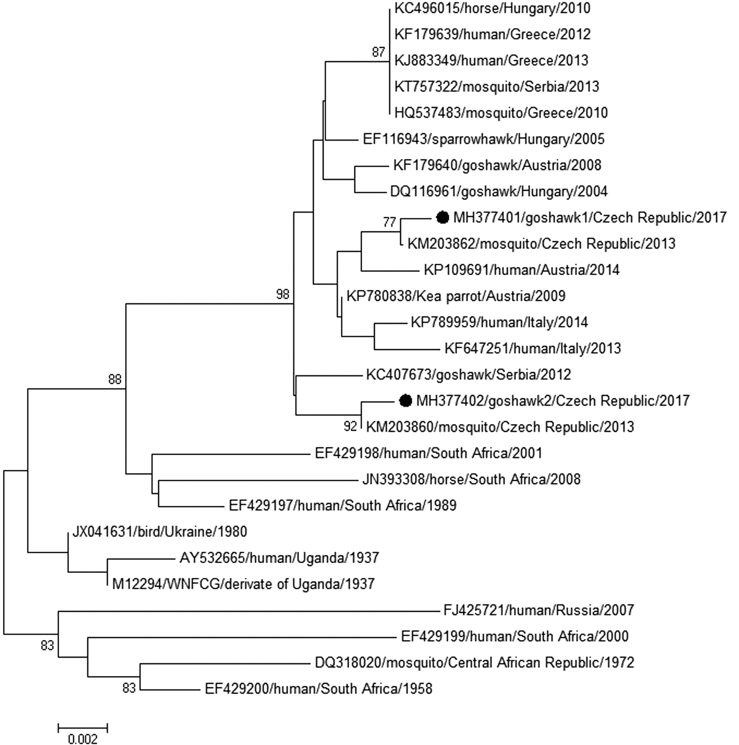
Relationships among nucleotide sequences of NSP5 fragment (745 bp) of two WNV strains isolated from goshawks in the Czech Republic (marked by black circles) with representative WNV lineage 2 strains by the neighbor-joining method (p-distance). GenBank accession numbers, isolation sources, countries of origins, and isolation years are indicated at the branches. Supporting (>70%) bootstrap values of 1000 replicates are displayed at the nodes. The horizontal bar show genetic distance. WNV, West Nile virus.
Discussion
The very first isolations of WNV from dead raptors were reported in North America: one Cooper's hawk (Accipiter cooperii) in Connecticut, 1999 (Anderson et al. 1999), and one red-tailed hawk (Buteo jamaicensis) in Westchester County, NY, in February 2000 (Garmendia et al. 2000). Wunschmann et al. (2004, 2005, 2014) later described pathological changes in a number of raptors naturally infected with WNV in USA, including red-tailed hawks, Cooper's hawks, and northern goshawks (A. gentilis). Joyner et al. (2006) tested 61 birds of prey admitted to the Wildlife Center of Virginia in 2003, and 40 birds were positive for WNV by RT-PCR; great horned owls (Bubo virginianus) and red-tailed hawks were most often affected.
Nemeth et al. (2006, 2007, 2009) studied natural and experimental WNV infection in several North American raptor species, including red-tailed hawks, American kestrels (Falco sparverius), and golden eagles (Aquila chrysaetos)–all these species were found to be susceptible to the disease (also even after per os inoculation). Saito et al. (2007) reported sick and dying raptors, mostly red-tailed hawks and great horned owls distributed over 12 US states: 71% of these birds were WNV positive. Pauli et al. (2007) recorded ocular (and other) lesions in red-tailed hawks and Cooper's hawks naturally infected with WNV. Ellis et al. (2007) detected WNV in a number of dead hawks in Georgia, USA: red-tailed hawks, sharp-shinned hawks (Accipiter striatus), and Cooper's hawks were most commonly affected.
WNV was the most frequent case of infectious disease for bald eagles (Haliaeetus leucocephalus) and peregrine falcons (Falco peregrinus) submitted to the Wildlife Center of Virginia (Harris and Sleeman 2007). Lund et al. (2017) reported a mass bird mortality event involving 15,000–20,000 eared grebes (Podiceps nigricollis) that occurred at the Great Salt Lake, Utah in 2013, which was followed by a mass mortality in bald eagles (more than 86 birds), and WNV was detected in carcasses of both the grebes and eagles. Interestingly, no activity of mosquitoes, the primary vectors of WNV, was detected in the area at that time.
In Europe, highly neurotropic lineage 2 WNV strains emerged lately in Hungary as described by Bakonyi et al. (2006, 2013) and dispersed quickly to neighboring Austria (Wodak et al. 2011), Moravia (Rudolf et al. 2014), and Slovakia (Csank et al. 2016). Several species of birds of prey were found to be susceptible to WNV in Europe. Jimenez-Clavero et al. (2008) reported isolation of WNV from two ill golden eagles (A. chrysaetos) in Spain. Hofle et al. (2008) detected RNA of WNV-1 by RT-PCR in 8 of 10 tested endemic Spanish imperial eagles (Aquila adalberti)—both in apparently clinically healthy birds and in animals that died with signs compatible with WNV disease.
Erdélyi et al. (2007) found a sparrow hawk and several goshawk fledglings succumbing to lineage 2 WNV encephalitis in southeast Hungary during the summers of 2004 and 2005. In 2008, fatal avian neuroinvasive WNV-2 disease was diagnosed all over Hungary, and included at least 25 goshawks and other birds of prey (Bakonyi et al. 2013).
Wodak et al. (2011) reported five goshawks and one gyrfalcon (Falco rusticolus) found dead in the eastern Austrian federal states (Lower Austria, Vienna and Styria): pathological and immunohistochemical findings suggested WNV infection, and WNV-2 was isolated, identified, and characterized using RT-PCR and sequencing. The Austrian WNV sequences exhibited nucleotide identities with the lineage 2 WNV sequences described in Hungary since 2004. Furthermore, three goshawks and one white-tailed eagle (Haliaetus albicilla) contained RNA of WNV-2 in Serbia, 2012 (Petrović et al. 2013). A WNV-2 strain was detected and isolated from a goshawk in Sardinia in August 2012 (Savini et al. 2013). Recently, RNA of WNV-2 was also detected in the brain of two goshawks that died after showing neurological signs in Nitra county, west Slovakia (Csank et al. 2016).
Summing up, raptors, in Europe mainly the goshawk, might serve as an example of early indicator species for the WNV emergence. Hungary, Austria, and Slovakia report similar cases in raptors preceding WNV disease in humans, which is reminiscent of the situation in North America with dying crows and the distribution of human WNV infection.
Conclusion
The results of this study indicate spread of WNV lineage 2 in Moravia (Czech Republic). While the WNV infection in goshawk 1 might have started in Slovakia, that of goshawk 2 is most probably autochthonous, of Moravian origin. Nonetheless, the death of goshawk 1 could also suggest local transmission, which is indicated by another goshawk (not examined by us), who died in the same falconry at the same time. The findings confirm previous data that WNV disease can be very serious in certain captive (and wild) raptors, including some endangered species worldwide, and at the same time indicate present WNV-2 activity in Czechland.
Footnotes
Acknowledgment
The study was supported by the grant project no. 16-20054S (Czech Science Foundation).
Authors Disclosure Statement
No competing financial interests exist.