
Abstract
A Flavivirus survey on 183 hunting dogs was conducted in Campania region, Southern Italy. The seroprevalence value of 40.43% (74/183, 95% confidence intervals [CIs] 33.37–47.49) detected in our study using a competitive enzyme-linked immunosorbent serologic assay (cELISA) proves a considerable level of Flavivirus exposition of these animals. Among the 74 cELISA-positive sera, seroneutralization (SN) test showed that 24 sera resulted positive for Usutu virus with an overall prevalence of 13.11% (24/183) (95% CI 8.27–17.95), but none of cELISA-positive samples resulted positive for West Nile virus. Data analysis showed a significant difference of cELISA seropositivity risk factors in case of presence of farm animals in contact with hunting dogs and for dogs living in a rural environment but not for gender, age, management, hunting season, and hunting abroad. A RT-PCR assay was performed to detect the Flavivirus RNA, but none of the blood samples tested positive. This study documents the first report regarding the circulation of Flavivirus in hunting dog in Southern Italy and suggests the dog as an interesting target to monitor Flavivirus circulation.
Introduction
T
WNV is a mosquito-borne Flavivirus. The circulation of this virus has been reported in most of the continents. In fact, WNV is enzootic in several countries (throughout Africa, Australia, Central and Western Asia, the Middle East), and is endemic in the Mediterranean area, with occasional outbreaks in Northern Europe and Siberia up to 55°N. The virus is carried by the migratory bird (Zeller et al. 2004, Rizzoli et al. 2007, Platonov et al. 2014) and is maintained in the environment in a mosquito–bird–mosquito transmission cycle (Hubálek and Halouzka 1999, Gould et al. 2001, Malkinson and Banet 2002), eventually involving accidental hosts such as humans and other mammals that express short-lived viremia and are therefore considered to be dead end-hosts, in which West Nile disease (WND) is usually asymptomatic but, sometimes, can cause severe disease. WNV circulation has been observed in several European Union states since the early 1950s, but large virus epidemic has only been documented starting from the mid-1990s (Bellini et al. 2014). Outbreaks of West Nile fever have been recorded in several European and Mediterranean countries during the last decade, including Romania, Russia, Serbia, Israel, and Morocco (Rizzoli et al. 2007, Llopis et al. 2015, Durand et al. 2016). In Italy, cases of WNV infection have been registered regularly since 2011, and have been distributed in several regions (Velati et al. 2017).
USUV is a mosquito-borne Flavivirus belonging to the Japanese Encephalitis Virus (JEV) serocomplex, and is thus related to JEV, Murray Valley encephalitis virus, and WNV (Kuno et al. 1998, Calisher and Gould 2003, Ashraf et al. 2015). USUV spread, since its first identification, has remained exclusively limited for decades to African continent where it was detected in different bird and mosquito species in several countries (Nikolay et al. 2011, Engel et al. 2016, Gaibani and Rossini 2017). Introductions of USUV in Europe from Africa were first described in Spain through migratory birds between 1950 and the 1960s, followed by other introductions between 1970 and 1980 in Italy, Austria and in Spain between 1984 and 2006 (Engel et al. 2016). Since then, USUV had been found in mosquitoes, birds, and bats in several European countries (Buckley et al. 2006, Bakonyi et al. 2007, 2014, Busquets et al. 2008, Jöst et al. 2011, Lupulovic et al. 2011, Steinmetz et al. 2011, Hubálek et al. 2014, Lecollinet et al. 2016, Rijks et al. 2016). In Italy, in the last 10 years, USUV has been identified in several regions of Northern Italy (Rizzoli et al. 2007, Lelli et al. 2008, Calzolari et al. 2015, Llopis et al. 2015). Moreover, some lethal USUV infections have been described in owls and blackbirds in the Lombardy region between 2006 and 2008 (Manarolla et al. 2010), and the zoonotic potential of the virus was evidenced in two cases of human encephalitis reported in Emilia Romagna region (Cavrini et al. 2009, Pecorari et al. 2009, Llopis et al. 2015). Furthermore, USUV has been found in healthy human blood in Austria, Italy, and Germany (Allering et al. 2012, Gaibani et al. 2012, Pierro et al. 2013, Weissenböck et al. 2013, Grottola et al. 2017).
The complex interactions of several factors involved make it hard to predict Flavivirus circulation and the consequent risk of epidemics. The viruses may remain undetected for long time, but during hot seasons and in places with appropriate ecological conditions, the virus circulation may increase with consequent increase of infected human and other mammals. As evidenced by these events, Italy plays a key role in Flavivirus infections at both the epidemiological and public health levels. The aim of this study was to evaluate Flavivirus circulation in a region where it has not been detected so far and to analyze the importance of hunting dogs as sentinels of Flavivirus spread in Campania region, Southern Italy. Serological and molecular assays were used to assess the presence of specific antibodies or RNAs in the blood samples.
Materials and Methods
Study area and sampling site
Campania region is located on the south of Italy, and stretches along the Tyrrhenian Sea and extends over an area of 13,595 km2 (41°00′00″N–14°30′00″E), with a mild Mediterranean climate on the seacoast and more continental in the mountain areas of the hinterland. The study was performed on dogs born and breed in the study area. The sample size was calculated using the formula suggested by Thrusfield (1995) for a theoretically “infinite” population inserting the following information: expected prevalence of Flavivirus (12.0%) (Durand et al. 2016), confidence interval (CI) (95%), and desired absolute precision (5%).
For this study, hunting associations of Campania region were contacted to assist in collecting biological samples from hunting dogs. Mainly hunters from Salerno province participated in the study, allowing us to collect samples from a total of 183 hunting dogs.
Blood samples were collected from autumn 2011 to winter 2012 from 183 hunting dogs in 47 municipalities in the Campania region with local veterinarian help, and informed consent of owners was required for inclusion. The complete history of the animals was obtained through a questionnaire The dogs were classified by age and divided into four groups: <1, 2–3, 3–6, and >6 years, 83 were females and 100 were males.
Clinical examination
Each dog underwent a complete clinical examination to reveal clinical signs related to neurological disorders.
Sample preparation and serological test
Blood samples were taken from the radial vein of each dog into sterile vacuum tubes (Vacutainer; Becton Dickinson) with and without anticoagulant; the serum samples were obtained by centrifugation at 1100 g for 10–15 min and stored at −20°C until analysis. Serological screening of serum sample was performed using a commercial competitive enzyme-linked immunoassay kit (ID Screen® West Nile Competition Multi-species, ID VET, France). This is a competitive enzyme-linked immunosorbent serologic assay (ELISA) kit for the detection of anti-pr-E antibodies in multiple species. The monoclonal antibody used in the kit cross-reacts with other JEVs and the tick-borne encephalitis virus.
The tests were conducted and interpreted according to the manufacturer's instructions. The cutoff value for categorizing a serum as positive was %OD sample/negative control <40% as recommended by the manufacturer. Although this assay is used to identify anti-WNV antibodies, cross-reactions with other Flaviviruses have been described, therefore a positive result has been interpreted as demonstration of the presence of anti-Flavivirus antibodies (WNV or other Flaviviruses).
To identify the circulating Flavivirus, WNV and USUV seroneutralization (SN) assays were performed on cELISA-positive samples. Neutralizing antibody titers against WNV and USUV were performed by SN assay on Vero cell. In brief, 25 μL each of several dilutions of the tested sample (from 1/5 to 1/640) was added to each test well of a sterile flat-bottomed microtiter plate and mixed with an equal volume of 100 TCID50 of WNV (Eg-101 strain) or USUV (Vienna 2001‐blackbird strain 939/01). Plates were incubated at 37°C in 5% CO2 in a humidified incubator. After 1 h of incubation, ∼104 Vero cells were added per well at a volume of 50 μL of medium containing antibiotics. Following incubation for 3–5 days, wells were scored for the degree of cytopathic effect (CPE) observed. A sample is considered positive when it shows >90% of CPE neutralization at the lowest dilution (1:10). The test was conducted at the Istituto Zooprofalitico Sperimentale dell'Abruzzo e del Molise “G. Caporale” (IZSAM), OIE reference laboratory for WND, under biosafety level 3 conditions as described (OIE 2012). The SN results were interpreted as suggested by Yeh et al. (2012): the neutralization titer for a specific virus must be at least fourfold greater than that for other Flaviviruses as a criterion for etiologic diagnosis.
Molecular detection of WNV and USUV
A RT-PCR was performed to test for the presence of USUV RNA using pan-Flavivirus primers according to Johnson et al. (2010).
Nucleic acids were purified from the blood samples of serological positive and negative dogs using Quiazol Lysis Reagent (Quiagen, Venlo, The Netherlands) following the manufacturer's instructions. The RT-PCR degenerate primers Flavi-For (sense) 5′-GCMATHTGGTWCATGTGG-3′ and Flavi-Rev (antisense) 5′-GTRTCCCAKCCDGCNGTRTC-3′ were used (Johnson et al. 2010); the amplicon from this primer pair is located toward the end of NS5 coding sequence and produces an amplicon of the expected size of 203 bp.
First-strand cDNA synthesis was performed using Superscript II reverse transcriptase (Life Technologies), and the mixture was incubated at 42°C for 50 min, followed by heating at 95°C for 5 min. Amplifications were performed in an T100 Thermal Cycler (Bio-rad) using 2.5 U Takara LA Taq (Takara BIO, Inc., Japan) with the amplification cycles consisting of an initial denaturation at 94°C for 3 min followed by 30 cycles of 94°C for 45 s, 50°C for 30 s, and 72°C for 90 s (Johnson et al. 2010). The amplification products were analyzed by 1.5% agarose gel electrophoresis in TBE (89 mM Tris-borate, 2 mM EDTA, pH 8.2) buffer using ChemiDoc gel scanner (Bio-Rad Laboratories).
Statistical analyses
Seroprevalence rates, related to the several classification variables (gender, age, management, contact with farm animals, environment, and hunting out the state), were computed, as well as their 95% CIs. To assess the effect of each risk factor, a two-tailed chi-squared test was used. The odds ratio (OR) and the relative 95% CI were calculated for the significant variable. Significance was set at p < 0.05. The statistical analyses were performed using MedCalc Statistical Software version 16.4.3 (MedCalc Software, Ostend, Belgium;
Results
One hundred eighty-three hunting dogs were included in this study, and a clinical examination was performed on all animals. Clinical examination of the seropositive dogs failed to identify clinical abnormalities related to neurological disorders.
Serological screening results demonstrated an overall cELISA prevalence rate of 40.43% (74/183, 95% CI 33.37–47.49). Figure 1 shows the hunting areas where Flavivirus-positive samples were found. Among the 74 cELISA-positive sera, USUV neutralizing antibodies were detected in 24 sera (32.43%, 95% CI 21.93–42.93), with an overall seroprevalence of 13.11% (24/183) (95% CI 8.27–17.95), and the distribution of SN antibody titers was 1:5 (n = 12), 1:10 (n = 7), 1:20 (n = 3), and 1:40 (n = 2); none of the cELISA-positive samples were positive for WNV.
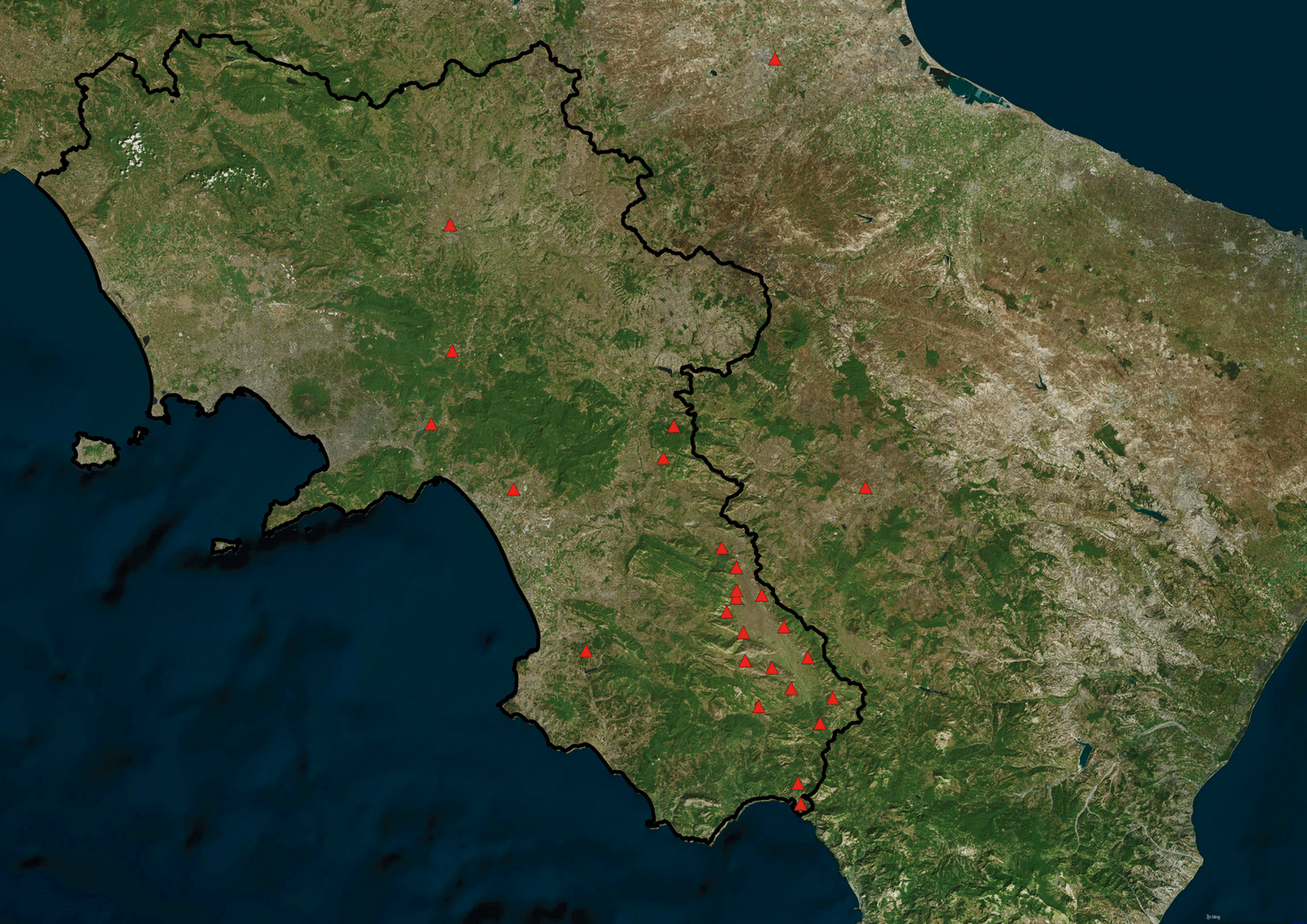
Map showing the distribution of Flavivirus positivity based on hunting areas. Color images available online
Table 1 shows the results of the seroprevalence analysis and p value in relation to the characteristics of the dog population hypothesized to be associated with the occurrence of Flavivirus. The analysis of risk factors evidenced that gender, age, management, hunting season, and hunting out the state showed no significant associations with seropositivity (p > 0.05); in fact, the OR values (OR <1.0) suggest that they may be epidemiologically not important for Flavivirus infection.
Values significantly different (p < 0.05) between groups are in bold and labelled with an asterisk.
CI, confidence interval.
Risk factor analysis revealed that OR, in dogs living in contact with farm animals, is 0.3764; this value indicates that odds of disease were 62.36% lower in dogs living in contact with farm animals compared with hunting dogs living alone. Similarly, dogs living in rural areas showed OR of 0.088, meaning that the odds of disease were 91.2% lower in dogs living in farming area compared with dogs living in urban environment (Table 2).
Values significantly different (p < 0.05) are in bold.
OR, odds ratio.
The 183 blood samples were further subjected to molecular detection of Flavivirus by RNA extraction, RT-PCR, followed by agarose gel electrophoresis. No sample resulted positive for Flavivirus by RT-PCR method.
Discussion
The Flaviviruses transmitted by mosquitoes represent a serious risk to public health worldwide, but in Central Europe the WNV and the USUV, both belonging to the group of JEVs (Flaviviridae), have become an emerging problem in the last 10 years (Rudolf et al. 2015).
This study documents the first report regarding the prevalence of Flavivirus in Southern Italy reporting USUV circulation in hunting dogs in Campania region. During our survey, 74 (40.43%, 95% CI 33.37–47.49) hunting dogs resulted positive for Flaviviruses by cELISA, and USUV neutralizing antibodies were detected in 24 samples with an overall prevalence of 13.11% (24/183) (95% CI 8.27–17.95). Detection of positive negative cELISA/VNT samples for USUV and WNV indicates the presence of non-neutralizing antibodies against these two viruses, this result could also indicate the presence of antibodies against other antigenically related Flaviviruses, although if only the USUV and WNV circulation has been detected in Italy (Rizzoli et al. 2007, Busani et al. 2011, Pauli et al. 2014, Calzolari et al. 2015, Llopis et al. 2015). USUV has been detected in various animal species and in different regions of Northern and Central Italy by virological and serological methods (Savini et al. 2011, Calzolari et al. 2015, Llopis et al. 2015); for example, antibodies against Flavivirus have been found in equine (21.5%; Savini et al. 2011), wild birds (1.34%; Llopis et al. 2015), chicken (10%; Rizzolie et al. 2007), cattle (2.8%), and dogs (27.7%; Busani et al. 2011). Interestingly, in Emilia Romagna (Northern Italy) in 2009, two cases of human encephalitis have been referred to USUV infection confirming the zoonotic potential of this virus (Pecorari et al. 2009).
The USUV prevalence rate observed in our study in hunting dogs is lower than that previously described by Busani et al. in 2011 in stray dogs in Veneto region (27.7%) but is higher than the prevalence previously described in horses in Friuli Venezia Giulia and in Tuscany region (2.9% and 5.45%, respectively) (Savini et al. 2011), similar to the prevalence rate observed, in the same species, in Liguria and Emilia Romagna regions (10.26% and 13.53%, respectively) and higher than that reported by Savini et al. (2011) in wild birds in Northern Italy (8.18%). However, it must be pointed out that comparing the prevalence of Flaviviruses among different studies is difficult because of the different diagnostic tests used, the epidemic/endemic context, the Flavivirus species detected, and the host species investigated (Jurado-Tarifa et al. 2016).
Our data showed that USUV-positive dogs are present (13.11%) in our region, with a seropositive rate in hunting dogs similar to the prevalence rate observed by Durand et al. (2016) in military dogs in Morocco (12.00%). We hypothesize that this could be related to the lifestyle of hunting and military dogs during the work activities, which have more opportunities for mosquito exposure than other dog categories (Busani et al., 2011).
In our study, molecular biology analyses performed on blood samples failed to detect Flavivirus RNA in the 183 hunting dogs tested. These results are similar to the results previously obtained by Gutierrez-Guzman et al. in wild boar, red foxes, and other mesomammals (2012); by Maggi et al. in humans (2015); and by Lelli et al. in horses (2008). In fact, humans and other mammals infected by most of the Flaviviruses circulating in Europe develop low-level and short-lived viremia (Bowen and Nemeth 2007, Colpitts et al. 2012). Thus, humans and other mammals are considered “dead-end” hosts, and usually they do not represent amplifying hosts, but their serological reaction allows their use as sentinels (Teehee et al. 2005, Wertheimer et al. 2010, Beck et al. 2013). Indeed, infections with WNV and USUV are often asymptomatic in ∼80% of infected people and mammals (Beck et al. 2013, Gaibani and Rossini 2017).
Therefore, the discrepancy between the results of virological (absence of RNA detection) and serological analyses is probably due to the nature of the tests used; in fact, serology is a measure of historical exposure, which may or may not be related to viremia at the time of sampling. However, serology remains an essential epidemiological tool to evaluate circulation and levels of exposure to a pathogen in a target population (Zottola et al. 2013).
In this study, risk factor analysis did not detect any statistically significant effect (p > 0.05) of age, management, hunting season, hunting abroad, and hunting areas on seropositivity, and this finding is similar to the results obtained by Durand et al. in 2016 in Morocco.
However, it must be pointed out that the interested hunting areas are near to a wetland area, where a significant number of waterfowls, including migratory bird species, are present and represent a possible risk of Flavivirus introduction.
However, it must be underlined that in a recent cross-sectional serosurvey performed in Iran in equine, the seropositivity risk of WNV increased with age (Ahmadnejad et al. 2011). Otherwise, analysis of risk factors evidenced the rural environment and the living with other animals in the farm as protective factors for seropositivity of hunting dogs to Flavivirus. This is probably due to the mosquito feeding preferences for animals other than dogs, probably easier to find in a rural environment (Rizzoli et al. 2015).
In conclusion, in this survey, we have shown the presence of Flavivirus antibodies in hunting dogs in the Campania region. To better understand the role of this species as a reservoir of these viruses, additional epidemiologic data are needed, as well as the molecular characterization of the circulating etiologic agents; finally, it would be interesting to continue monitoring several dog categories to obtain more information about the circulation of USUV and WNV in Italy in this species.
Footnotes
Acknowledgments
The authors gratefully acknowledge the technical contribution of G. Marzatico and of the hunting associations of Campania region.
Author Disclosure Statement
No competing interests exist.